AII Amacrine Cells on:
[Wikipedia]
[Google]
[Amazon]
 AII (A2) amacrine cells are a subtype of
 Morphology
AII amacrine cells are round or oval, and include
  The Retina
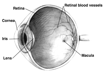
* The retina is a region of tissue at the back of the eye. It is light sensitive, which is why the mentioned light activated cells are located here. It is responsible for collecting light information through the eye, turning it to electric information, and transmitting it to the optic nerve.
The Optic Nerve
* The optic nerve is a bundle of
  *When photons of light reach the retina, they are absorbed by the chromophore (retinal). This causes a change of configuration in the retinal, which causes a signaling pathway that results in the closure of membrane sodium channels. This change triggers the cell membrane to become charged negatively through the process of hyperpolarization.
**As soon as a photon of light stimulates a rod, the AII amacrine cells are stimulated.
*The chromophore and opsin detach, reverting the chromophore to its original conformation.
**This step is the main difference between this
  * Now, the information must travel from the photoreceptors to the
 AII (A2) amacrine cells are a subtype of
 amacrine cells
In the anatomy of the eye, amacrine cells are interneurons in the retina. They are named , because of their short neuronal processes. Amacrine cells are inhibitory neurons which project their dendritic arbors onto the inner plexiform layer ...
. Amacrine cells are neurons that exist in the retina
The retina (; or retinas) is the innermost, photosensitivity, light-sensitive layer of tissue (biology), tissue of the eye of most vertebrates and some Mollusca, molluscs. The optics of the eye create a focus (optics), focused two-dimensional ...
of mammals
A mammal () is a vertebrate animal of the class Mammalia (). Mammals are characterised by the presence of milk-producing mammary glands for feeding their young, a broad neocortex region of the brain, fur or hair, and three middle e ...
to assist in interpreting photoreceptive signals. AII amacrine cells serve the critical role of transferring light signals from rod photoreceptors to the retinal ganglion cells
A retinal ganglion cell (RGC) is a type of neuron located near the inner surface (the ganglion cell layer) of the retina of the eye. It receives visual information from photoreceptors via two intermediate neuron types: bipolar cells and retina ...
(which contain the axons
An axon (from Greek ἄξων ''áxōn'', axis) or nerve fiber (or nerve fibre: see spelling differences) is a long, slender projection of a nerve cell, or neuron, in vertebrates, that typically conducts electrical impulses known as action pot ...
of the optic nerve
In neuroanatomy, the optic nerve, also known as the second cranial nerve, cranial nerve II, or simply CN II, is a paired cranial nerve that transmits visual system, visual information from the retina to the brain. In humans, the optic nerve i ...
).
The AII amacrine cells are unique because they work primarily with the vertical transmission of information, meaning they connect the bipolar and ganglion cells
Introduction
In neurophysiology, a ganglion cell is a cell found in a ganglion (a cluster of neurons in the peripheral nervous system). Depending on their location and function, ganglion cells can be categorized into several major groups:
* ...
. Other amacrine cells primarily assist with horizontal pathways, meaning they connect similar types of neurons
A neuron (American English), neurone (British English), or nerve cell, is an membrane potential#Cell excitability, excitable cell (biology), cell that fires electric signals called action potentials across a neural network (biology), neural net ...
. Amacrine II cells also work very similarly to rod photoreceptors in terms of threshold
Threshold may refer to:
Science Biology
* Threshold (reference value)
* Absolute threshold
* Absolute threshold of hearing
* Action potential
* Aerobic threshold
* Anaerobic threshold
* Dark adaptation threshold
* Epidemic threshold
* Flicke ...
(amount of stimulation needed to begin performing), saturation level (how densely they exist, and where), and spectral sensitivities (how sensitive the cell is to changes in stimulation levels). However, the Amacrine II cell works faster than the rod photoreceptors.
 Morphology
AII amacrine cells are round or oval, and include
 dendrites
A dendrite (from Greek δένδρον ''déndron'', "tree") or dendron is a branched cytoplasmic process that extends from a nerve cell that propagates the electrochemical stimulation received from other neural cells to the cell body, or soma ...
which connect together to create a systematic mosaic. They have two main forms, which differ in their dendritic trees (dendritic formations). The first form is made of one dendrite with multiple short, thin arms that end in circular appendages. The second form has multiple thin dendrites with attached spines, and extensive branching. Amacrine II cells are found most densely in the central retina, but are found in the surrounding retinal areas as well.
Development
The study of the development of amacrine cells is relatively recent. A recent study found that, in mice, amacrine cells develop during the Peri-Eye-Opening Period (during 7-28 days post birth). During this time, the cells are developing dendrites and dendritic spines, shifting resting membrane potentials (RMPs), developing synaptic activity, and developing Potassium
Potassium is a chemical element; it has Symbol (chemistry), symbol K (from Neo-Latin ) and atomic number19. It is a silvery white metal that is soft enough to easily cut with a knife. Potassium metal reacts rapidly with atmospheric oxygen to ...
currents (K+).
 The Retina
* The retina is a region of tissue at the back of the eye. It is light sensitive, which is why the mentioned light activated cells are located here. It is responsible for collecting light information through the eye, turning it to electric information, and transmitting it to the optic nerve.
The Optic Nerve
* The optic nerve is a bundle of
 nerves
A nerve is an enclosed, cable-like bundle of nerve fibers (called axons). Nerves have historically been considered the basic units of the peripheral nervous system. A nerve provides a common pathway for the electrochemical nerve impulses called ...
that transmits one-way electrical information from the retina to the brain. Each eye has an optic nerve.
Classical Rod Pathway
To understand the role of AII amacrine cells in the mammalian retina
The retina (; or retinas) is the innermost, photosensitivity, light-sensitive layer of tissue (biology), tissue of the eye of most vertebrates and some Mollusca, molluscs. The optics of the eye create a focus (optics), focused two-dimensional ...
, we must understand the Classical Rod Pathway. This can be summarized as follows:
* Scotopic
In the study of visual perception, scotopic vision (or scotopia) is the vision of the eye under low-light conditions. The term comes from the Greek ''skotos'', meaning 'darkness', and ''-opia'', meaning 'a condition of sight'. In the human eye, co ...
lighting is dim lighting. The mammalian eye
Mammals normally have a pair of eyes. Although mammalian vision is not as excellent as bird vision, it is at least dichromatic for most of mammalian species, with certain families (such as Hominidae) possessing a trichromatic color perception. ...
primarily utilizes its rod shaped photoreceptors to interact with this light.
** Photoreceptors contain photopigments
Photopigments are unstable pigments that undergo a chemical change when they absorb light. The term is generally applied to the non-protein chromophore moiety of photosensitive chromoproteins, such as the pigments involved in photosynthesis and p ...
. These units contain one protein, known as opsin
Animal opsins are G-protein-coupled receptors and a group of proteins made light-sensitive via a chromophore, typically retinal. When bound to retinal, opsins become retinylidene proteins, but are usually still called opsins regardless. Most pro ...
, and one molecule, known as a chromophore
A chromophore is the part of a molecule responsible for its color. The word is derived .
The color that is seen by our eyes is that of the light not Absorption (electromagnetic radiation), absorbed by the reflecting object within a certain wavele ...
. The chromophore of vertebrate animals is retinal
Retinal (also known as retinaldehyde) is a polyene chromophore. Retinal, bound to proteins called opsins, is the chemical basis of visual phototransduction, the light-detection stage of visual perception (vision).
Some microorganisms use ret ...
, a form of Vitamin A
Vitamin A is a fat-soluble vitamin that is an essential nutrient. The term "vitamin A" encompasses a group of chemically related organic compounds that includes retinol, retinyl esters, and several provitamin (precursor) carotenoids, most not ...
.
*** Humans have four types of opsin. One exists in rod photoreceptors and is responsible for low light vision (as in the classical rod pathway). The rest exist in cone photoreceptors and work together to provide colored vision. The amino acid composition of each opsin causes them to be used specifically when the environment calls for them. vertebrate
Vertebrates () are animals with a vertebral column (backbone or spine), and a cranium, or skull. The vertebral column surrounds and protects the spinal cord, while the cranium protects the brain.
The vertebrates make up the subphylum Vertebra ...
pathway and the invertebrate
Invertebrates are animals that neither develop nor retain a vertebral column (commonly known as a ''spine'' or ''backbone''), which evolved from the notochord. It is a paraphyletic grouping including all animals excluding the chordata, chordate s ...
pathway. In invertebrates, the chromophore is rarely reused. Instead, new chromophores are generated for the next use.  * Now, the information must travel from the photoreceptors to the
 optic nerve
In neuroanatomy, the optic nerve, also known as the second cranial nerve, cranial nerve II, or simply CN II, is a paired cranial nerve that transmits visual system, visual information from the retina to the brain. In humans, the optic nerve i ...
. This process takes place in a three layered network of synapsing retinal cells. The photoreceptors exist at the back of the retina. The photoreceptors synapse (connect) with bipolar cells
A bipolar neuron, or bipolar cell, is a type of neuron characterized by having both an axon and a dendrite extending from the soma (cell body) in opposite directions. These neurons are predominantly found in the retina and olfactory system. The em ...
. The signals are transmitted along the network through the processes of depolarization
In biology, depolarization or hypopolarization is a change within a cell (biology), cell, during which the cell undergoes a shift in electric charge distribution, resulting in less negative charge inside the cell compared to the outside. Depolar ...
and hyperpolarization. The photoreceptors first depolarize, meaning the cell becomes less negatively charged than its surroundings. This causes them to release glutamate
Glutamic acid (symbol Glu or E; known as glutamate in its anionic form) is an α-amino acid that is used by almost all living beings in the biosynthesis of proteins. It is a Essential amino acid, non-essential nutrient for humans, meaning that ...
, a neurotransmitter
A neurotransmitter is a signaling molecule secreted by a neuron to affect another cell across a Chemical synapse, synapse. The cell receiving the signal, or target cell, may be another neuron, but could also be a gland or muscle cell.
Neurotra ...
. Bipolar cells possess glutamate receptors, meaning they recognize the neurotransmitter and respond to the stimulus. They respond by hyperpolarizing, meaning their cells become more negative than their surroundings (opposite of photoreceptors). The bipolar cells then synapse with the ganglion cells
Introduction
In neurophysiology, a ganglion cell is a cell found in a ganglion (a cluster of neurons in the peripheral nervous system). Depending on their location and function, ganglion cells can be categorized into several major groups:
* ...
that make up the optic nerve in the region known as the inner plexiform layer
The inner plexiform layer is an area of the retina that is made up of a dense reticulum of fibrils formed by interlaced dendrites
A dendrite (from Greek δένδρον ''déndron'', "tree") or dendron is a branched cytoplasmic process that ex ...
(IPL). When the system is 'ON', signals are interpreted by the inner half of the IPL. When the system is 'OFF', signals are interpreted by the outer half of the IPL. Bipolar, amacrine, and ganglion cells interact in both regions.
**This is where the Amacrine II cells come into play. They assist in the transfer of information between the rod photoreceptors and the ganglion cells. These cells create a complex system that controls the connections between each layer, and ensures that the activity of these connections stays within acceptable bounds. This control system ensure that even when light is varying strongly, the system is not overwhelmed.
***The Amacrine II cells also create contrast between signals, allowing them to be differentiated.
***This system allows for small, but important, signals to be amplified, while large, but irrelevant signals, may be muted.
***Multiple types of amacrine cells exist here, but only the Amacrine II cells form vertical pathways.
***The amacrine cells were originally thought to be methodically placed in this system. However, it has been discovered that the placement of amacrine cells, especially amacrine II cells, to each other and to bipolar cells is largely random in most vertebrates. It is not yet known why this is. It is hypothesized that amacrine cells are able to migrate within the retina, causing a random formation. It is also hypothesized that the reasoning for the randomness is to promote more even coverage and connectivity that a rigid system would not allow.
**The amacrine II cells transfer signals bidirectionally, allowing for impressive synchronization of responses from this network.
**From this network, the bipolar cells each turn on or off to control the intensity of light perceived, and to contrast between colors.
Interconnectivity between Amacrine II cells
* Dopamine decreases the interconnectivity of Amacrine II cells.
* The base level of interconnectivity between Amacrine II cells is currently unknown, as different methods of study have produced different results. One model has suggested that each Amacrine II cell connects to an average of three others, while other models have suggested that each cell may be connected to between 20-300 others. Connectivity may depend on the region of the retina, or the level of light the region is accustomed to.
''Note: A small proportion of rods contact the cone bipolar cells directly.''
Footnotes
{{reflist, group=noteReferences
Cells