|
Volume CT
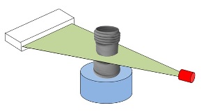
Flat-panel Volume CT is a technique under development to make computed tomography images with improved performance (in particular, with improved spatial resolution). The key difference between volume CT and traditional CT is that volume CT uses a two-dimensional x-ray detector orientation (usually in a square panel orientation), to take multiple two-dimensional images. On the other hand, the conventional CT uses a one-dimensional x-ray detector orientation (a row of detectors) to take one-dimensional x-ray images. A CT machine consists of an x-ray source, an x-ray detector, a series of moving stages (Gantry) and computers to assemble the x-ray data into an image. The x-ray beam used in volume CT is cone shaped, in contrast to the fan shaped beam of the regular CT scanner. This cone shape allows the beam to cover the two-dimensional detector panel. See also * Industrial CT Scanning * Radiology * Computed Tomography * Medical Imaging * Cone beam reconstruction In microtomography ... [...More Info...] [...Related Items...] OR: [Wikipedia] [Google] [Baidu] |
Computed Tomography
A computed tomography scan (CT scan; formerly called computed axial tomography scan or CAT scan) is a medical imaging technique used to obtain detailed internal images of the body. The personnel that perform CT scans are called radiographers or radiology technologists. CT scanners use a rotating X-ray tube and a row of detectors placed in a gantry to measure X-ray attenuations by different tissues inside the body. The multiple X-ray measurements taken from different angles are then processed on a computer using tomographic reconstruction algorithms to produce tomographic (cross-sectional) images (virtual "slices") of a body. CT scans can be used in patients with metallic implants or pacemakers, for whom magnetic resonance imaging (MRI) is contraindicated. Since its development in the 1970s, CT scanning has proven to be a versatile imaging technique. While CT is most prominently used in medical diagnosis, it can also be used to form images of non-living objects. The 1979 N ... [...More Info...] [...Related Items...] OR: [Wikipedia] [Google] [Baidu] |
X-ray
X-rays (or rarely, ''X-radiation'') are a form of high-energy electromagnetic radiation. In many languages, it is referred to as Röntgen radiation, after the German scientist Wilhelm Conrad Röntgen, who discovered it in 1895 and named it ''X-radiation'' to signify an unknown type of radiation.Novelline, Robert (1997). ''Squire's Fundamentals of Radiology''. Harvard University Press. 5th edition. . X-ray wavelengths are shorter than those of ultraviolet rays and longer than those of gamma rays. There is no universally accepted, strict definition of the bounds of the X-ray band. Roughly, X-rays have a wavelength ranging from 10 nanometers to 10 picometers, corresponding to frequencies in the range of 30 petahertz to 30 exahertz ( to ) and photon energies in the range of 100 eV to 100 keV, respectively. X-rays can penetrate many solid substances such as construction materials and living tissue, so X-ray radiography is widely used in medi ... [...More Info...] [...Related Items...] OR: [Wikipedia] [Google] [Baidu] |
Gantry (medical)
In a medical facility, such as a hospital or clinic, a gantry holds radiation detectors and/or a radiation source used to diagnose or treat a patient's illness. Radiation sources may produce gamma radiation, x-rays, electromagnetic radiation, or magnetic fields depending on the purpose of the device. CT scanner The gantry of a computed tomography scanner (CT) is a ring or cylinder, into which a patient is placed. The x-ray tube and x-ray detector spin rapidly in the gantry, as the patient is moved in and out of the gantry. The CT scanner produces 3-dimensional x-ray images of the patient. Nuclear magnetic resonance imaging An MRI gantry remains fixed, and contains cryogenically cooled superconducting electromagnets and radio transmitters that flip protons in hydrogen atoms in the human body via proton nuclear magnetic resonance. The machine then listens and processes the signals given off by the hydrogen atoms as the protons flip back in order to produce a 3D image of the ... [...More Info...] [...Related Items...] OR: [Wikipedia] [Google] [Baidu] |
Industrial CT Scanning
Industrial computed tomography (CT) scanning is any computer-aided tomographic process, usually X-ray computed tomography, that uses irradiation to produce three-dimensional internal and external representations of a scanned object. Industrial CT scanning has been used in many areas of industry for internal inspection of components. Some of the key uses for industrial CT scanning have been flaw detection, failure analysis, metrology, assembly analysis and reverse engineering applications. Just as in medical imaging, industrial imaging includes both nontomographic radiography ( industrial radiography) and computed tomographic radiography (computed tomography). Types of scanners ''Line beam scanning'' is the traditional process of industrial CT scanning.Hofmann, J., Flisch, A., Obrist, A., Adaptive CT scanning-mesh based optimisation methods for industrial X-ray computer tomography applications. NDT&E International (37), 2004, pp. 271–278. X-rays are produced and the beam is ... [...More Info...] [...Related Items...] OR: [Wikipedia] [Google] [Baidu] |
Radiology
Radiology ( ) is the medical discipline that uses medical imaging to diagnose diseases and guide their treatment, within the bodies of humans and other animals. It began with radiography (which is why its name has a root referring to radiation), but today it includes all imaging modalities, including those that use no electromagnetic radiation (such as ultrasonography and magnetic resonance imaging), as well as others that do, such as computed tomography (CT), fluoroscopy, and nuclear medicine including positron emission tomography (PET). Interventional radiology is the performance of usually minimally invasive medical procedures with the guidance of imaging technologies such as those mentioned above. The modern practice of radiology involves several different healthcare professions working as a team. The radiologist is a medical doctor who has completed the appropriate post-graduate training and interprets medical images, communicates these findings to other physicians b ... [...More Info...] [...Related Items...] OR: [Wikipedia] [Google] [Baidu] |
Computed Tomography
A computed tomography scan (CT scan; formerly called computed axial tomography scan or CAT scan) is a medical imaging technique used to obtain detailed internal images of the body. The personnel that perform CT scans are called radiographers or radiology technologists. CT scanners use a rotating X-ray tube and a row of detectors placed in a gantry to measure X-ray attenuations by different tissues inside the body. The multiple X-ray measurements taken from different angles are then processed on a computer using tomographic reconstruction algorithms to produce tomographic (cross-sectional) images (virtual "slices") of a body. CT scans can be used in patients with metallic implants or pacemakers, for whom magnetic resonance imaging (MRI) is contraindicated. Since its development in the 1970s, CT scanning has proven to be a versatile imaging technique. While CT is most prominently used in medical diagnosis, it can also be used to form images of non-living objects. The 1979 N ... [...More Info...] [...Related Items...] OR: [Wikipedia] [Google] [Baidu] |
Medical Imaging
Medical imaging is the technique and process of imaging the interior of a body for clinical analysis and medical intervention, as well as visual representation of the function of some organs or tissues ( physiology). Medical imaging seeks to reveal internal structures hidden by the skin and bones, as well as to diagnose and treat disease. Medical imaging also establishes a database of normal anatomy and physiology to make it possible to identify abnormalities. Although imaging of removed organs and tissues can be performed for medical reasons, such procedures are usually considered part of pathology instead of medical imaging. Measurement and recording techniques that are not primarily designed to produce images, such as electroencephalography (EEG), magnetoencephalography (MEG), electrocardiography (ECG), and others, represent other technologies that produce data susceptible to representation as a parameter graph versus time or maps that contain data about the measurement ... [...More Info...] [...Related Items...] OR: [Wikipedia] [Google] [Baidu] |
Cone Beam Reconstruction
In microtomography X-ray scanners, cone beam reconstruction is one of two common scanning methods, the other being Fan beam reconstruction. Cone beam reconstruction uses a 2-dimensional approach for obtaining projection data. Instead of utilizing a single row of detectors, as fan beam methods do, a cone beam systems uses a standard charge-coupled device camera, focused on a scintillator material. The scintillator converts X-ray radiation to visible light, which is picked up by the camera and recorded. The method has enjoyed widespread implementation in microtomography, and is also used in several larger-scale systems. An X-ray source is positioned across from the detector, with the object being scanned in between. (This is essentially the same setup used for an ordinary X-ray fluoroscope). Projections from different angles are obtained in one of two ways. In one method, the object being scanned is rotated. This has the advantage of simplicity in implementation; a rotating ... [...More Info...] [...Related Items...] OR: [Wikipedia] [Google] [Baidu] |


