|
Uvea
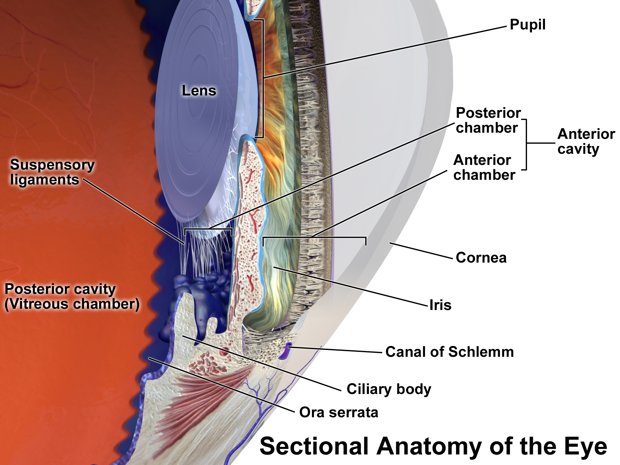
The uvea (; derived from meaning "grape"), also called the uveal layer, uveal coat, uveal tract, vascular tunic or vascular layer, is the pigmented middle layer of the three concentric layers that make up an eye, precisely between the inner retina and the outer fibrous layer composed of the sclera and cornea The cornea is the transparency (optics), transparent front part of the eyeball which covers the Iris (anatomy), iris, pupil, and Anterior chamber of eyeball, anterior chamber. Along with the anterior chamber and Lens (anatomy), lens, the cornea .... History and etymology The originally medieval Latin term comes from the Latin word ''uva'' ("grape") and is a reference to its grape-like appearance (reddish-blue or almost black colour, wrinkled appearance and grape-like size and shape when stripped intact from a cadaveric eye). In fact, it is a partial loan translation of the Ancient Greek term for the choroid, which literally means “covering resembling a grape ... [...More Info...] [...Related Items...] OR: [Wikipedia] [Google] [Baidu] |
Uveal Melanoma
Uveal melanoma is a type of eye cancer in the uvea of the eye. It is traditionally classed as originating in the iris, choroid, and ciliary body, but can also be divided into class I (low metastatic risk) and class II (high metastatic risk). Symptoms include blurred vision, loss of vision, and photopsia, but there may be no symptoms. Tumors arise from the pigment cells that reside within the uvea and give color to the eye. These melanocytes are distinct from the retinal pigment epithelium cells underlying the retina that do not form melanomas. When eye melanoma is spread to distant parts of the body, the five-year survival rate is about 15%.Eye Cancer Survival Rates American Cancer Society, Last Medical Review: December 9, 2014 Last Revised: February 5, 2016 It is the most co ... [...More Info...] [...Related Items...] OR: [Wikipedia] [Google] [Baidu] |
Choroid
The choroid, also known as the choroidea or choroid coat, is a part of the uvea, the vascular layer of the eye. It contains connective tissues, and lies between the retina and the sclera. The human choroid is thickest at the far extreme rear of the eye (at 0.2 mm), while in the outlying areas it narrows to 0.1 mm. The choroid provides oxygen and nourishment to the outer layers of the retina. Along with the ciliary body and iris, the choroid forms the uveal tract. The structure of the choroid is generally divided into four layers (classified in order of furthest away from the retina to closest): *Haller's layer – outermost layer of the choroid consisting of larger diameter blood vessels; * Sattler's layer – layer of medium diameter blood vessels; * Choriocapillaris – layer of capillaries; and * Bruch's membrane (synonyms: Lamina basalis, Complexus basalis, Lamina vitra) – innermost layer of the choroid. Blood supply There are two circulations of the eye: ... [...More Info...] [...Related Items...] OR: [Wikipedia] [Google] [Baidu] |
Parasympathetic Nervous System
The parasympathetic nervous system (PSNS) is one of the three divisions of the autonomic nervous system, the others being the sympathetic nervous system and the enteric nervous system. The autonomic nervous system is responsible for regulating the body's unconscious actions. The parasympathetic system is responsible for stimulation of "rest-and-digest" or "feed-and-breed" activities that occur when the body is at rest, especially after eating, including sexual arousal, salivation, lacrimation (tears), urination, digestion, and defecation. Its action is described as being complementary to that of the sympathetic nervous system, which is responsible for stimulating activities associated with the fight-or-flight response. Nerve fibres of the parasympathetic nervous system arise from the central nervous system. Specific nerves include several cranial nerves, specifically the oculomotor nerve, facial nerve, glossopharyngeal nerve, and vagus nerve. Three spinal nerves ... [...More Info...] [...Related Items...] OR: [Wikipedia] [Google] [Baidu] |
Anterior Uveitis
Uveitis () is inflammation of the uvea, the pigmented layer of the eye between the inner retina and the outer fibrous layer composed of the sclera and cornea. The uvea consists of the middle layer of pigmented vascular structures of the eye and includes the iris, ciliary body, and choroid. Uveitis is described anatomically, by the part of the eye affected, as anterior, intermediate or posterior, or panuveitic if all parts are involved. Anterior uveitis ( iridocyclitis) is the most common, with the incidence of uveitis overall affecting approximately 1:4500, most commonly those between the ages of 20–60. Symptoms include eye pain, eye redness, floaters and blurred vision, and ophthalmic examination may show dilated ciliary blood vessels and the presence of cells in the anterior chamber. Uveitis may arise spontaneously, have a genetic component, or be associated with an autoimmune disease or infection. While the eye is a relatively protected environment, its immune mechanisms ... [...More Info...] [...Related Items...] OR: [Wikipedia] [Google] [Baidu] |
Choroiditis
Chorioretinitis is an inflammation of the choroid (thin pigmented vascular coat of the eye) and retina of the eye. It is a form of posterior uveitis. Inflammation of these layers can lead to vision-threatening complications. If only the choroid is inflamed, not the retina, the condition is termed choroiditis. The ophthalmologist's goal in treating these potentially blinding conditions is to eliminate the inflammation and minimize the potential risk of therapy to the patient. Symptoms Symptoms may include the presence of floating black spots, blurred vision, pain or redness in the eye, sensitivity to light, or excessive tearing. Causes Chorioretinitis is often caused by toxoplasmosis and cytomegalovirus infections (mostly seen in immunodeficient subjects such as people with HIV/AIDS or on immunosuppressant drugs). Congenital toxoplasmosis via transplacental transmission can also lead to sequelae such as chorioretinitis along with hydrocephalus and cerebral calcifications. Other ... [...More Info...] [...Related Items...] OR: [Wikipedia] [Google] [Baidu] |
Uveitis
Uveitis () is inflammation of the uvea, the pigmented layer of the eye between the inner retina and the outer fibrous layer composed of the sclera and cornea. The uvea consists of the middle layer of pigmented vascular structures of the eye and includes the iris, ciliary body, and choroid. Uveitis is described anatomically, by the part of the eye affected, as anterior, intermediate or posterior, or panuveitic if all parts are involved. Anterior uveitis ( iridocyclitis) is the most common, with the incidence of uveitis overall affecting approximately 1:4500, most commonly those between the ages of 20–60. Symptoms include eye pain, eye redness, floaters and blurred vision, and ophthalmic examination may show dilated ciliary blood vessels and the presence of cells in the anterior chamber. Uveitis may arise spontaneously, have a genetic component, or be associated with an autoimmune disease or infection. While the eye is a relatively protected environment, its immune mecha ... [...More Info...] [...Related Items...] OR: [Wikipedia] [Google] [Baidu] |
Antigens
In immunology, an antigen (Ag) is a molecule, moiety, foreign particulate matter, or an allergen, such as pollen, that can bind to a specific antibody or T-cell receptor. The presence of antigens in the body may trigger an immune response. Antigens can be proteins, peptides (amino acid chains), polysaccharides (chains of simple sugars), lipids, or nucleic acids. Antigens exist on normal cells, cancer cells, parasites, viruses, fungi, and bacteria. Antigens are recognized by antigen receptors, including antibodies and T-cell receptors. Diverse antigen receptors are made by cells of the immune system so that each cell has a specificity for a single antigen. Upon exposure to an antigen, only the lymphocytes that recognize that antigen are activated and expanded, a process known as clonal selection. In most cases, antibodies are ''antigen-specific'', meaning that an antibody can only react to and bind one specific antigen; in some instances, however, antibodies may cross-react ... [...More Info...] [...Related Items...] OR: [Wikipedia] [Google] [Baidu] |
Sympathetic Ophthalmia
Sympathetic ophthalmia (SO), also called spared eye injury, is a diffuse granulomatous inflammation of the uveal layer of both eyes following trauma to one eye. It can leave the affected person completely blind. Symptoms may develop from days to several years after a penetrating eye injury. It typically results from a delayed hypersensitivity reaction. Signs and symptoms Eye floaters and loss of accommodation are among the earliest symptoms. The disease may progress to severe inflammation of the uveal layer of the eye (uveitis) with pain and sensitivity of the eyes to light. The affected eye often remains relatively painless while the inflammatory disease spreads through the uvea, where characteristic focal infiltrates in the choroid named Dalén–Fuchs nodules can be seen. The retina, however, usually remains uninvolved, although perivascular cuffing of the retinal vessels with inflammatory cells may occur. Swelling of the optic disc (papilledema), secondary glaucoma, vi ... [...More Info...] [...Related Items...] OR: [Wikipedia] [Google] [Baidu] |
Glaucoma
Glaucoma is a group of eye diseases that can lead to damage of the optic nerve. The optic nerve transmits visual information from the eye to the brain. Glaucoma may cause vision loss if left untreated. It has been called the "silent thief of sight" because the loss of vision usually occurs slowly over a long period of time. A major risk factor for glaucoma is increased pressure within the eye, known as Intraocular pressure, intraocular pressure (IOP). It is associated with old age, a family history of glaucoma, and certain medical conditions or the use of some medications. The word ''glaucoma'' comes from the Ancient Greek word (), meaning 'gleaming, blue-green, gray'. Of the different types of glaucoma, the most common are called open-angle glaucoma and closed-angle glaucoma. Inside the eye, a liquid called Aqueous humour, aqueous humor helps to maintain shape and provides nutrients. The aqueous humor normally drains through the trabecular meshwork. In open-angle glaucoma, ... [...More Info...] [...Related Items...] OR: [Wikipedia] [Google] [Baidu] |


