 |
Triple Test
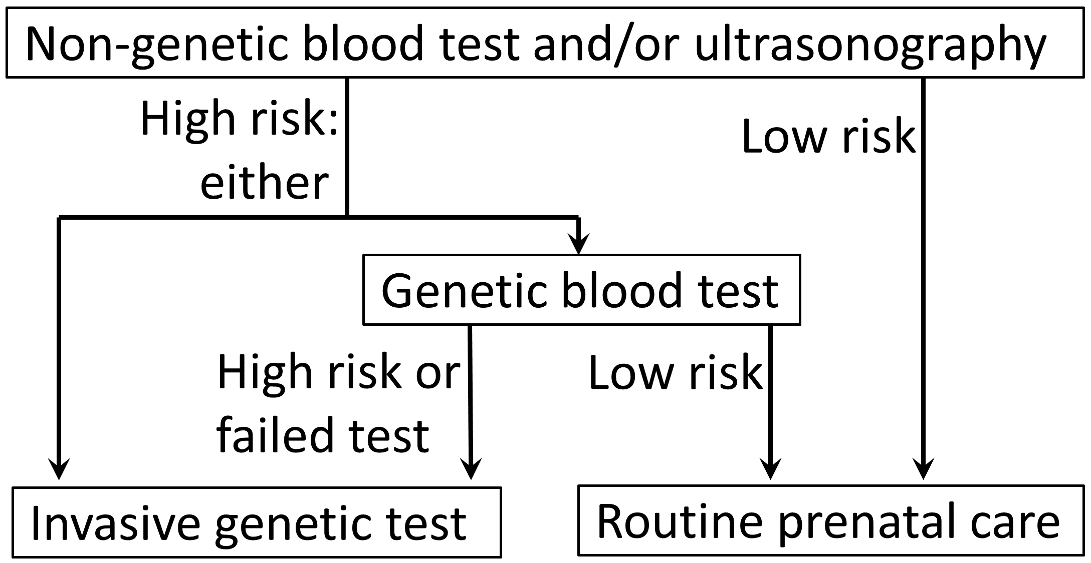
The triple test, also called triple screen, the Kettering test or the Bart's test, is an investigation performed during pregnancy in the second trimester to classify a patient as either high-risk or low-risk for chromosomal abnormalities (and neural tube defects). The term "multiple-marker screening test" is sometimes used instead. This term can encompass the "double test" and "quadruple test" (described below). The Triple screen measures serum levels of AFP, estriol, and beta-hCG, with a 70% sensitivity and 5% false-positive rate. It is complemented in some regions of the United States, as the ''Quad screen'' (adding inhibin A to the panel, resulting in an 81% sensitivity and 5% false-positive rate for detecting Down syndrome when taken at 15–18 weeks of gestational age) and other prenatal diagnosis techniques, although it remains widely used in Canada and other countries. A positive screen indicates an increased risk of chromosomal abnormalities (and neural tube defec ... [...More Info...] [...Related Items...] OR: [Wikipedia] [Google] [Baidu] |
 |
Triple Test Score
The triple test score is a diagnostic tool for examining potentially breast cancer, cancerous breasts. Diagnostic accuracy of the triple test score is nearly 100%. Scoring includes using the procedures of physical examination, mammography and needle biopsy. If the results of a triple test score are greater than five, an excisional biopsy is indicated. The term ''triple test scoring'' (TSS) was first noted in 1975 as a means of rapidly diagnosing and examining breast malignancies. TSS developed as a useful and accurate clinical tool for breast masses because it was cheaper and it cut down on the diagnosis time. Scoring To obtain the triple test score, a number from 1 through 3 is assigned to each one of the procedures. A score of 1 is assigned to a benign test result, 2 applies to a suspicious test result, and 3 applies to a malignant result. The sum of the scores of all three procedures is the triple test score. If the total summed score from the three tests is 3 to 4 then the d ... [...More Info...] [...Related Items...] OR: [Wikipedia] [Google] [Baidu] |
 |
Turner Syndrome
Turner syndrome (TS), commonly known as 45,X, or 45,X0,Also written as 45,XO. is a chromosomal disorder in which cells of females have only one X chromosome instead of two, or are partially missing an X chromosome (sex chromosome monosomy) leading to the complete or partial deletion of the pseudoautosomal regions (PAR1, PAR2) in the affected X chromosome. Typically, people have two sex chromosomes (XX for females or XY for males). The chromosomal abnormality is often present in just some cells, in which case it is known as Turner syndrome with mosaicism. 45,X0 with mosaicism can occur in males or females, but Turner syndrome without mosaicism only occurs in females. Signs and symptoms vary among those affected but often include additional skin folds on the neck, arched palate, low-set ears, low hairline at the nape of the neck, short stature, and lymphedema of the hands and feet. Those affected do not normally develop menstrual periods or mammary glands without hormone trea ... [...More Info...] [...Related Items...] OR: [Wikipedia] [Google] [Baidu] |
 |
Gastroschisis
Gastroschisis is a birth defect in which the baby's intestines extend outside of the abdomen through a hole next to the belly button. The size of the hole is variable, and other organs including the stomach and liver may also occur outside the baby's body. Complications may include feeding problems, prematurity, intestinal atresia, and intrauterine growth restriction. The cause is typically unknown. Rates are higher in babies born to mothers who smoke, drink alcohol, or are younger than 20 years old. Ultrasounds during pregnancy may make the diagnosis. Otherwise, diagnosis occurs at birth. It differs from omphalocele in that there is no covering membrane over the intestines. Treatment involves surgery. This typically occurs shortly after birth. In those with large defects, the exposed organs may be covered with a special material and slowly moved back into the abdomen. This condition affects about 4 per 10,000 newborns. Rates of the condition appear to be increasing. Sig ... [...More Info...] [...Related Items...] OR: [Wikipedia] [Google] [Baidu] |
 |
Omphalocele
An omphalocele or omphalocoele, also known as an exomphalos, is a rare abdominal wall defect. Beginning at the 6th week of development, rapid elongation of the gut and increased liver size reduces intra abdominal space, which pushes intestinal loops out of the abdominal cavity. Around 10th week, the intestine returns to the abdominal cavity and the process is completed by the 12th week. Persistence of intestine or the presence of other abdominal viscera (e.g. stomach, liver) in the umbilical cord results in an omphalocele. Omphalocele occurs in 1 in 4,000 births and is associated with a high rate of mortality (25%) and severe malformations, such as cardiac anomalies (50%), neural tube defect (40%), exstrophy of the bladder and Beckwith–Wiedemann syndrome. Approximately 15% of live-born infants with omphalocele have chromosomal abnormalities. About 30% of infants with an omphalocele have other congenital abnormalities. Signs and symptoms The sac, which is formed from an out ... [...More Info...] [...Related Items...] OR: [Wikipedia] [Google] [Baidu] |
 |
Acetylcholinesterase
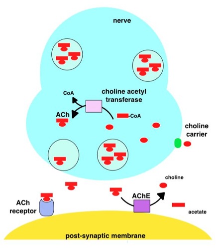
Acetylcholinesterase (HUGO Gene Nomenclature Committee, HGNC symbol ACHE; EC 3.1.1.7; systematic name acetylcholine acetylhydrolase), also known as AChE, AChase or acetylhydrolase, is the primary cholinesterase in the body. It is an enzyme that catalysis, catalyzes the breakdown of acetylcholine and some other choline esters that function as neurotransmitters: : acetylcholine + H2O = choline + acetate It is found at mainly neuromuscular junctions and in chemical synapses of the cholinergic type, where its activity serves to terminate cholinergic neurotransmission, synaptic transmission. It belongs to the carboxylesterase family of enzymes. It is the primary target of inhibition by organophosphorus compounds such as nerve agents and pesticides. Enzyme structure and mechanism AChE is a hydrolase that hydrolyzes choline esters. It has a very high catalytic activity—each molecule of AChE degrades about 5,000 molecules of acetylcholine (ACh) per second, approaching the limit ... [...More Info...] [...Related Items...] OR: [Wikipedia] [Google] [Baidu] |
 |
Spina Bifida
Spina bifida (SB; ; Latin for 'split spine') is a birth defect in which there is incomplete closing of the vertebral column, spine and the meninges, membranes around the spinal cord during embryonic development, early development in pregnancy. There are three main types: spina bifida occulta, meningocele and myelomeningocele. Meningocele and myelomeningocele may be grouped as spina bifida cystica. The most common location is the Lumbar vertebrae, lower back, but in rare cases it may be in the Thoracic vertebrae, middle back or Cervical vertebrae, neck. Occulta has no or only mild signs, which may include a hairy patch, dimple, dark spot or swelling on the back at the site of the gap in the spine. Meningocele typically causes mild problems, with a sac of fluid present at the gap in the spine. Myelomeningocele, also known as open spina bifida, is the most severe form. Problems associated with this form include poor ability to walk, impaired Neurogenic bladder dysfunction, bladder o ... [...More Info...] [...Related Items...] OR: [Wikipedia] [Google] [Baidu] |
|
Edward's Syndrome
Trisomy 18, also known as Edwards syndrome, is a genetic disorder caused by the presence of a third copy of all or part of chromosome 18. Many parts of the body are affected. Babies are often born small and have heart defects. Other features include a small head, small jaw, clenched fists with overlapping fingers, and severe intellectual disability. Most cases of trisomy 18 occur due to problems during the formation of the reproductive cells or during early development. The chance of this condition occurring increases with the mother's age. Rarely, cases may be inherited. Occasionally, not all cells have the extra chromosome, known as mosaic trisomy, and symptoms in these cases may be less severe. An ultrasound during pregnancy can increase suspicion for the condition, which can be confirmed by amniocentesis. Treatment is supportive. After having one child with the condition, the risk of having a second is typically around one percent. It is the second-most common conditi ... [...More Info...] [...Related Items...] OR: [Wikipedia] [Google] [Baidu] |
|
 |
Human Chorionic Gonadotropin
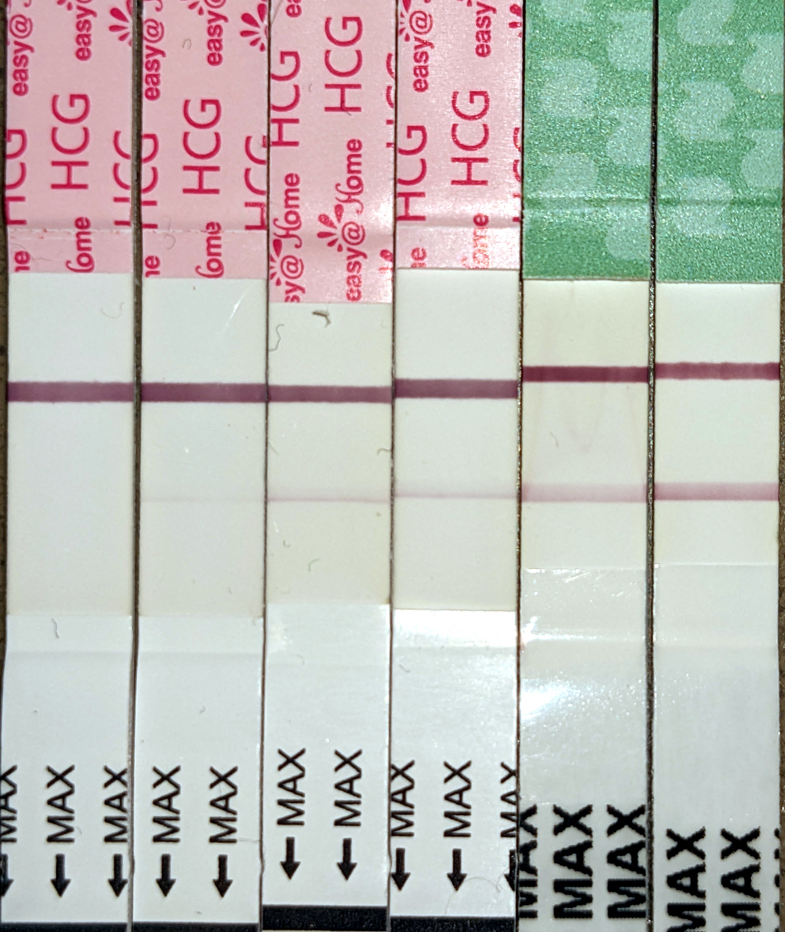
Human chorionic gonadotropin (hCG) is a hormone for the maternal recognition of pregnancy produced by trophoblast cells that are surrounding a growing embryo (syncytiotrophoblast initially), which eventually forms the placenta after implantation. The presence of hCG is detected in some pregnancy tests (HCG pregnancy strip tests). Some cancerous tumors produce this hormone; therefore, elevated levels measured when the patient is not pregnant may lead to a cancer diagnosis and, if high enough, paraneoplastic syndromes, however, it is unknown whether this production is a contributing cause or an effect of carcinogenesis. The pituitary analog of hCG, known as luteinizing hormone (LH), is produced in the pituitary gland of males and females of all ages. Beta-hCG is initially secreted by the syncytiotrophoblast. Structure Human chorionic gonadotropin is a glycoprotein composed of 237 amino acids with a molecular mass of 36.7 kDa, approximately 14.5kDa αhCG and 22.2kDa βhCG ... [...More Info...] [...Related Items...] OR: [Wikipedia] [Google] [Baidu] |
|
Alpha-fetoprotein
Alpha-fetoprotein (AFP, α-fetoprotein; also sometimes called alpha-1-fetoprotein, alpha-fetoglobulin, or alpha fetal protein) is a protein that in humans is encoded by the ''AFP'' gene. The ''AFP'' gene is located on the ''q'' arm of chromosome 4 (4q13.3). Maternal AFP serum level is used to screen for Down syndrome, neural tube defects, and other chromosomal abnormalities. AFP is a major plasma protein produced by the yolk sac and the fetal liver during fetal development. It is thought to be the fetal analog of serum albumin. AFP binds to copper, nickel, fatty acids and bilirubin and is found in monomeric, dimeric and trimeric forms. Structure AFP is a glycoprotein of 591 amino acids and a carbohydrate moiety. Function The function of AFP in adult humans is unknown. AFP is the most abundant plasma protein found in the human fetus. In the fetus, AFP is produced by both the liver and the yolk sac. It is believed to function as a carrier protein (similar to albumin) ... [...More Info...] [...Related Items...] OR: [Wikipedia] [Google] [Baidu] |
|
 |
Steroid Sulfatase Deficiency
A steroid is an organic compound with four fused rings (designated A, B, C, and D) arranged in a specific molecular configuration. Steroids have two principal biological functions: as important components of cell membranes that alter membrane fluidity; and as signaling molecules. Examples include the lipid cholesterol, sex hormones estradiol and testosterone, anabolic steroids, and the anti-inflammatory corticosteroid drug dexamethasone. Hundreds of steroids are found in fungi, plants, and animals. All steroids are manufactured in cells from a sterol: cholesterol (animals), lanosterol (opisthokonts), or cycloartenol (plants). All three of these molecules are produced via cyclization of the triterpene squalene. Structure The steroid nucleus ( core structure) is called gonane (cyclopentanoperhydrophenanthrene). It is typically composed of seventeen carbon atoms, bonded in four fused rings: three six-member cyclohexane rings (rings A, B and C in the first illustration) and ... [...More Info...] [...Related Items...] OR: [Wikipedia] [Google] [Baidu] |
 |
Smith–Lemli–Opitz Syndrome
Smith–Lemli–Opitz syndrome is an inborn error of metabolism, inborn error of cholesterol synthesis. It is an autosome, autosomal recessive (genetics), recessive, multiple malformation syndrome caused by a mutation in the enzyme 7-Dehydrocholesterol reductase encoded by the DHCR7 gene. It causes a broad spectrum of effects, ranging from mild intellectual disability and behavioural problems to lethal malformations. Signs and symptoms SLOS can present itself differently in different cases, depending on the severity of the mutation and other factors. Originally, SLOS patients were classified into two categories (classic and severe) based on physical and mental characteristics, alongside other clinical features. Since the discovery of the specific biochemical defect responsible for SLOS, patients are given a severity score based on their levels of cerebral, ocular, oral, and genital defects. It is then used to classify patients as having mild, classical, or severe SLOS. Physical ... [...More Info...] [...Related Items...] OR: [Wikipedia] [Google] [Baidu] |