|
Transverse Tubule
T-tubules (transverse tubules) are extensions of the cell membrane that penetrate into the center of skeletal and cardiac muscle cells. With membranes that contain large concentrations of ion channels, transporters, and pumps, T-tubules permit rapid transmission of the action potential into the cell, and also play an important role in regulating cellular calcium concentration. Through these mechanisms, T-tubules allow heart muscle cells to contract more forcefully by synchronising calcium release from the sarcoplasmic reticulum throughout the cell. T-tubule structure and function are affected beat-by-beat by cardiomyocyte contraction, as well as by diseases, potentially contributing to heart failure and arrhythmias. Although these structures were first seen in 1897, research into T-tubule biology is ongoing. Structure T-tubules are tubules formed from the same phospholipid bilayer as the surface membrane or sarcolemma of skeletal or cardiac muscle cells. They connect directly ... [...More Info...] [...Related Items...] OR: [Wikipedia] [Google] [Baidu] |
Skeletal Muscle
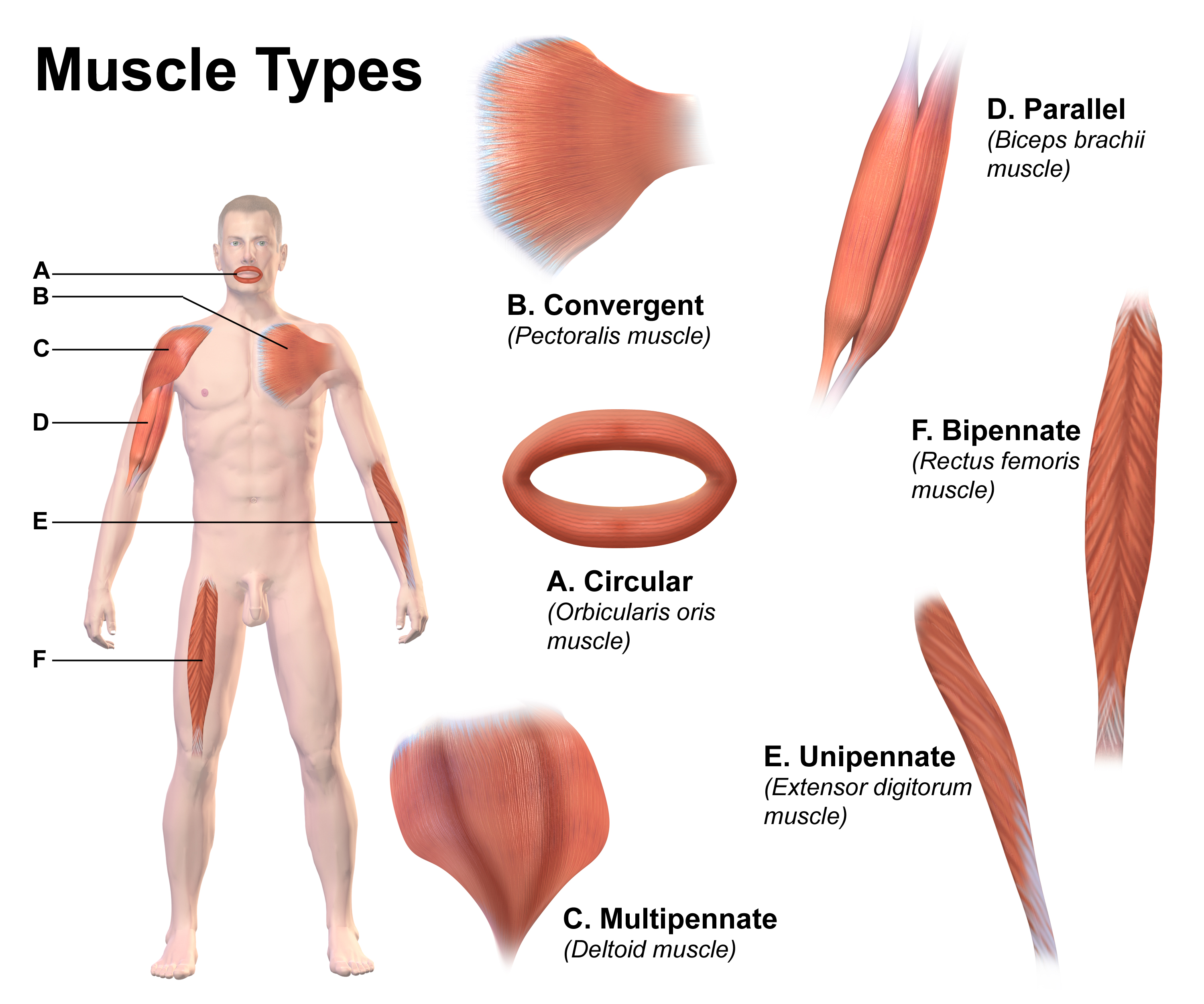
Skeletal muscle (commonly referred to as muscle) is one of the three types of vertebrate muscle tissue, the others being cardiac muscle and smooth muscle. They are part of the somatic nervous system, voluntary muscular system and typically are attached by tendons to bones of a skeleton. The skeletal muscle cells are much longer than in the other types of muscle tissue, and are also known as ''muscle fibers''. The tissue of a skeletal muscle is striated muscle tissue, striated – having a striped appearance due to the arrangement of the sarcomeres. A skeletal muscle contains multiple muscle fascicle, fascicles – bundles of muscle fibers. Each individual fiber and each muscle is surrounded by a type of connective tissue layer of fascia. Muscle fibers are formed from the cell fusion, fusion of developmental myoblasts in a process known as myogenesis resulting in long multinucleated cells. In these cells, the cell nucleus, nuclei, termed ''myonuclei'', are located along the inside ... [...More Info...] [...Related Items...] OR: [Wikipedia] [Google] [Baidu] |
Cardiac Muscle Cell
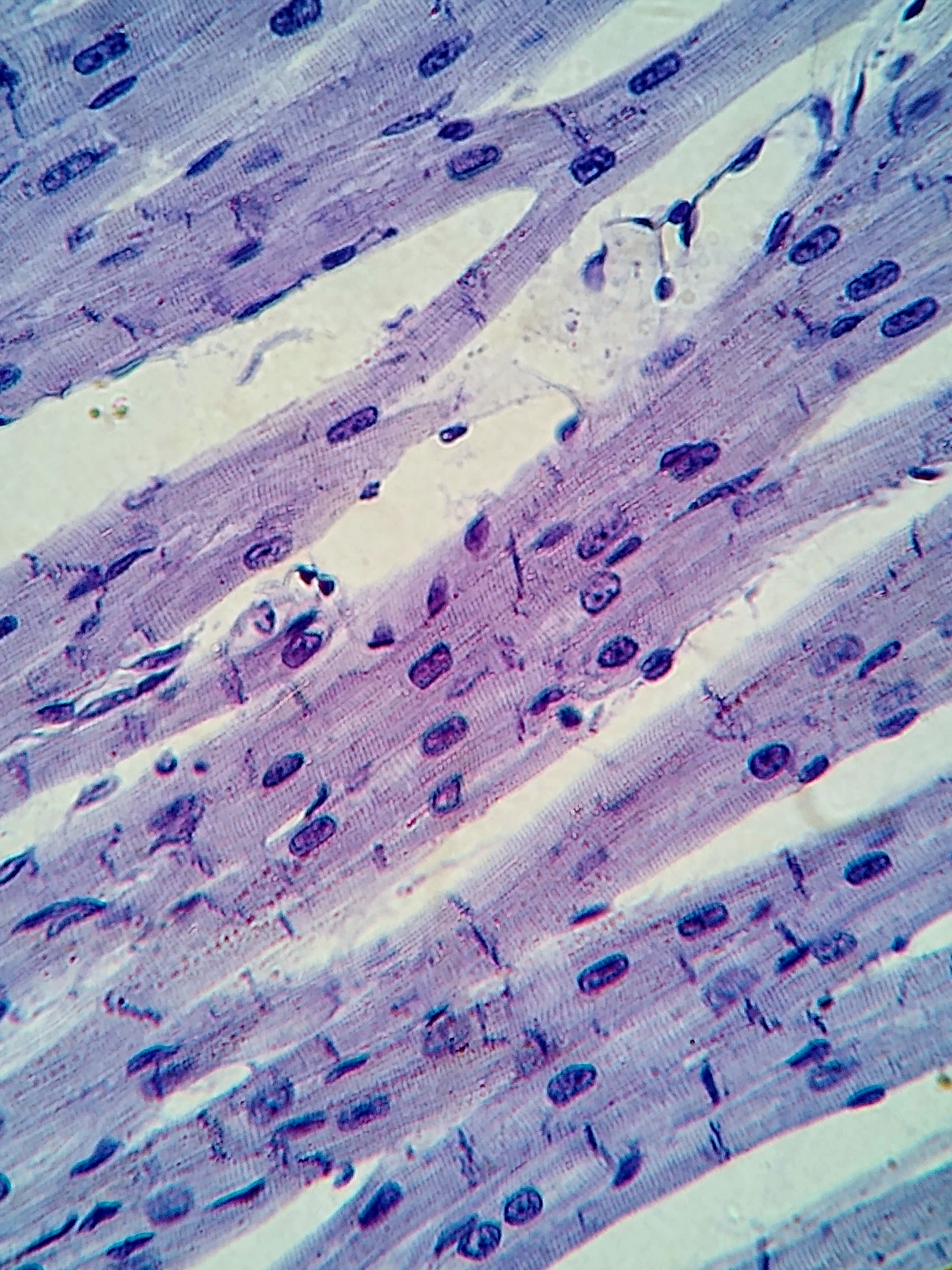
Cardiac muscle (also called heart muscle or myocardium) is one of three types of vertebrate muscle tissues, the others being skeletal muscle and smooth muscle. It is an involuntary, striated muscle that constitutes the main tissue of the Heart#Wall, wall of the heart. The cardiac muscle (myocardium) forms a thick middle layer between the outer layer of the heart wall (the pericardium) and the inner layer (the endocardium), with blood supplied via the coronary circulation. It is composed of individual cardiac muscle cells joined by intercalated discs, and encased by collagen fibers and other substances that form the extracellular matrix. Cardiac muscle Muscle contraction, contracts in a similar manner to skeletal muscle, although with some important differences. Electrical stimulation in the form of a cardiac action potential triggers the release of calcium from the cell's internal calcium store, the sarcoplasmic reticulum. The rise in calcium causes the cell's myofilaments to ... [...More Info...] [...Related Items...] OR: [Wikipedia] [Google] [Baidu] |
Cardiac Action Potential
Unlike the action potential in skeletal muscle cells, the cardiac action potential is not initiated by nervous activity. Instead, it arises from a group of specialized cells known as pacemaker cells, that have automatic action potential generation capability. In healthy hearts, these cells form the cardiac pacemaker and are found in the sinoatrial node in the right atrium. They produce roughly 60–100 action potentials every minute. The action potential passes along the cell membrane causing the cell to contract, therefore the activity of the sinoatrial node results in a resting heart rate of roughly 60–100 beats per minute. All cardiac muscle cells are electrically linked to one another, by intercalated discs which allow the action potential to pass from one cell to the next. This means that all atrial cells can contract together, and then all ventricular cells. Rate dependence of the action potential is a fundamental property of cardiac cells and alterations can lead to se ... [...More Info...] [...Related Items...] OR: [Wikipedia] [Google] [Baidu] |
TCAP (gene)
Telethonin, also known as Tcap, is a protein that in humans is encoded by the ''TCAP'' gene. Telethonin is expressed in cardiac and skeletal muscle at Z-discs and functions to regulate sarcomere assembly, T-tubule function and apoptosis. Telethonin has been implicated in several diseases, including limb-girdle muscular dystrophy, hypertrophic cardiomyopathy, dilated cardiomyopathy and idiopathic cardiomyopathy. Structure Telethonin is a 19.0 kDa protein composed of 167 amino acids. Telethonin has a unique β-sheet structure, which enables antiparallel association with the Titin Z1-Z2 domains in cardiac and skeletal muscle. Structural analysis of full-length Telethonin with the N-terminal region of Titin indicate that the C-terminus of Telethonin is critical for the dimerization of two Telethonin/Titin complexes into a higher oligomeric structure. Function Telethonin expression is developmentally regulated in both cardiac and skeletal muscle and is thought to be critical to sar ... [...More Info...] [...Related Items...] OR: [Wikipedia] [Google] [Baidu] |
Telethonin
Telethonin, also known as Tcap, is a protein that in humans is encoded by the ''TCAP'' gene. Telethonin is expressed in cardiac and skeletal muscle at Z-discs and functions to regulate sarcomere assembly, T-tubule function and apoptosis. Telethonin has been implicated in several diseases, including limb-girdle muscular dystrophy, hypertrophic cardiomyopathy, dilated cardiomyopathy and idiopathic cardiomyopathy. Structure Telethonin is a 19.0 kDa protein composed of 167 amino acids. Telethonin has a unique β-sheet structure, which enables antiparallel association with the Titin Z1-Z2 domains in cardiac and skeletal muscle. Structural analysis of full-length Telethonin with the N-terminal region of Titin indicate that the C-terminus of Telethonin is critical for the dimerization of two Telethonin/ Titin complexes into a higher oligomeric structure. Function Telethonin expression is developmentally regulated in both cardiac and skeletal muscle and is thought to be criti ... [...More Info...] [...Related Items...] OR: [Wikipedia] [Google] [Baidu] |
Excitation Contraction Coupling
Excitation, excite, exciting, or excitement may refer to: * Excitation (magnetic), provided with an electrical generator or alternator * ''Exite'', a series of racing video games published by Nintendo starting with '' Excitebike'' * Excite (web portal), web portal owned by IAC * Excite Ballpark, located in San Jose, California * Electron excitation, the transfer of an electron to a higher atomic orbital ** More generally, the transfer of energy to a normal mode * ''Excitement'' (film), a lost 1924 silent comedy by Robert F. Hill * Sexual excitation * Stimulation Stimulation is the encouragement of development or the cause of activity in general. For example, "The press provides stimulation of political discourse." An interesting or fun activity can be described as "stimulating", regardless of its physic ... or excitation or excitement, the action of various agents on nerves, muscles, or a sensory end organ, by which activity is evoked * "Exciting", a song by Hieroglyphics ... [...More Info...] [...Related Items...] OR: [Wikipedia] [Google] [Baidu] |
JPH2
Junctophilin 2, also known as JPH2, is a protein which in humans is encoded by the ''JPH2'' gene. Alternative splicing has been observed at this locus and two variants encoding distinct isoforms are described. Function Junctional complexes between the plasma membrane and endoplasmic/ sarcoplasmic reticulum are a common feature of all excitable cell types and mediate cross talk between cell surface and intracellular ion channels. The protein encoded by this gene is a component of junctional complexes and is composed of a C-terminal hydrophobic segment spanning the endoplasmic/sarcoplasmic reticulum membrane and a remaining cytoplasmic membrane occupation and recognition nexus (MORN) domain that shows specific affinity for the plasma membrane. JPH2 is a member of the junctophilin gene family (the other members of the family are JPH1, JPH3, and JPH4) and is the predominant isoform in cardiac tissue, but is also expressed with JPH1 in skeletal muscle. The JPH2 protein product pl ... [...More Info...] [...Related Items...] OR: [Wikipedia] [Google] [Baidu] |
BIN1
Myc box-dependent-interacting protein 1, also known as Bridging Integrator-1 and Amphiphysin-2 is a protein that in humans is encoded by the ''BIN1'' gene. This gene encodes several isoforms of a nucleocytoplasmic adaptor protein, one of which was initially identified as a MYC-interacting protein with features of a tumor suppressor. Isoforms that are expressed in the central nervous system may be involved in synaptic vesicle endocytosis and may interact with dynanim, synaptojanin, endophilin, and clathrin. Isoforms that are expressed in muscle and ubiquitously expressed isoforms localize to the cytoplasm and nucleus and activate a caspase-independent apoptotic process. Studies in mouse suggest that this gene plays an important role in cardiac muscle development. Alternate splicing of the gene results in ten transcript variants encoding different isoforms. Aberrant splice variants expressed in tumor cell lines have also been described. Clinical significance In humans, mutatio ... [...More Info...] [...Related Items...] OR: [Wikipedia] [Google] [Baidu] |
Amphiphysin
Amphiphysin is a protein that in humans is encoded by the ''AMPH'' gene. Function This gene encodes a protein associated with the cytoplasmic surface of synaptic vesicles. A subset of patients with stiff person syndrome who were also affected by breast cancer are positive for autoantibodies against this protein. Alternate splicing of this gene results in two transcript variants encoding different isoforms. Additional splice variants have been described, but their full length sequences have not been determined. Amphiphysin is a brain-enriched protein with an N-terminal lipid interaction, dimerisation and cell membrane, membrane bending BAR domain, a middle clathrin and adaptor binding domain and a C-terminal SH3 domain. In the brain, its primary function is thought to be the recruitment of dynamin to sites of clathrin-mediated endocytosis. There are 2 mammalian amphiphysins with similar overall structure. A ubiquitous splice form of amphiphysin-2 (BIN1) that does not contain cla ... [...More Info...] [...Related Items...] OR: [Wikipedia] [Google] [Baidu] |
Triad (anatomy)
In the histology of skeletal muscle, a triad is the structure formed by a T tubule with a sarcoplasmic reticulum (SR) known as the terminal cisterna on either side. Each skeletal muscle fiber has many thousands of triads, visible in muscle fibers that have been sectioned longitudinally. (This property holds because T tubules run perpendicular to the longitudinal axis of the muscle fiber.) In mammals, triads are typically located at the A-I junction; that is, the junction between the A and I bands of the sarcomere, which is the smallest unit of a muscle fiber. Triads form the anatomical basis of excitation-contraction coupling, whereby a stimulus excites the muscle and causes it to contract. A stimulus, in the form of positively charged current, is transmitted from the neuromuscular junction down the length of the T tubules, activating dihydropyridine receptors (DHPRs). Their activation causes 1) a negligible influx of calcium and 2) a mechanical interaction with calc ... [...More Info...] [...Related Items...] OR: [Wikipedia] [Google] [Baidu] |
Terminal Cisternae
Terminal cisternae (singular: terminal cisterna) are enlarged areas of the sarcoplasmic reticulum surrounding the transverse tubules. Function Terminal cisternae are discrete regions within the muscle cell. They store calcium (increasing the capacity of the sarcoplasmic reticulum to release calcium) and release it when an action potential courses down the transverse tubules, eliciting muscle contraction. Because terminal cisternae ensure rapid calcium delivery, they are well developed in muscles that contract quickly, such as fast twitch skeletal muscle. Terminal cisternae then go on to release calcium, which binds to troponin. This releases tropomyosin, exposing active sites of the thin filament, actin. There are several mechanisms directly linked to the terminal cisternae which facilitate excitation-contraction coupling. When excitation of the membrane arrives at the T-tubule nearest the muscle fiber, a dihydropyridine channel ( DHP channel) is activated. This is simil ... [...More Info...] [...Related Items...] OR: [Wikipedia] [Google] [Baidu] |
Diad
Within the muscle tissue of animals and humans, contraction and relaxation of the muscle cells (myocytes) is a highly regulated and rhythmic process. In cardiomyocytes, or cardiac muscle cells, muscular contraction takes place due to movement at a structure referred to as the diad, sometimes spelled "dyad." The dyad is the connection of transverse- tubules (t-tubules) and the junctional sarcoplasmic reticulum (jSR). Like skeletal muscle contractions, Calcium (Ca2+) ions are required for polarization and depolarization through a voltage-gated calcium channel. The rapid influx of calcium into the cell signals for the cells to contract. When the calcium intake travels through an entire muscle, it will trigger a united muscular contraction. This process is known as excitation-contraction coupling. This contraction pushes blood inside the heart and from the heart to other regions of the body. Myocyte Anatomy Myocytes are incredibly specialized cells with only a select number of differe ... [...More Info...] [...Related Items...] OR: [Wikipedia] [Google] [Baidu] |


