Diad on:
[Wikipedia]
[Google]
[Amazon]
Within the muscle tissue of animals and humans, contraction and relaxation of the muscle cells (
 Myocytes are incredibly specialized cells with only a select number of different organelle types. A myocyte is composed of multiple
 Within the sarcolemma of the myocyte, there are specific invaginations referred to as transverse- tubules. These structures attached to the sarcomere z-lines help to promote interaction between the
myocytes
A muscle cell, also known as a myocyte, is a mature contractile cell in the muscle of an animal. In humans and other vertebrates there are three types: skeletal, smooth, and cardiac (cardiomyocytes). A skeletal muscle cell is long and threadli ...
) is a highly regulated and rhythmic process. In cardiomyocytes, or cardiac muscle cells, muscular contraction takes place due to movement at a structure referred to as the diad, sometimes spelled "dyad." The dyad is the connection of transverse- tubules (t-tubules
T-tubules (transverse tubules) are extensions of the cell membrane that penetrate into the center of skeletal and cardiac muscle cells. With membranes that contain large concentrations of ion channels, transporters, and pumps, T-tubules permit ...
) and the junctional sarcoplasmic reticulum
The sarcoplasmic reticulum (SR) is a membrane-bound structure found within muscle cells that is similar to the smooth endoplasmic reticulum in other cells. The main function of the SR is to store calcium ions (Ca2+). Calcium ion levels are kep ...
(jSR). Like skeletal muscle contractions, Calcium (Ca2+) ions are required for polarization and depolarization through a voltage-gated calcium channel
Voltage-gated calcium channels (VGCCs), also known as voltage-dependent calcium channels (VDCCs), are a group of voltage-gated ion channels found in the membrane of excitable cells (''e.g.'' muscle, glial cells, neurons) with a permeability to ...
. The rapid influx of calcium into the cell signals for the cells to contract. When the calcium intake travels through an entire muscle, it will trigger a united muscular contraction. This process is known as excitation-contraction coupling
Muscle contraction is the activation of tension-generating sites within muscle cells. In physiology, muscle contraction does not necessarily mean muscle shortening because muscle tension can be produced without changes in muscle length, such as ...
. This contraction pushes blood inside the heart and from the heart to other regions of the body.
Myocyte Anatomy
 Myocytes are incredibly specialized cells with only a select number of different organelle types. A myocyte is composed of multiple
 myofibril
A myofibril (also known as a muscle fibril or sarcostyle) is a basic rod-like organelle of a muscle cell. Skeletal muscles are composed of long, tubular cells known as Skeletal muscle#Skeletal muscle cells, muscle fibers, and these cells contain ...
s, which contain the “contractile units” of the muscle known as a sarcomere
A sarcomere (Greek σάρξ ''sarx'' "flesh", μέρος ''meros'' "part") is the smallest functional unit of striated muscle tissue. It is the repeating unit between two Z-lines. Skeletal striated muscle, Skeletal muscles are composed of tubular ...
. These sarcomeres are arranged in adjacent formations along the myofibrils. Similarly to the plasma membrane of other cells, the sarcolemma
The sarcolemma (''sarco'' (from ''sarx'') from Greek; flesh, and ''lemma'' from Greek; sheath), also called the myolemma, is the cell membrane surrounding a skeletal muscle fibre or a cardiomyocyte.
It consists of a lipid bilayer and a thin ...
protects and surrounds the myocytes. The two cellular components that perform the “sliding filament” contraction are myosin
Myosins () are a Protein family, family of motor proteins (though most often protein complexes) best known for their roles in muscle contraction and in a wide range of other motility processes in eukaryotes. They are adenosine triphosphate, ATP- ...
and actin
Actin is a family of globular multi-functional proteins that form microfilaments in the cytoskeleton, and the thin filaments in muscle fibrils. It is found in essentially all eukaryotic cells, where it may be present at a concentration of ...
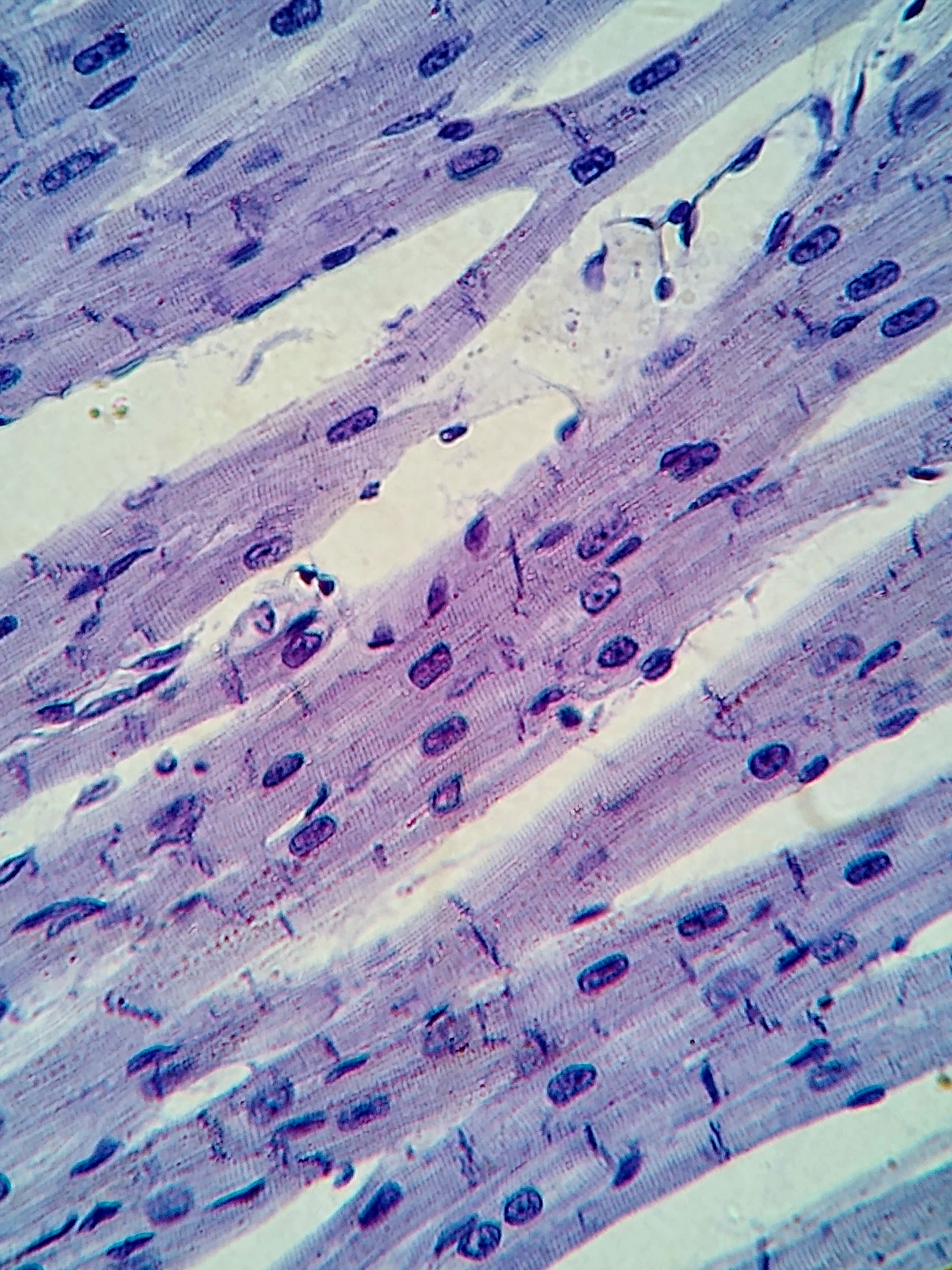
, also referred to as the thick and thin filaments respectively The striations viewed using microscopy of the cardiac muscle are a result of the contrast between the thick and thin filaments. The z-line defines the borders of each sarcomere and act as the connection point between the thin filaments. The t-tubules and sarcoplasmic reticulum are used in conjunction to receive and direct the calcium ions and cause contraction. Once contracted, the clear H-zone between the actin filaments disappears as the filaments move towards each other.
Cardiomyocytes are a particular form of myocyte, only present in heart tissue. Along with the basic myocyte elements, these cells also contain one to four nuclei and a large amount of Adenosine Triphosphate
Adenosine triphosphate (ATP) is a nucleoside triphosphate that provides energy to drive and support many processes in living cell (biology), cells, such as muscle contraction, nerve impulse propagation, and chemical synthesis. Found in all known ...
(ATP). These additions aid in the heart's resistance to fatigue to consistently pump blood throughout the body to deliver oxygen. Most muscle cells contain a triad, which is a joining of 2 terminal cisternae of the sarcoplasmic reticulum and one t- tubule. However, cardiac muscle cells contain a diad, which is a linking of only one sarcoplasmic reticulum with its respective t-tubule. Another notable distinction between all muscle cells and cardiac muscle cells is the presence of intercalated disc
Intercalated discs or lines of Eberth are microscopic identifying features of cardiac muscle. Cardiac muscle consists of individual heart muscle cells (cardiomyocytes) connected by intercalated discs to work as a single functional Syncytium#Cardia ...
s. These tight connections between the cardiomyocytes allows for the accelerated sending of action potential signals to perform the rapid, rhythmic contraction of the heart muscle.
One of the most incredible attributes of cardiac muscle is the ability to automatically beat. This means that even when isolated, for example on a petri dish in an in- vitro setting, the tissue is able to contract and release. This is due to the presence of “ pacemaker cells,” which originate from the sinoatrial node
The sinoatrial node (also known as the sinuatrial node, SA node, sinus node or Keith–Flack node) is an ellipse, oval shaped region of special cardiac muscle in the upper back wall of the right atrium made up of Cell (biology), cells known as pa ...
. This structure allows for spontaneous depolarization, sending signals throughout the tissue.
Transverse-tubules
 Within the sarcolemma of the myocyte, there are specific invaginations referred to as transverse- tubules. These structures attached to the sarcomere z-lines help to promote interaction between the
 extracellular space
Extracellular space refers to the part of a multicellular organism outside the cells, usually taken to be outside the plasma membranes, and occupied by fluid. This is distinguished from intracellular space, which is inside the cells.
The composit ...
and the interior of the cell. Connecting these tubules to the Z line allows for a closer range of excitation- contraction coupling within the cell. Within the t-tubules, distinct ion channels and cellular proteins are present within the t- tubule bilayer that allow movement of calcium influx from the extracellular space into the myocyte to initiate depolarization and contraction. Once traveling through the t- tubules, the calcium arrives at the sarcoplasmic reticulum.
Sarcoplasmic Reticulum
Within the lumen of the cardiac myocyte, the sarcoplasmic reticulum serves as the area of controlling the amount of calcium influx into the interior of the cell. After traveling through the t- tubule, the calcium is stored in the sarcoplasmic reticulum to maintain low concentration of calcium inside the lumen. Upon contraction of this muscle, the cell is depolarized and the calcium is released into the lumen to create theexcitation-contraction coupling
Muscle contraction is the activation of tension-generating sites within muscle cells. In physiology, muscle contraction does not necessarily mean muscle shortening because muscle tension can be produced without changes in muscle length, such as ...
. Once the initial calcium is released, a wave of additional calcium is discharged from the sarcoplasmic reticulum to maintain the contraction integrity.
The tension felt in muscles from prolonged contraction can be attributed to extended release of calcium ions through the sarcoplasmic reticulum. By absorbing calcium ions after contraction, the sarcoplasmic reticulum can regulate muscle fatigue and prevent overuse damage within the body.
Voltage-Gated Calcium Channels
Voltage- gated calcium channels play a critical role in controlling the influx of calcium ions into the myocyte in response to the changingaction potential
An action potential (also known as a nerve impulse or "spike" when in a neuron) is a series of quick changes in voltage across a cell membrane. An action potential occurs when the membrane potential of a specific Cell (biology), cell rapidly ri ...
of the sarcoplasmic membrane. The increase in action potential of the cell indicates depolarization of the cell, directly opening the ion channels to cause muscular contraction. When the action potential decreases, the ion channels close, preventing any calcium influx and further muscular contraction. This fluctuation within the myocyte contributes to the rhythmic “pacemaking” of the cardiac tissue.
There are two classes of voltage- gated calcium channels, L- type and T- type. L-type calcium channel
The L-type calcium channel (also known as the dihydropyridine channel, or DHP channel) is part of the high-voltage activated family of voltage-dependent calcium channel.
"L" stands for long-lasting referring to the length of activation. This ...
s are more commonly found in myocardial tissue throughout the heart whereas T-type calcium channels are more concentrated in the pacemaker cells of the sinoatrial node. These channels also have slightly different activation levels. The L- type responds to a more positive action potential while the T- type channels are triggered at a more negative action potential. Discrepancies and/ or malfunctioning of these gates can contribute to a number of cardiac conditions, such as bradycardia.
Cardiac Conditions Due to Diad Defects
Because the structural organization of the myocyte is very complex and specific, changes to their arrangement and/ or function can cause cardiac illnesses or defects. For example, a leading cause ofheart failure
Heart failure (HF), also known as congestive heart failure (CHF), is a syndrome caused by an impairment in the heart's ability to Cardiac cycle, fill with and pump blood.
Although symptoms vary based on which side of the heart is affected, HF ...
can be attributed to the lack of t- tubule and sarcoplasmic reticulum junctions or a decreased distance between the structures. This change in structure causes the excitation- coupling response in the myocyte to either lessen significantly or be completely diminished. Therefore, few to no heart contractions would take place causing heart failure. On the contrary, an increase in the distance of the junction can create an increased excitation- coupling response, shown in both hypertension
Hypertension, also known as high blood pressure, is a Chronic condition, long-term Disease, medical condition in which the blood pressure in the artery, arteries is persistently elevated. High blood pressure usually does not cause symptoms i ...
and cardiomyopathy
Cardiomyopathy is a group of primary diseases of the heart muscle. Early on there may be few or no symptoms. As the disease worsens, shortness of breath, feeling tired, and swelling of the legs may occur, due to the onset of heart failure. A ...
. These conditions are proof that even the smallest changes in a complex structure can have long- range consequences.
References
{{improve categories, date=November 2022 Cardiac anatomy Muscular system sr:Дијада