|
Tingible Body Macrophage
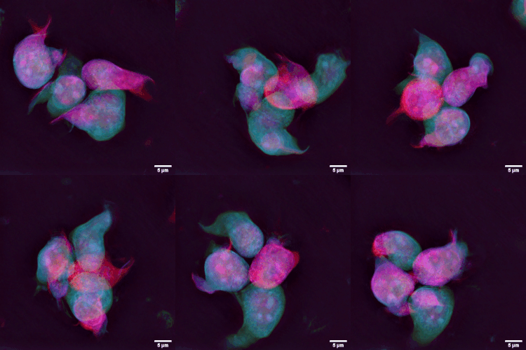
A tingible body macrophage (TBM) is a type of macrophage predominantly found in germinal centers of lymph nodes. They contain many phagocytized, apoptotic cells in various states of degradation, referred to as tingible bodies (''tingible'' meaning stainable). Tingible body macrophages contain condensed chromatin fragments. TBMs are licensed (empowered) for phagocytosis by follicular dendritic cells (FDCs). FDCs provide TBMs with MFGE8 protein, which is a phosphatidylserine-binding "eat me" signal for removal of apoptotic germinal center B cells. It is thought that they may play a role in downregulating the germinal center reaction by the release of prostaglandins and hence a reduced B-cell induction of IL-2. Macrophages that contain debris from ingested lymphocyte A lymphocyte is a type of white blood cell (leukocyte) in the immune system of most vertebrates. Lymphocytes include T cells (for cell-mediated and cytotoxic adaptive immunity), B cells (for humoral, antib ... [...More Info...] [...Related Items...] OR: [Wikipedia] [Google] [Baidu] |
Trends (journals)
''Trends'' is a series of 16 review journals in a range of areas of biology and chemistry published under its Cell Press imprint by Elsevier Elsevier ( ) is a Dutch academic publishing company specializing in scientific, technical, and medical content. Its products include journals such as ''The Lancet'', ''Cell (journal), Cell'', the ScienceDirect collection of electronic journals, .... The publisher in lieu is Danielle Loughlin. The ''Trends'' series was established in 1976 with ''Trends in Biochemical Sciences'', rapidly followed by ''Trends in Neurosciences'', ''Trends in Pharmacological Sciences'', and ''Immunology Today''. ''Immunology Today'', ''Parasitology Today'', and ''Molecular Medicine Today'' changed their names to ''Trends in...'' in 2001. '' Drug Discovery Today'' was spun off as an independent brand. Titles The current set of ''Trends'' journals are all published monthly: References External links * Academic journal series Cell Press academic jo ... [...More Info...] [...Related Items...] OR: [Wikipedia] [Google] [Baidu] |
Lymphadenopathy
Lymphadenopathy or adenopathy is a disease of the lymph nodes, in which they are abnormal in size or consistency. Lymphadenopathy of an inflammatory type (the most common type) is lymphadenitis, producing swollen or enlarged lymph nodes. In clinical practice, the distinction between lymphadenopathy and lymphadenitis is rarely made and the words are usually treated as synonymous. Inflammation of the lymphatic vessels is known as lymphangitis. Infectious lymphadenitis affecting lymph nodes in the neck is often called scrofula. Lymphadenopathy is a common and nonspecific sign. Common causes include infections (from minor causes such as the common cold and post-vaccination swelling to serious ones such as HIV/AIDS), autoimmune diseases, and cancer. Lymphadenopathy is frequently idiopathic and self-limiting. Causes Lymph node enlargement is recognized as a common sign of infectious, autoimmune, or malignant disease. Examples may include: * Reactive: acute infection (e.g., ... [...More Info...] [...Related Items...] OR: [Wikipedia] [Google] [Baidu] |
Lymphocyte
A lymphocyte is a type of white blood cell (leukocyte) in the immune system of most vertebrates. Lymphocytes include T cells (for cell-mediated and cytotoxic adaptive immunity), B cells (for humoral, antibody-driven adaptive immunity), and innate lymphoid cells (ILCs; "innate T cell-like" cells involved in mucosal immunity and homeostasis), of which natural killer cells are an important subtype (which functions in cell-mediated, cytotoxic innate immunity). They are the main type of cell found in lymph, which prompted the name "lymphocyte" (with ''cyte'' meaning cell). Lymphocytes make up between 18% and 42% of circulating white blood cells. Types The three major types of lymphocyte are T cells, B cells and natural killer (NK) cells. They can also be classified as small lymphocytes and large lymphocytes based on their size and appearance. Lymphocytes can be identified by their large nucleus. T cells and B cells T cells (thymus cells) and B cells ( bone marrow- ... [...More Info...] [...Related Items...] OR: [Wikipedia] [Google] [Baidu] |
Interleukin 2
Interleukin-2 (IL-2) is an interleukin, which is a type of cytokine signaling molecule forming part of the immune system. It is a 15.5–16 Dalton (unit), kDa protein that regulates the activities of white blood cells (leukocytes, often lymphocytes) that are responsible for immunity. IL-2 is part of the body's immune response, natural response to microbial infection, and in discriminating between foreign ("non-self") and "self". IL-2 mediates its effects by binding to IL-2 receptors, which are expressed by lymphocytes. The major sources of IL-2 are activated T helper cell, CD4+ T cells and activated CD8+ T cells, CD8+ T cells. Put shortly the function of IL-2 is to stimulate the growth of helper, cytotoxic and regulatory T cells. IL-2 receptor IL-2 is a member of a specific family of cytokines, each member of which has a Helix bundle#Four-helix bundles, four alpha helix bundle; this cytokine family also includes Interleukin-4, IL-4, Interleukin 7, IL-7, Interleukin 9 ... [...More Info...] [...Related Items...] OR: [Wikipedia] [Google] [Baidu] |
Prostaglandin
Prostaglandins (PG) are a group of physiology, physiologically active lipid compounds called eicosanoids that have diverse hormone-like effects in animals. Prostaglandins have been found in almost every Tissue (biology), tissue in humans and other animals. They are derived enzymatically from the fatty acid arachidonic acid. Every prostaglandin contains 20 carbon atoms, including a carbon ring, 5-carbon ring. They are a subclass of eicosanoids and of the prostanoid class of fatty acid derivatives. The structural differences between prostaglandins account for their different biological activities. A given prostaglandin may have different and even opposite effects in different tissues in some cases. The ability of the same prostaglandin to stimulate a reaction in one tissue and inhibit the same reaction in another tissue is determined by the type of receptor (biochemistry), receptor to which the prostaglandin binds. They act as autocrine or paracrine factors with their target cells ... [...More Info...] [...Related Items...] OR: [Wikipedia] [Google] [Baidu] |
B Cell
B cells, also known as B lymphocytes, are a type of the lymphocyte subtype. They function in the humoral immunity component of the adaptive immune system. B cells produce antibody molecules which may be either secreted or inserted into the plasma membrane where they serve as a part of B-cell receptors. When a naïve or memory B cell is activated by an antigen, it proliferates and differentiates into an antibody-secreting effector cell, known as a plasmablast or plasma cell. In addition, B cells Antigen presentation, present antigens (they are also classified as professional Antigen-presenting cell, antigen-presenting cells, APCs) and secrete cytokines. In mammals B cells Cellular differentiation, mature in the bone marrow, which is at the core of most bones. In birds, B cells mature in the bursa of Fabricius, a lymphoid organ where they were first discovered by Chang and Glick, which is why the ''B'' stands for ''bursa'' and not ''bone marrow'', as commonly believed. B cells, unl ... [...More Info...] [...Related Items...] OR: [Wikipedia] [Google] [Baidu] |
Phosphatidylserine
Phosphatidylserine (abbreviated Ptd-L-Ser or PS) is a phospholipid and is a component of the cell membrane. It plays a key role in cell cycle signaling, specifically in relation to apoptosis. It is a key pathway for viruses to enter cells via apoptotic mimicry. Its exposure on the outer surface of a membrane marks the cell for destruction via apoptosis. Structure Phosphatidylserine is a phospholipid—more specifically a glycerophospholipid—which consists of two fatty acids attached in ester linkage to the first and second carbon of glycerol and serine attached through a phosphodiester linkage to the third carbon of the glycerol. Phosphatidylserine sourced from plants differs in fatty acid composition from that sourced from animals. It is commonly found in the inner (cytoplasmic) leaflet of biological membranes. It is almost entirely found in the inner monolayer of the membrane with only less than 10% of it in the outer monolayer. Biosynthesis Phosphatidylserine (PS) is ... [...More Info...] [...Related Items...] OR: [Wikipedia] [Google] [Baidu] |
MFGE8
Milk fat globule-EGF factor 8 protein (Mfge8), also known as lactadherin, is a protein that in humans is encoded by the ''MFGE8'' gene. Species distribution Mfge8 is a secreted protein found in vertebrates, including mammals as well as birds. Function MFGE8 may function as a cell adhesion protein to connect smooth muscle to elastic fibers in arteries. An amyloid fragment of MFGE8 known as medin accumulates in the aorta with aging. MFGE8 in the vasculature of adults can induce recovery from ischemia by facilitating angiogenesis. It has been suggested that antagonizing MFGE8-induced angiogenesis could be a way of fighting cancer. MFGE8 contains a phosphatidylserine (PS) binding domain, as well as an arginine-glycine-aspartic acid motif, which enables the binding to integrins. MFGE8 binds PS, which is exposed on the surface of apoptotic cells. Opsonization Opsonins are extracellular proteins that, when bound to substances or cells, induce phagocytes to phagocytose the subs ... [...More Info...] [...Related Items...] OR: [Wikipedia] [Google] [Baidu] |
Follicular Dendritic Cells
Follicular dendritic cells (FDC) are cells of the immune system found in primary and secondary lymph follicles (lymph nodes) of the B cell areas of the lymphoid tissue. Unlike dendritic cells (DC), FDCs are not derived from the bone-marrow hematopoietic stem cell, but are of mesenchymal origin. Possible functions of FDC include: organizing lymphoid tissue's cells and microarchitecture, capturing antigen to support B cell, promoting debris removal from germinal centers, and protecting against autoimmunity. Disease processes that FDC may contribute include primary FDC-tumor, chronic inflammatory conditions, HIV-1 infection development, and neuroinvasive scrapie. Location and molecular markers Follicular DCs are a non-migratory population found in primary and secondary follicles of the B cell areas of lymph nodes, spleen, and mucosa-associated lymphoid tissue (MALT). They form a stable network due to intercellular connections between FDCs processes and intimate interaction with ... [...More Info...] [...Related Items...] OR: [Wikipedia] [Google] [Baidu] |
Histopathology Of Follicular Cervicitis
Histopathology (compound of three Greek words: 'tissue', 'suffering', and ''-logia'' 'study of') is the microscopic examination of tissue in order to study the manifestations of disease. Specifically, in clinical medicine, histopathology refers to the examination of a biopsy or surgical specimen by a pathologist, after the specimen has been processed and histological sections have been placed onto glass slides. In contrast, cytopathology examines free cells or tissue micro-fragments (as "cell blocks "). Collection of tissues Histopathological examination of tissues starts with surgery, biopsy, or autopsy. The tissue is removed from the body or plant, and then, often following expert dissection in the fresh state, placed in a fixative which stabilizes the tissues to prevent decay. The most common fixative is 10% neutral buffered formalin (corresponding to 3.7% w/v formaldehyde in neutral buffered water, such as phosphate buffered saline). Preparation for histology Th ... [...More Info...] [...Related Items...] OR: [Wikipedia] [Google] [Baidu] |
Chromatin
Chromatin is a complex of DNA and protein found in eukaryote, eukaryotic cells. The primary function is to package long DNA molecules into more compact, denser structures. This prevents the strands from becoming tangled and also plays important roles in reinforcing the DNA during cell division, preventing DNA repair#DNA damage, DNA damage, and regulating gene expression and DNA replication. During mitosis and meiosis, chromatin facilitates proper segregation of the chromosomes in anaphase; the characteristic shapes of chromosomes visible during this stage are the result of DNA being coiled into highly condensed chromatin. The primary protein components of chromatin are histones. An octamer of two sets of four histone cores (Histone H2A, Histone H2B, Histone H3, and Histone H4) bind to DNA and function as "anchors" around which the strands are wound.Maeshima, K., Ide, S., & Babokhov, M. (2019). Dynamic chromatin organization without the 30 nm fiber. ''Current opinion in cell biolog ... [...More Info...] [...Related Items...] OR: [Wikipedia] [Google] [Baidu] |


