|
Sternothyroid Muscle
The sternothyroid muscle (or sternothyroideus) is an infrahyoid muscle of the neck. It acts to depress the hyoid bone. Structure The two muscles are in contact with each other proximally (close to their origin), but diverge distally (towards their insertions). Origin The sternothyroid arises from the posterior surface of the manubrium of the sternum from the midline to the notch for the first rib (inferior to the origin of the sternohyoid muscle), and the posterior margin of the first costal cartilage. Insertion It inserts onto the oblique line of the lamina of thyroid cartilage. Innervation The sternothyroid muscle receives motor innervation from branches of the ansa cervicalis (ultimately derived from cervical spinal nerves C1-C3). Relations The sternothyroid muscle is shorter and wider than the sternohyoid muscle and is situated deep to and partially medial to it. Variations The muscle may be absent or doubled. It may issue accessory slips to the thyrohyoid ... [...More Info...] [...Related Items...] OR: [Wikipedia] [Google] [Baidu] |
Manubrium
The sternum (: sternums or sterna) or breastbone is a long flat bone located in the central part of the chest. It connects to the ribs via cartilage and forms the front of the rib cage, thus helping to protect the heart, human lung, lungs, and major blood vessels from injury. Shaped roughly like a necktie, it is one of the largest and longest flat bones of the body. Its three regions are the manubrium, the body, and the xiphoid process. The word ''sternum'' originates from Ancient Greek στέρνον (''stérnon'') 'chest'. Structure The sternum is a narrow, flat bone, forming the middle portion of the front of the chest. The top of the sternum supports the clavicles (collarbones) and its edges join with the costal cartilages of the first two pairs of ribs. The inner surface of the sternum is also the attachment of the sternopericardial ligaments. Its top is also connected to the sternocleidomastoid muscle. The sternum consists of three main parts, listed from the top: * Manubr ... [...More Info...] [...Related Items...] OR: [Wikipedia] [Google] [Baidu] |
Costal Cartilage
Costal cartilage, also known as rib cartilage, are bars of hyaline cartilage that serve to prolong the ribs forward and contribute to the elasticity of the walls of the thorax. Costal cartilage is only found at the anterior ends of the ribs, providing medial extension. Differences from ribs 1-12 The first seven pairs are connected with the sternum; the next three are each articulated with the lower border of the cartilage of the preceding rib; the last two have pointed extremities, which end in the wall of the abdomen. Like the ribs, the costal cartilages vary in their length, breadth, and direction. They increase in length from the first to the seventh, then gradually decrease to the twelfth. Their breadth, as well as that of the intervals between them, diminishes from the first to the last. They are broad at their attachments to the ribs, and taper toward their sternal extremities, excepting the first two, which are of the same breadth throughout, and the sixth, seventh, an ... [...More Info...] [...Related Items...] OR: [Wikipedia] [Google] [Baidu] |
Waynesburg College
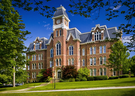
Waynesburg University is a private Christian university in Waynesburg, Pennsylvania, United States. It was established in 1850 and offers undergraduate and graduate programs in more than 70 academic concentrations. The university enrolls around 1,400 students, including approximately 1,100 undergraduates. History In honor of General Anthony Wayne, the university was founded in 1849 as Waynesburg College by the Cumberland Presbyterian Church and was officially established with a charter by the Commonwealth of Pennsylvania in 1850. Waynesburg University is located on a contemporary campus on the hills of southwestern Pennsylvania, and also has facilities in the Greater Pittsburgh Region. Miller Hall and Hanna Hall are listed on the National Register of Historic Places. Academics Waynesburg University offers bachelor's and master's degrees in up to 70 majors and minors. It is accredited by the Middle States Commission on Higher Education. Graduate and professional studies ... [...More Info...] [...Related Items...] OR: [Wikipedia] [Google] [Baidu] |
Goitre
A goitre (British English), or goiter (American English), is a swelling in the neck resulting from an enlarged thyroid gland. A goitre can be associated with a thyroid that is not functioning properly. Worldwide, over 90% of goitre cases are caused by iodine deficiency. The term is from the Latin ''gutturia'', meaning throat. Most goitres are not cancerous ( benign), though they may be potentially harmful. Signs and symptoms A goitre can present as a palpable or visible enlargement of the thyroid gland at the base of the neck. A goitre, if associated with hypothyroidism or hyperthyroidism, may be present with symptoms of the underlying disorder. For hyperthyroidism, the most common symptoms are associated with adrenergic stimulation: tachycardia (increased heart rate), palpitations, nervousness, tremor, increased blood pressure and heat intolerance. Clinical manifestations are often related to hypermetabolism (increased metabolism), excessive thyroid hormone, an in ... [...More Info...] [...Related Items...] OR: [Wikipedia] [Google] [Baidu] |
Pitch (music)
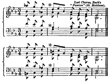
Pitch is a perception, perceptual property that allows sounds to be ordered on a frequency-related scale (music), scale, or more commonly, pitch is the quality that makes it possible to judge sounds as "higher" and "lower" in the sense associated with musical melody, melodies. Pitch is a major auditory system, auditory attribute of musical tones, along with duration (music), duration, loudness, and timbre. Pitch may be quantified as a frequency, but pitch is not a purely objective physical property; it is a subjective Psychoacoustics, psychoacoustical attribute of sound. Historically, the study of pitch and pitch perception has been a central problem in psychoacoustics, and has been instrumental in forming and testing theories of sound representation, processing, and perception in the auditory system. Perception Pitch and frequency Pitch is an auditory sensation in which a listener assigns musical tones to relative positions on a musical scale based primarily on their percep ... [...More Info...] [...Related Items...] OR: [Wikipedia] [Google] [Baidu] |
Carotid Sheath
The carotid sheath is a condensation of the deep cervical fascia enveloping multiple vital neurovascular structures of the neck, including the common and internal carotid arteries, the internal jugular vein, the vagus nerve (CN X), and ansa cervicalis. The carotid sheath helps protects the structures contained therein. Anatomy One carotid sheath is situated on each side of the neck, extending between the base of the skull superiorly and the thorax inferiorly. Superiorly, the carotid sheath encircles the margins of the carotid canal and jugular foramen. Inferiorly, it terminates at the arch of the aorta; it is continuous inferiorly with the axillary sheath at the venous angle. Its inferior end occurs at the level of the first rib and sternum inferiorly (varying between the levels of C7 and T4). Structure The carotid sheath is a fibrous connective tissue formation surrounding several important structures of the neck. It is thicker around the arteries than around the ... [...More Info...] [...Related Items...] OR: [Wikipedia] [Google] [Baidu] |
Inferior Pharyngeal Constrictor
The inferior pharyngeal constrictor muscle is a skeletal muscle of the neck. It is the thickest of the three outer pharyngeal muscles. It arises from the sides of the cricoid cartilage and the thyroid cartilage. It is supplied by the vagus nerve (CN X). It is active during swallowing, and partially during breathing and speech. It may be affected by Zenker's diverticulum. Structure The inferior pharyngeal constrictor muscle is composed of two parts. The first part (and more superior) arises from the thyroid cartilage (thyropharyngeal part), and the second part arises from the cricoid cartilage (cricopharyngeal part). * On the ''thyroid cartilage'', it arises from the oblique line on the side of the lamina, from the surface behind this nearly as far as the posterior border and from the inferior horn of the thyroid cartilage. * From the ''cricoid cartilage'', it arises in the interval between the cricothyroid muscle in front, and the articular facet for the inferior ho ... [...More Info...] [...Related Items...] OR: [Wikipedia] [Google] [Baidu] |
Thyrohyoid
The thyrohyoid muscle is a small skeletal muscle of the neck. Above, it attaches onto the greater cornu of the hyoid bone; below, it attaches onto the oblique line of the thyroid cartilage. It is innervated by fibres derived from the cervical spinal nerve 1 that run with the hypoglossal nerve (CN XII) to reach this muscle. The thyrohyoid muscle depresses the hyoid bone and elevates the larynx during swallowing. By controlling the position and shape of the larynx, it aids in making sound. Structure The thyrohyoid muscle is a small, broad and short muscle. It is quadrilateral in shape. It may be considered a superior-ward continuation of sternothyroid muscle. It belongs to the infrahyoid muscles group and the outer laryngeal muscle group. Attachments Its superior attachment is the inferior border of the greater cornu of the hyoid bone and adjacent portions of the body of hyoid bone. Its inferior attachment is the oblique line of the thyroid cartilage (alongside the ster ... [...More Info...] [...Related Items...] OR: [Wikipedia] [Google] [Baidu] |
Cervical Spinal Nerve
A spinal nerve is a mixed nerve, which carries motor, sensory, and autonomic signals between the spinal cord and the body. In the human body there are 31 pairs of spinal nerves, one on each side of the vertebral column. These are grouped into the corresponding cervical, thoracic, lumbar, sacral and coccygeal regions of the spine. There are eight pairs of cervical nerves, twelve pairs of thoracic nerves, five pairs of lumbar nerves, five pairs of sacral nerves, and one pair of coccygeal nerves. The spinal nerves are part of the peripheral nervous system. Structure Each spinal nerve is a mixed nerve, formed from the combination of nerve root fibers from its dorsal and ventral roots. The dorsal root is the afferent sensory root and carries sensory information to the brain. The ventral root is the efferent motor root and carries motor information from the brain. The spinal nerve emerges from the spinal column through an opening (intervertebral foramen) between adjacent ... [...More Info...] [...Related Items...] OR: [Wikipedia] [Google] [Baidu] |
Ansa Cervicalis
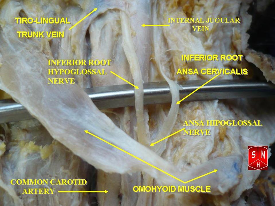
The ansa cervicalis (or ansa hypoglossi in older literature) is a loop formed by muscular branches of the cervical plexus formed by branches of cervical spinal nerves C1-C3. The ansa cervicalis has two roots - a superior root (formed by branch of C1) and an inferior root (formed by union of branches of C2 and C3) - that unite distally, forming a loop. It is situated anterior to the carotid sheath. Branches of the ansa cervicalis innervate three of the four infrahyoid muscles: the sternothyroid, sternohyoid, and omohyoid muscles (note that the thyrohyoid muscle is the one infrahyoid muscle not innervated by the ansa cervicalis - it is instead innervated by cervical spinal nerve 1 via a separate thyrohyoid branch). Its name means "handle of the neck" in Latin. Anatomy The ansa cervicalis is typically embedded within the anterior wall of the carotid sheath anterior to the internal jugular vein. Superior root The superior root of the ansa cervicalis (formerly known as d ... [...More Info...] [...Related Items...] OR: [Wikipedia] [Google] [Baidu] |
First Rib
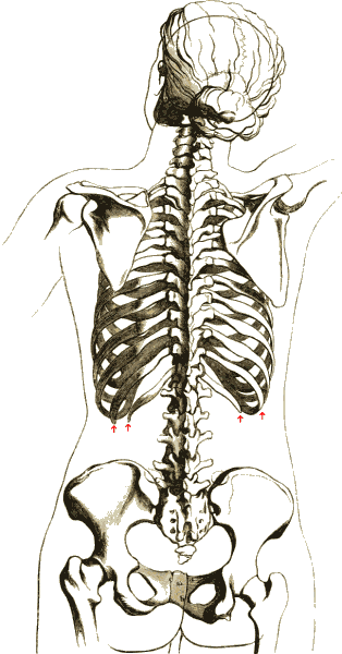
The rib cage or thoracic cage is an endoskeletal enclosure in the thorax of most vertebrates that comprises the ribs, vertebral column and sternum, which protect the vital organs of the thoracic cavity, such as the heart, lungs and great vessels and support the shoulder girdle to form the core part of the axial skeleton. A typical human thoracic cage consists of 12 pairs of ribs and the adjoining costal cartilages, the sternum (along with the manubrium and xiphoid process), and the 12 thoracic vertebrae articulating with the ribs. The thoracic cage also provides attachments for extrinsic skeletal muscles of the neck, upper limbs, upper abdomen and back, and together with the overlying skin and associated fascia and muscles, makes up the thoracic wall. In tetrapods, the rib cage intrinsically holds the muscles of respiration ( diaphragm, intercostal muscles, etc.) that are crucial for active inhalation and forced exhalation, and therefore has a major ventilatory function in th ... [...More Info...] [...Related Items...] OR: [Wikipedia] [Google] [Baidu] |


