|
SPECT
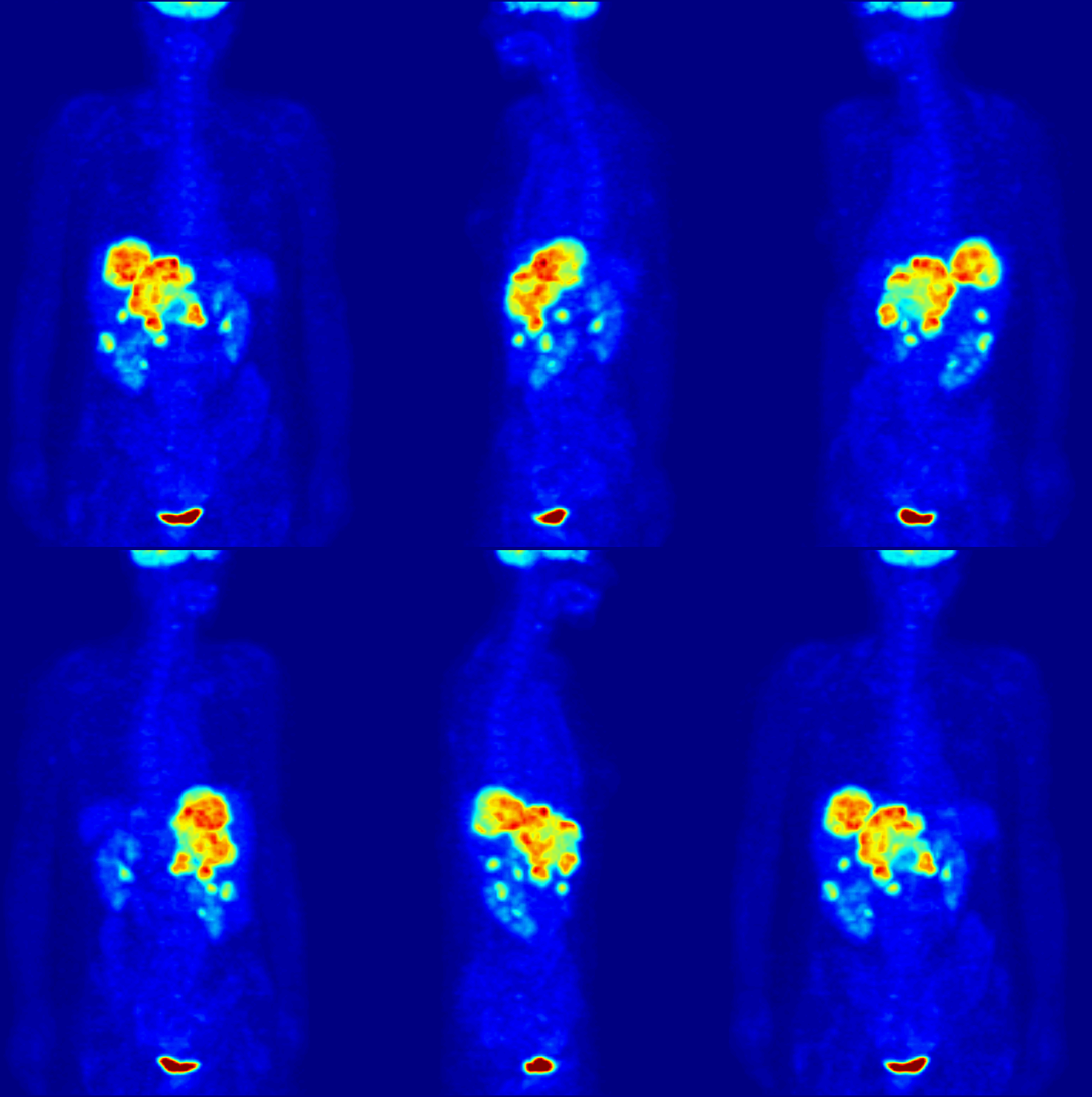
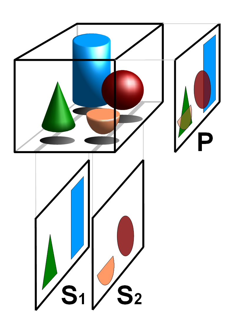
Single-photon emission computed tomography (SPECT, or less commonly, SPET) is a nuclear medicine tomographic imaging technique using gamma rays. It is very similar to conventional nuclear medicine planar imaging using a gamma camera (that is, scintigraphy), but is able to provide true 3D information. This information is typically presented as cross-sectional slices through the patient, but can be freely reformatted or manipulated as required. The technique needs delivery of a gamma-emitting radioisotope (a radionuclide) into the patient, normally through injection into the bloodstream. On occasion, the radioisotope is a simple soluble dissolved ion, such as an isotope of gallium(III). Usually, however, a marker radioisotope is attached to a specific ligand to create a radioligand, whose properties bind it to certain types of tissues. This marriage allows the combination of ligand and radiopharmaceutical to be carried and bound to a place of interest in the body, where th ... [...More Info...] [...Related Items...] OR: [Wikipedia] [Google] [Baidu] |
Radioligand
A radioligand is a microscopic particle which consists of a Radiopharmaceutical, therapeutic radioactive isotope and the cell-targeting compound - the ligand. The ligand is the target binding site, it may be on the surface of the targeted cancer cell for therapeutic purposes. Radioisotopes can occur naturally or be synthesized and produced in a cyclotron/nuclear reactor. The different types of radioisotopes include Y-90, H-3, C-11, Lu-177, Ac-225, Ra-223, In-111, I-131, I-125, etc. Thus, radioligands must be produced in special nuclear reactors for the radioisotope to remain stable. Radioligands can be used to analyze/characterize receptors, to perform binding assays, to help in diagnostic imaging, and to provide targeted cancer therapy. Radiation is a novel method of treating cancer and is effective in short distances along with being unique/personalizable and causing minimal harm to normal surrounding cells. Furthermore, radioligand binding can provide information about receptor-l ... [...More Info...] [...Related Items...] OR: [Wikipedia] [Google] [Baidu] |
Nuclear Medicine
Nuclear medicine (nuclear radiology, nucleology), is a medical specialty involving the application of radioactivity, radioactive substances in the diagnosis and treatment of disease. Nuclear imaging is, in a sense, ''radiology done inside out'', because it records radiation radiant exitance, emitted from within the body rather than radiation that is transmittance, transmitted through the body from external sources like X-ray generators. In addition, nuclear medicine scans differ from radiology, as the emphasis is not on imaging anatomy, but on the function. For such reason, it is called a Functional imaging, physiological imaging modality. Single photon emission computed tomography (SPECT) and positron emission tomography (PET) scans are the two most common imaging modalities in nuclear medicine. Diagnostic medical imaging Diagnostic In nuclear medicine imaging, radiopharmaceuticals are taken internally, for example, through inhalation, intravenously, or orally. Then, externa ... [...More Info...] [...Related Items...] OR: [Wikipedia] [Google] [Baidu] |
Bone Scintigraphy
A bone scan or bone scintigraphy is a nuclear medicine imaging technique used to help diagnose and assess different bone diseases. These include cancer of the bone or metastasis, location of bone inflammation and fractures (that may not be visible in traditional X-ray images), and bone infection (osteomyelitis). Nuclear medicine provides functional imaging and allows visualisation of bone metabolism or bone remodeling, which most other imaging techniques (such as X-ray computed tomography, CT) cannot. Bone scintigraphy competes with positron emission tomography (PET) for imaging of abnormal metabolism in bones, but is considerably less expensive. Bone scintigraphy has higher sensitivity but lower specificity than CT or MRI for diagnosis of scaphoid fractures following negative plain radiography. History Some of the earliest investigations into skeletal metabolism were carried out by George de Hevesy in the 1930s, using phosphorus-32 and by Charles Pecher in the 1940s. ... [...More Info...] [...Related Items...] OR: [Wikipedia] [Google] [Baidu] |
Positron Emission Tomography
Positron emission tomography (PET) is a functional imaging technique that uses radioactive substances known as radiotracers to visualize and measure changes in metabolic processes, and in other physiological activities including blood flow, regional chemical composition, and absorption. Different tracers are used for various imaging purposes, depending on the target process within the body, such as: * Fluorodeoxyglucose ( 18F">sup>18FDG or FDG) is commonly used to detect cancer; * 18Fodium fluoride">sup>18Fodium fluoride (Na18F) is widely used for detecting bone formation; * Oxygen-15 (15O) is sometimes used to measure blood flow. PET is a common imaging technique, a medical scintillography technique used in nuclear medicine. A radiopharmaceutical—a radioisotope attached to a drug—is injected into the body as a tracer. When the radiopharmaceutical undergoes beta plus decay, a positron is emitted, and when the positron interacts with an ordinary electron, the tw ... [...More Info...] [...Related Items...] OR: [Wikipedia] [Google] [Baidu] |
Gated SPECT
Gated SPECT is a nuclear medicine imaging technique, typically for the heart in myocardial perfusion imagery. An electrocardiogram (ECG) guides the image acquisition, and the resulting set of single-photon emission computed tomography (SPECT) images shows the heart as it contracts over the interval from one R wave to the next. Gated myocardial perfusion imaging has been shown to have high prognostic value and sensitivity for critical stenosis. The acquisition computer defines the number of time bins or frames to divide the R to R interval of the patient's electrocardiogram. A "window" may be set which discards data from R to R intervals which deviate from some amount from the patient's average R to R wave duration. This discards preventricular contractions and arrhythmias from the acquisition and improves the quality of the resulting study. The gamma camera will take a series of pictures around the patient, dividing each 'step' of the camera head's motion into the predetermin ... [...More Info...] [...Related Items...] OR: [Wikipedia] [Google] [Baidu] |
Gamma Camera
A gamma camera (γ-camera), also called a scintillation camera or Anger camera, is a device used to image gamma radiation emitting radioisotopes, a technique known as scintigraphy. The applications of scintigraphy include early drug development and nuclear medical imaging to view and analyse images of the human body or the distribution of medically injected, inhaled, or ingested radionuclides emitting gamma rays. Imaging techniques Scintigraphy ("scint") is the use of gamma cameras to capture emitted radiation from internal radioisotopes to create two-dimensional images. SPECT (single photon emission computed tomography) imaging, as used in nuclear cardiac stress testing, is performed using gamma cameras. Usually one, two or three detectors or heads, are slowly rotated around the patient. Construction A gamma camera consists of one or more flat crystal planes (or detectors) optically coupled to an array of photomultiplier tubes in an assembly known as a "head", mounted ... [...More Info...] [...Related Items...] OR: [Wikipedia] [Google] [Baidu] |
Radionuclide Angiography
Radionuclide angiography is an area of nuclear medicine which specialises in imaging to show the functionality of the right and left ventricles of the heart, thus allowing informed diagnostic intervention in heart failure. It involves use of a radiopharmaceutical, injected into a patient, and a gamma camera for acquisition. A MUGA scan (multigated acquisition) involves an acquisition triggered (gated) at different points of the cardiac cycle. MUGA scanning is also called equilibrium radionuclide angiocardiography, radionuclide ventriculography (RNVG), or gated blood pool imaging, as well as SYMA scanning (synchronized multigated acquisition scanning). This mode of imaging uniquely provides a cine type of image of the beating heart, and allows the interpreter to determine the efficiency of the individual heart valves and chambers. MUGA/Cine scanning represents a robust adjunct to the now more common echocardiogram. Mathematics regarding acquisition of cardiac output (''Q' ... [...More Info...] [...Related Items...] OR: [Wikipedia] [Google] [Baidu] |
Scintigraphy
Scintigraphy (from Latin ''scintilla'', "spark"), also known as a gamma scan, is a diagnostic test in nuclear medicine, where radioisotopes attached to drugs that travel to a specific organ or tissue (radiopharmaceuticals) are taken internally and the emitted gamma ray, gamma radiation is captured by gamma cameras, which are external detectors that form two-dimensional images in a process similar to the capture of radiography, x-ray images. In contrast, Single photon emission computed tomography, SPECT and positron emission tomography (PET) form 3-dimensional images and are therefore classified as separate techniques from scintigraphy, although they also use gamma cameras to detect internal radiation. Scintigraphy is unlike a diagnostic X-ray where external radiation is passed through the body to form an image. Process Scintillography is an Molecular imaging, imaging method of nuclear events provoked by collisions or charged current interactions among nuclear particles or ioniz ... [...More Info...] [...Related Items...] OR: [Wikipedia] [Google] [Baidu] |
Technetium Exametazime
Technetium (99mTc) exametazime is a radiopharmaceutical sold under the trade name Ceretec, and is used by nuclear medicine physicians for the detection of altered regional cerebral perfusion in stroke and other cerebrovascular diseases. It can also be used for the labelling of leukocytes to localise intra-abdominal infections and inflammatory bowel disease. Exametazime (the part without technetium) is sometimes referred to as ''hexamethylpropylene amine oxime'' or ''HMPAO'', although correct chemical names are: *(NE)-N-[(3R)-3-3-(2R,3E)-3-hydroxyiminobutan-2-yl]amino]-2,2-dimethylpropyl]amino]butan-2-ylidene]hydroxylamine *or 3,3'-((2,2,-dimethyl-1,3-propanediyl)diimino)bis-2-butanone dioxime. Chemistry The drug consists of exametazime as a chelating agent for the radioisotope technetium-99m. Both enantiomeric forms of exametazime are used—the drug is racemic. The third stereoisomer of this structure, the meso form, ''meso'' form, is not included. References External l ... [...More Info...] [...Related Items...] OR: [Wikipedia] [Google] [Baidu] |
Tomography
Tomography is imaging by sections or sectioning that uses any kind of penetrating wave. The method is used in radiology, archaeology, biology, atmospheric science, geophysics, oceanography, plasma physics, materials science, cosmochemistry, astrophysics, quantum information Quantum information is the information of the state of a quantum system. It is the basic entity of study in quantum information theory, and can be manipulated using quantum information processing techniques. Quantum information refers to both t ..., and other areas of science. The word ''tomography'' is derived from Ancient Greek τόμος ''tomos'', "slice, section" and γράφω ''graphō'', "to write" or, in this context as well, "to describe." A device used in tomography is called a tomograph, while the image produced is a tomogram. In many cases, the production of these images is based on the mathematical procedure tomographic reconstruction, such as X-ray computed tomography technically being pro ... [...More Info...] [...Related Items...] OR: [Wikipedia] [Google] [Baidu] |
Gamma Ray
A gamma ray, also known as gamma radiation (symbol ), is a penetrating form of electromagnetic radiation arising from high energy interactions like the radioactive decay of atomic nuclei or astronomical events like solar flares. It consists of the shortest wavelength electromagnetic waves, typically shorter than those of X-rays. With frequencies above 30 exahertz () and wavelengths less than 10 picometers (), gamma ray photons have the highest photon energy of any form of electromagnetic radiation. Paul Villard, a French chemist and physicist, discovered gamma radiation in 1900 while studying radiation emitted by radium. In 1903, Ernest Rutherford named this radiation ''gamma rays'' based on their relatively strong penetration of matter; in 1900, he had already named two less penetrating types of decay radiation (discovered by Henri Becquerel) alpha rays and beta rays in ascending order of penetrating power. Gamma rays from radioactive decay are in the energy range ... [...More Info...] [...Related Items...] OR: [Wikipedia] [Google] [Baidu] |


