|
Speckle Variance Optical Coherence Tomography
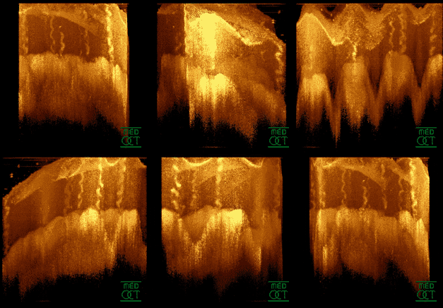
Speckle variance optical coherence tomography (SV-OCT) is an imaging algorithm for functional optical imaging. Optical coherence tomography is an imaging modality that uses low-coherence interferometry to obtain high resolution, depth-resolved volumetric images. OCT can be used to capture functional images of blood flow, a technique known as optical coherence tomography angiography (OCT-A). SV-OCT is one method for OCT-A that uses the variance of consecutively acquired images to detect flow at the micron scale. SV-OCT can be used to measure the microvasculature of tissue. In particular, it is useful in ophthalmology for visualizing blood flow in retinal and choroidal regions of the eye, which can provide information on the pathophysiology of diseases. Introduction Color fundus photography, fluorescein angiography (FA) and indocyanine green angiography (ICGA) are methods for imaging retinal microvasculature networks. However, these methods have drawbacks in that they require the use ... [...More Info...] [...Related Items...] OR: [Wikipedia] [Google] [Baidu] |
Optical Coherence Tomography
Optical coherence tomography (OCT) is an imaging technique that uses low-coherence light to capture micrometer-resolution, two- and three-dimensional images from within optical scattering media (e.g., biological tissue). It is used for medical imaging and industrial nondestructive testing (NDT). Optical coherence tomography is based on low-coherence interferometry, typically employing near-infrared light. The use of relatively long wavelength light allows it to penetrate into the scattering medium. Confocal microscopy, another optical technique, typically penetrates less deeply into the sample but with higher resolution. Depending on the properties of the light source (superluminescent diodes, ultrashort pulsed lasers, and supercontinuum lasers have been employed), optical coherence tomography has achieved sub-micrometer resolution (with very wide-spectrum sources emitting over a ~100 nm wavelength range). Optical coherence tomography is one of a class of optical tom ... [...More Info...] [...Related Items...] OR: [Wikipedia] [Google] [Baidu] |
Optical Coherence Tomography Angiography
Optical coherence tomography angiography (OCTA) is a non-invasive imaging technique based on optical coherence tomography (OCT) developed to visualize vascular networks in the human retina, choroid, skin and various animal models. OCTA may make use of speckle variance optical coherence tomography. OCTA uses low-coherence interferometry to measure changes in backscattered signal to differentiate areas of blood flow from areas of static tissue. To correct for patient movement during scanning, bulk tissue changes in the axial direction are eliminated, ensuring that all detected changes are due to red blood cell movement. This form of OCT requires a very high sampling density in order to achieve the resolution needed to detect the tiny capillaries found in the retina. Recent advancements in OCT acquisition speed have made it possible the required sampling density to obtain a high enough resolution for OCTA. This has allowed OCTA to become widely used clinically to diagnose a va ... [...More Info...] [...Related Items...] OR: [Wikipedia] [Google] [Baidu] |
Ophthalmology
Ophthalmology ( ) is a surgical subspecialty within medicine that deals with the diagnosis and treatment of eye disorders. An ophthalmologist is a physician who undergoes subspecialty training in medical and surgical eye care. Following a medical degree, a doctor specialising in ophthalmology must pursue additional postgraduate residency training specific to that field. This may include a one-year integrated internship that involves more general medical training in other fields such as internal medicine or general surgery. Following residency, additional specialty training (or fellowship) may be sought in a particular aspect of eye pathology. Ophthalmologists prescribe medications to treat eye diseases, implement laser therapy, and perform surgery when needed. Ophthalmologists provide both primary and specialty eye care - medical and surgical. Most ophthalmologists participate in academic research on eye diseases at some point in their training and many include research as p ... [...More Info...] [...Related Items...] OR: [Wikipedia] [Google] [Baidu] |
Indocyanine Green Angiography
Indocyanine green angiography (ICGA) is a diagnostic procedure used to examine choroidal blood flow and associated pathology. Indocyanine green (ICG) is a water soluble cyanine dye which shows fluorescence in near-infrared (790–805 nm) range, with peak spectral absorption of 800-810 nm in blood. The near infrared light used in ICGA penetrates ocular pigments such as melanin and xanthophyll, as well as exudates and thin layers of sub-retinal vessels. Age-related macular degeneration is the third main cause of blindness worldwide, and it is the leading cause of blindness in industrialized countries. Indocyanine green angiography is widely used to study choroidal neovascularization in patients with exudative age-related macular degeneration. In nonexudative AMD, ICGA is used in classification of drusen and associated subretinal deposits. Indications Indications for indocyanine green angiography include: * Choroidal neovascularisation (CNV):Indocyanine green angiography is widely used ... [...More Info...] [...Related Items...] OR: [Wikipedia] [Google] [Baidu] |
Diabetic Retinopathy
Diabetic retinopathy (also known as diabetic eye disease), is a medical condition in which damage occurs to the retina due to diabetes mellitus. It is a leading cause of blindness in developed countries. Diabetic retinopathy affects up to 80 percent of those who have had both type 1 and type 2 diabetes for 20 years or more. In at least 90% of new cases, progression to more aggressive forms of sight threatening retinopathy and maculopathy could be reduced with proper treatment and monitoring of the eyes The longer a person has diabetes, the higher his or her chances of developing diabetic retinopathy. Each year in the United States, diabetic retinopathy accounts for 12% of all new cases of blindness. It is also the leading cause of blindness in people aged 20 to 64. Signs and symptoms Nearly all people with diabetes develop some degree of retina damage ("retinopathy") over several decades with the disease. For many, that damage can only be detected by a retinal exam, and h ... [...More Info...] [...Related Items...] OR: [Wikipedia] [Google] [Baidu] |

