|
Smooth Muscles
Smooth muscle is one of the three major types of vertebrate muscle tissue, the others being skeletal and cardiac muscle. It can also be found in invertebrates and is controlled by the autonomic nervous system. It is non- striated, so-called because it has no sarcomeres and therefore no striations (''bands'' or ''stripes''). It can be divided into two subgroups, ''single-unit'' and ''multi-unit'' smooth muscle. Within single-unit muscle, the whole bundle or sheet of smooth muscle cells contracts as a syncytium. Smooth muscle is found in the walls of hollow organs, including the stomach, intestines, bladder and uterus. In the walls of blood vessels, and lymph vessels, (excluding blood and lymph capillaries) it is known as vascular smooth muscle. There is smooth muscle in the tracts of the respiratory, urinary, and reproductive systems. In the eyes, the ciliary muscles, iris dilator muscle, and iris sphincter muscle are types of smooth muscles. The iris dilator and sphincter muscles ... [...More Info...] [...Related Items...] OR: [Wikipedia] [Google] [Baidu] |
Vertebrate
Vertebrates () are animals with a vertebral column (backbone or spine), and a cranium, or skull. The vertebral column surrounds and protects the spinal cord, while the cranium protects the brain. The vertebrates make up the subphylum Vertebrata with some 65,000 species, by far the largest ranked grouping in the phylum Chordata. The vertebrates include mammals, birds, amphibians, and various classes of fish and reptiles. The fish include the jawless Agnatha, and the jawed Gnathostomata. The jawed fish include both the Chondrichthyes, cartilaginous fish and the Osteichthyes, bony fish. Bony fish include the Sarcopterygii, lobe-finned fish, which gave rise to the tetrapods, the animals with four limbs. Despite their success, vertebrates still only make up less than five percent of all described animal species. The first vertebrates appeared in the Cambrian explosion some 518 million years ago. Jawed vertebrates evolved in the Ordovician, followed by bony fishes in the Devonian. T ... [...More Info...] [...Related Items...] OR: [Wikipedia] [Google] [Baidu] |
Vascular Smooth Muscle
Vascular smooth muscle is the type of smooth muscle that makes up most of the walls of blood vessels. Structure Vascular smooth muscle refers to the particular type of smooth muscle found within, and composing the majority of the wall of blood vessels. Nerve supply Vascular smooth muscle is innervated primarily by the sympathetic nervous system through adrenergic receptors (adrenoceptors). The three types present are: alpha-1, alpha-2 and beta-2 adrenergic receptors, . The main endogenous agonist of these cell receptors is norepinephrine (NE). The adrenergic receptors exert opposite physiologic effects in the vascular smooth muscle under activation: * alpha-1 receptors. Under NE binding alpha-1 receptors cause vasoconstriction (contraction of the vascular smooth muscle cells decreasing the diameter of the vessels). These receptors are activated in response to shock or low blood pressure as a defensive reaction trying to restore the normal blood pressure. Antagonists o ... [...More Info...] [...Related Items...] OR: [Wikipedia] [Google] [Baidu] |
Arrector Pili
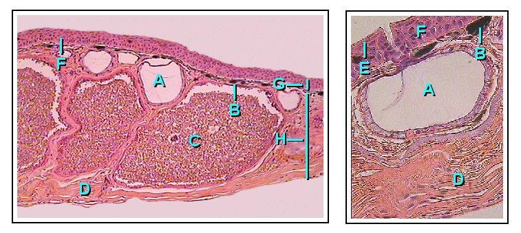
The arrector pili muscles, also known as hair erector muscles, are small muscles attached to hair follicles in mammals. Contraction of these muscles causes the hairs to stand on end, known colloquially as goose bumps (piloerection). Structure Each arrector pili is composed of a bundle of smooth muscle fibres which attach to several follicles (a follicular unit). Each is innervated by the sympathetic division of the autonomic nervous system. The muscle attaches to the follicular stem cell niche in the follicular bulge, splitting at their deep end to encircle the follicle. Function The contraction of the muscle is involuntary. Stresses such as cold, fear etc. may stimulate the sympathetic nervous system, and thus cause muscle contraction. Thermal insulation Contraction of arrector pili muscles have a principal function in the majority of mammals of providing thermal insulation. Air becomes trapped between the erect hairs, helping the animal retain heat. Self defence M ... [...More Info...] [...Related Items...] OR: [Wikipedia] [Google] [Baidu] |
Skin
Skin is the layer of usually soft, flexible outer tissue covering the body of a vertebrate animal, with three main functions: protection, regulation, and sensation. Other animal coverings, such as the arthropod exoskeleton, have different developmental origin, structure and chemical composition. The adjective cutaneous means "of the skin" (from Latin ''cutis'' 'skin'). In mammals, the skin is an organ of the integumentary system made up of multiple layers of ectodermal tissue and guards the underlying muscles, bones, ligaments, and internal organs. Skin of a different nature exists in amphibians, reptiles, and birds. Skin (including cutaneous and subcutaneous tissues) plays crucial roles in formation, structure, and function of extraskeletal apparatus such as horns of bovids (e.g., cattle) and rhinos, cervids' antlers, giraffids' ossicones, armadillos' osteoderm, and os penis/ os clitoris. All mammals have some hair on their skin, even marine mammals like whales, ... [...More Info...] [...Related Items...] OR: [Wikipedia] [Google] [Baidu] |
Accommodation (vertebrate Eye)
Accommodation is the process by which the vertebrate eye changes optical power to maintain a clear image or focus on an object as its distance varies. In this, distances vary for individuals from the far point—the maximum distance from the eye for which a clear image of an object can be seen, to the near point—the minimum distance for a clear image. Accommodation usually acts like a reflex, including part of the accommodation-convergence reflex, but it can also be consciously controlled. The main ways animals may change focus are: * Changing the shape of the lens. * Changing the position of the lens relative to the retina. * Changing the axial length of the eyeball. * Changing the shape of the cornea. Focusing mechanisms Focusing the light scattered by objects in a three dimensional environment into a two dimensional collection of individual bright points of light requires the light to be bent. To get a good image of these points of light on a defined area requires a p ... [...More Info...] [...Related Items...] OR: [Wikipedia] [Google] [Baidu] |
Lens (anatomy)
The lens, or crystalline lens, is a Transparency and translucency, transparent Biconvex lens, biconvex structure in most land vertebrate eyes. Relatively long, thin fiber cells make up the majority of the lens. These cells vary in architecture and are arranged in concentric layers. New layers of cells are recruited from a thin epithelium at the front of the lens, just below the basement membrane surrounding the lens. As a result the vertebrate lens grows throughout life. The surrounding lens membrane referred to as the lens capsule also grows in a systematic way, ensuring the lens maintains an optically suitable shape in concert with the underlying fiber cells. Thousands of suspensory ligaments are embedded into the capsule at its largest diameter which suspend the lens within the eye. Most of these lens structures are derived from the epithelium of the embryo before birth. Along with the cornea, aqueous humour, aqueous, and vitreous humours, the lens Refraction, refracts light, Fo ... [...More Info...] [...Related Items...] OR: [Wikipedia] [Google] [Baidu] |
Miosis
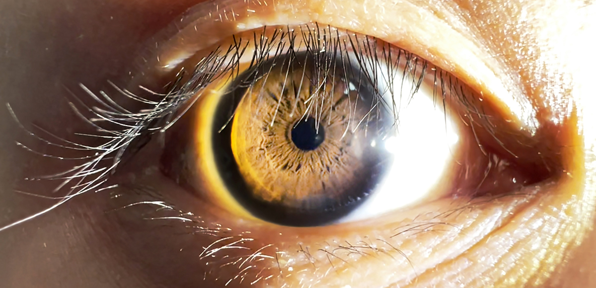
Miosis, or myosis (), is excessive constriction of the pupil. citing: Mosby's Medical Dictionary, 8th ed. The opposite condition, mydriasis, is the dilation of the pupil. Anisocoria is the condition of one pupil being more dilated than the other. Causes Age * Senile miosis (a reduction in the size of a person's pupil in old age)Diseases *[...More Info...] [...Related Items...] OR: [Wikipedia] [Google] [Baidu] |
Mydriasis
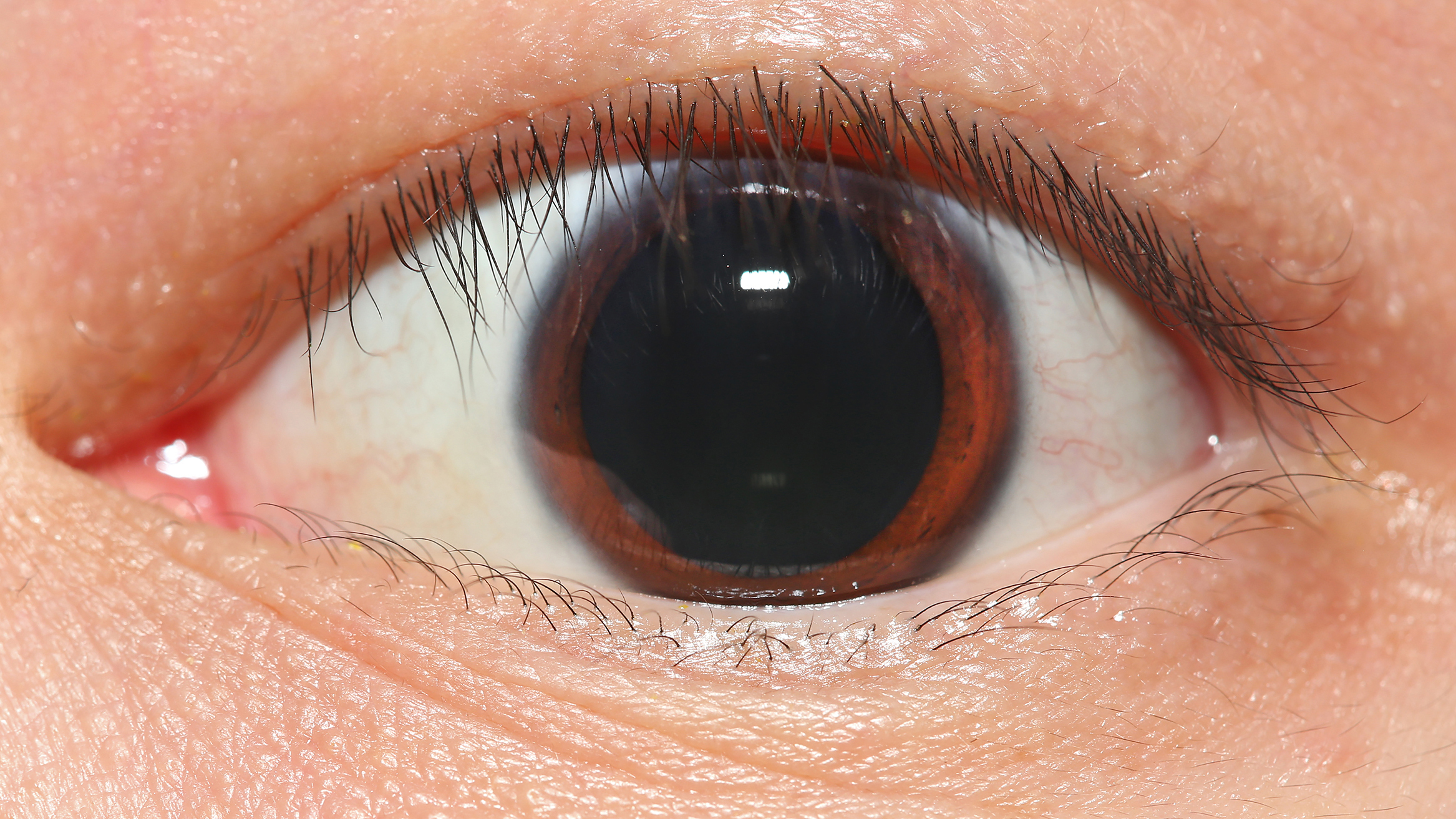
Mydriasis is the Pupillary dilation, dilation of the pupil, usually having a non-physiological cause, or sometimes a physiological pupillary response. Non-physiological causes of mydriasis include disease, Physical trauma, trauma, or the use of certain types of drug, drugs. It may also be of unknown cause. Normally, as part of the pupillary light reflex, the pupil dilates in the dark and miosis, constricts in the light to respectively improve vividity at night and to protect the retina from sunlight damage during the day. A ''mydriatic'' pupil will remain excessively large even in a bright environment. The excitation of the radial fibres of the iris which increases the pupillary aperture is referred to as a mydriasis. More generally, mydriasis also refers to the natural dilation of pupils, for instance in low light conditions or under sympathetic stimulation. Mydriasis is frequently induced by drugs for certain Ophthalmology, ophthalmic examinations and procedures, particularly th ... [...More Info...] [...Related Items...] OR: [Wikipedia] [Google] [Baidu] |
Iris Sphincter Muscle
The iris sphincter muscle (pupillary sphincter, pupillary constrictor, circular muscle of iris, circular fibers) is a muscle in the part of the eye called the iris. It encircles the pupil of the iris, appropriate to its function as a constrictor of the pupil. The ciliary muscle, pupillary sphincter muscle and pupillary dilator muscle sometimes are called intrinsic ocular muscles or intraocular muscles. Comparative anatomy This structure is found in vertebrates and in some cephalopods. General structure All the myocytes are of the smooth muscle type. Its dimensions are about 0.75 mm wide by 0.15 mm thick. Mode of action In humans, it functions to constrict the pupil in bright light (pupillary light reflex) or during accommodation. In , the muscle cells themselves are photosensitive causing iris action without brain input. Innervation It is controlled by parasympathetic postganglionic fibers releasing acetylcholine acting primarily on the muscarinic acetylc ... [...More Info...] [...Related Items...] OR: [Wikipedia] [Google] [Baidu] |
Iris Dilator Muscle
The iris dilator muscle (pupil dilator muscle, pupillary dilator, radial muscle of iris, radiating fibers), is a smooth muscle of the eye, running radially in the iris and therefore fit as a dilator. The pupillary dilator consists of a spokelike arrangement of modified contractile cells called myoepithelial cells. These cells are stimulated by the sympathetic nervous system. When stimulated, the cells contract, widening the pupil and allowing more light to enter the eye. The ciliary muscle, pupillary sphincter muscle and pupillary dilator muscle sometimes are called intrinsic ocular muscles or intraocular muscles. Structure Innervation It is innervated by the sympathetic system, which acts by releasing noradrenaline, which acts on α1-receptors. Thus, when presented with a threatening stimulus that activates the fight-or-flight response, this innervation contracts the muscle and dilates the pupil, thus temporarily letting more light reach the retina. The dilator muscl ... [...More Info...] [...Related Items...] OR: [Wikipedia] [Google] [Baidu] |
Ciliary Muscle
The ciliary muscle is an intrinsic muscle of the eye formed as a ring of smooth muscleSchachar, Ronald A. (2012). "Anatomy and Physiology." (Chapter 4) . in the eye's middle layer, the uvea ( vascular layer). It controls accommodation for viewing objects at varying distances and regulates the flow of aqueous humor into Schlemm's canal. It also changes the shape of the lens within the eye but not the size of the pupil which is carried out by the sphincter pupillae muscle and dilator pupillae. The ciliary muscle, pupillary sphincter muscle and pupillary dilator muscle sometimes are called intrinsic ocular muscles or intraocular muscles. Structure Development The ciliary muscle develops from mesenchyme within the choroid and is considered a cranial neural crest derivative. Nerve supply The ciliary muscle receives parasympathetic fibers from the short ciliary nerves that arise from the ciliary ganglion. The parasympathetic postganglionic fibers are part of cranial n ... [...More Info...] [...Related Items...] OR: [Wikipedia] [Google] [Baidu] |
Organ System
An organ system is a biological system consisting of a group of organ (biology), organs that work together to perform one or more bodily functions. Each organ has a specialized role in an organism body, and is made up of distinct Tissue (biology), tissues. Humans There are 11 distinct organ systems in human beings, which form the basis of human body, human anatomy and physiology. The 11 organ systems: the respiratory system, digestive and excretory system, circulatory system, urinary system, integumentary system, skeletal system, muscular system, endocrine system, lymphatic system, nervous system, and reproductive system. There are other systems in the body that are not organ systems—for example, the immune system protects the organism from infection, but it is not an organ system since it is not composed of organs. Some organs are in more than one system—for example, the nose is in the respiratory system and also serves as a sensory organ in the nervous system; the test ... [...More Info...] [...Related Items...] OR: [Wikipedia] [Google] [Baidu] |


