|
Sacrospinous Ligament
The sacrospinous ligament (small or anterior sacrosciatic ligament) is a thin, triangular ligament in the human pelvis. The base of the ligament is attached to the outer edge of the sacrum and coccyx, and the tip of the ligament attaches to the ischial spine, spine of the ischium, a bony protuberance on the human pelvis. Its fibres are intermingled with the sacrotuberous ligament. Structure The sacrotuberous ligament passes behind the sacrospinous ligament. In its entire length, the sacrospinous ligament covers the equally triangular coccygeus muscle, to which its closely connected.Gray's Anatomy 1918 Function The presence of the ligament in the greater sciatic notch creates an opening (:wikt:foramen, foramen), the greater sciatic foramen, and also converts the lesser sciatic notch into the lesser sciatic foramen.Platzer (2004), p 188 The greater sciatic foramen lies above the ligament, and the lesser sciatic foramen lies below it. The pudendal vessels and pudendal nerve, nerv ... [...More Info...] [...Related Items...] OR: [Wikipedia] [Google] [Baidu] |
Ischial Spine
The ischial spine is part of the posterior border of the body of the ischium bone of the pelvis. It is a thin and pointed triangular eminence, more or less elongated in different subjects. Structure The pudendal nerve travels close to the ischial spine. Clinical significance The ischial spine can serve as a landmark in pudendal anesthesia, as the pudendal nerve The pudendal nerve is the main nerve of the perineum. It is a Mixed nerve, mixed (motor and sensory) nerve and also conveys Sympathetic nervous system, sympathetic Autonomic nervous system, autonomic fibers. It carries sensation from the exter ... lies close to the ischial spine. Additional images File:Sciatic notches.png, Right hip bone, external surface, showing the greater and lesser sciatic notches, separated by the ischial spine File:Gray319.png, Articulations of pelvis. Anterior view. File:Slide3ADA.JPG, Pelvis. Anterior view. File:Ischial spine - animation02-1.gif, Animation showing the ischial spine (highl ... [...More Info...] [...Related Items...] OR: [Wikipedia] [Google] [Baidu] |
Lesser Sciatic Foramen
The lesser sciatic foramen is an opening (foramen) between the pelvis and the back of the thigh. The foramen is formed by the sacrotuberous ligament which runs between the sacrum and the ischial tuberosity and the sacrospinous ligament which runs between the sacrum and the ischial spine. Structure The lesser sciatic foramen has the following boundaries: * Anterior: the tuberosity of the ischium * Superior: the spine of the ischium and sacrospinous ligament * Posterior: the sacrotuberous ligament Alternatively, the foramen can be defined by the boundaries of the lesser sciatic notch and the two ligaments. Function The following pass through the foramen: * the tendon of the obturator internus * internal pudendal vessels * pudendal nerve * nerve to the obturator internus See also *Greater sciatic foramen The greater sciatic foramen is an opening (:wikt:foramen, foramen) in the posterior human pelvis. It is formed by the sacrotuberous ligament, sacrotuberous and sacrospino ... [...More Info...] [...Related Items...] OR: [Wikipedia] [Google] [Baidu] |
Prolapse
In medicine, prolapse is a condition in which organ (anatomy), organs fall down or slip out of place. It is used for organs protruding through the vagina, rectum, or for the misalignment of the valves of the heart. A spinal disc herniation is also sometimes called "disc prolapse". Prolapse means "to fall out of place", from the Latin ' meaning "to fall out". Relating to the uterus, prolapse condition results in an inferior extension of the organ into the vagina, caused by weakened pelvic muscles. Humans Heart valve prolapse The main type of prolapse of heart valves in humans is mitral valve prolapse (MVP), which is a valvular heart disease characterized by the displacement of an abnormally thickened mitral valve leaflet into the left atrium during systole. ''Tricuspid valve prolapse'' can cause tricuspid regurgitation.Page 41 in: Rectal prolapse Rectal prolapse is a condition in which part of the wall or the entire wall of the rectum falls out of place. Rectal prolapse ca ... [...More Info...] [...Related Items...] OR: [Wikipedia] [Google] [Baidu] |
Vagina
In mammals and other animals, the vagina (: vaginas or vaginae) is the elastic, muscular sex organ, reproductive organ of the female genital tract. In humans, it extends from the vulval vestibule to the cervix (neck of the uterus). The #Vaginal opening and hymen, vaginal introitus is normally partly covered by a thin layer of mucous membrane, mucosal tissue called the hymen. The vagina allows for Copulation (zoology), copulation and birth. It also channels Menstruation (mammal), menstrual flow, which occurs in humans and closely related primates as part of the menstrual cycle. To accommodate smoother penetration of the vagina during sexual intercourse or other sexual activity, vaginal moisture increases during sexual arousal in human females and other female mammals. This increase in moisture provides vaginal lubrication, which reduces friction. The texture of the vaginal walls creates friction for the penis during sexual intercourse and stimulates it toward ejaculation, en ... [...More Info...] [...Related Items...] OR: [Wikipedia] [Google] [Baidu] |
Uterine Prolapse
Uterine prolapse is a form of pelvic organ prolapse in which the uterus and a portion of the upper vagina protrude into the vaginal canal and, in severe cases, through the opening of the vagina. It is most often caused by injury or damage to structures that hold the uterus in place within the pelvic cavity. Symptoms may include vaginal fullness, pain with sexual intercourse, difficulty urinating, and urinary incontinence. Risk factors include older age, pregnancy, vaginal childbirth, obesity, chronic constipation, and chronic cough. Prevalence, based on physical exam alone, is estimated to be approximately 14%. Diagnosis is based on a symptom history and physical examination, including pelvic examination. Preventive efforts include managing medical risk factors, such as chronic lung conditions, smoking cessation, and maintaining a healthy weight. Management of mild cases of uterine prolapse include pelvic floor therapy and pessaries. More severe cases may require surgical interv ... [...More Info...] [...Related Items...] OR: [Wikipedia] [Google] [Baidu] |
Vaginal Prolapse
Pelvic organ prolapse (POP) is characterized by descent of pelvic organs from their normal positions into the vagina. In women, the condition usually occurs when the pelvic floor collapses after gynecological cancer treatment, childbirth or heavy lifting. Injury incurred to fascia membranes and other connective structures can result in cystocele, rectocele or both. Treatment can involve dietary and lifestyle changes, physical therapy, or surgery. Types * Anterior vaginal wall prolapse ** Cystocele (bladder into vagina) ** Urethrocele (urethra into vagina) ** Cystourethrocele (both bladder and urethra) * Posterior vaginal wall prolapse ** Enterocele (small intestine into vagina) ** Rectocele (rectum into vagina) ** Sigmoidocele * Apical vaginal prolapse ** Uterine prolapse (uterus into vagina) ** Vaginal vault prolapse (descent of the roof of vagina) – after surgical removal of the uterus hysterectomy Grading Pelvic organ prolapses are graded either via the Baden–Walker ... [...More Info...] [...Related Items...] OR: [Wikipedia] [Google] [Baidu] |
Ilium (bone)
The ilium () (: ilia) is the uppermost and largest region of the coxal bone, and appears in most vertebrates including mammals and birds, but not bony fish. All reptiles have an ilium except snakes, with the exception of some snake species which have a tiny bone considered to be an ilium. The ilium of the human is divisible into two parts, the body and the wing; the separation is indicated on the top surface by a curved line, the arcuate line, and on the external surface by the margin of the acetabulum. The name comes from the Latin ('' ile'', ''ilis''), meaning "groin" or "flank". Structure The ilium consists of the body and wing. Together with the ischium and pubis, to which the ilium is connected, these form the pelvic bone, with only a faint line indicating the place of union. The body () forms less than two-fifths of the acetabulum; and also forms part of the acetabular fossa. The internal surface of the body is part of the wall of the lesser pelvis and gives o ... [...More Info...] [...Related Items...] OR: [Wikipedia] [Google] [Baidu] |
Sciatic Nerve
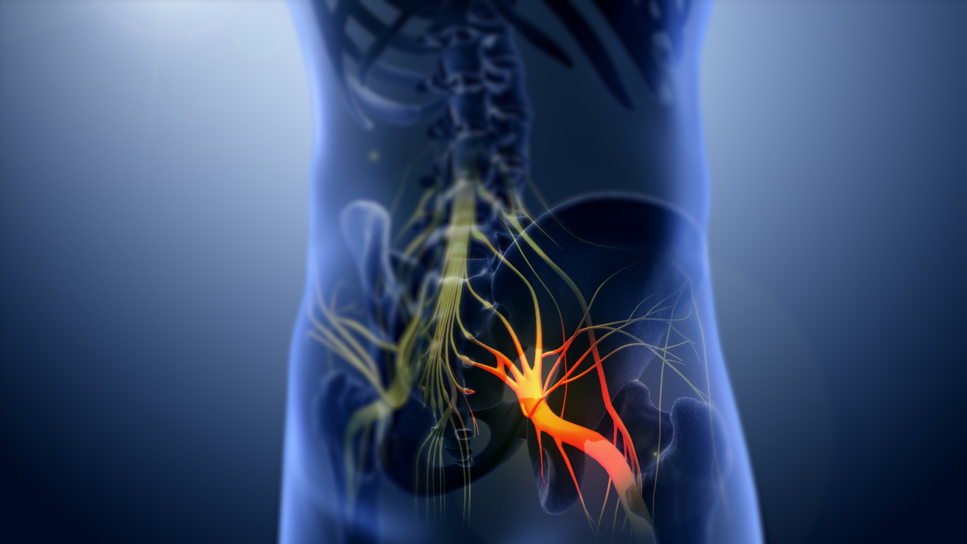
The sciatic nerve, also called the ischiadic nerve, is a large nerve in humans and other vertebrate animals. It is the largest branch of the sacral plexus and runs alongside the hip joint and down the right lower limb. It is the longest and widest single nerve in the human body, going from the top of the leg to the foot on the posterior aspect. The sciatic nerve has no cutaneous branches for the thigh. This nerve provides the connection to the nervous system for the skin of the lateral leg and the whole foot, the muscles of the back of the thigh, and those of the leg and foot. It is derived from Spinal nerve, spinal nerves Lumbar spinal nerve 4, L4 to Sacral spinal nerve 3, S3. It contains Axon, fibres from both the anterior and posterior divisions of the lumbosacral plexus. Structure In humans, the sciatic nerve is formed from the L4 to S3 segments of the sacral plexus, a collection of nerve fibres that emerge from the Sacrum, sacral part of the spinal cord. The lumbosacral trunk ... [...More Info...] [...Related Items...] OR: [Wikipedia] [Google] [Baidu] |
Internal Iliac Artery
The internal iliac artery (formerly known as the hypogastric artery) is the main artery of the pelvis. Structure The internal iliac artery supplies the walls and viscera of the pelvis, the buttock, the reproductive organs, and the medial compartment of the thigh. The vesicular branches of the internal iliac arteries supply the bladder. It is a short, thick vessel, smaller than the external iliac artery, and about 3 to 4 cm in length. Course The internal iliac artery arises at the bifurcation of the common iliac artery, opposite the lumbosacral articulation, and, passing downward to the upper margin of the greater sciatic foramen, divides into two large trunks, an anterior and a posterior. It is posterior to the ureter, anterior to the internal iliac vein, anterior to the lumbosacral trunk, and anterior to the piriformis muscle. Near its origin, it is medial to the external iliac vein, which lies between it and the psoas major muscle. It is above the obturator ... [...More Info...] [...Related Items...] OR: [Wikipedia] [Google] [Baidu] |
Inferior Gluteal Artery
The inferior gluteal artery (sciatic artery) is a terminal branch of the anterior trunk of the internal iliac artery. It exits the pelvis through the greater sciatic foramen. It is distributed chiefly to the buttock and the back of the thigh. Anatomy Origin It is the smaller of the two terminal branches of the anterior trunk of the internal iliac artery. Course It passes posterior-ward within parietal pelvic fascia. It travels in between the S1 nerve and S2 (or S2-S3) nerve(s). It descends upon the nerves of the sacral plexus and the piriformis muscle, posterior to the internal pudendal artery. It passes through the inferior part of the greater sciatic foramen. It exits the pelvis inferior to the piriformis muscle, between piriformis muscle and coccygeus muscle. It then descends in the interval between the greater trochanter of the femur and tuberosity of the ischium. It is accompanied by the sciatic nerve and the posterior femoral cutaneous nerves, and covered by the glu ... [...More Info...] [...Related Items...] OR: [Wikipedia] [Google] [Baidu] |
Pudendal Nerve
The pudendal nerve is the main nerve of the perineum. It is a Mixed nerve, mixed (motor and sensory) nerve and also conveys Sympathetic nervous system, sympathetic Autonomic nervous system, autonomic fibers. It carries sensation from the external genitalia of both sexes and the skin around the Human anus, anus and perineum, as well as the Motor neuron, motor supply to various pelvic muscles, including the external sphincter muscle of male urethra, male or external sphincter muscle of female urethra, female external urethral sphincter and the external anal sphincter. If damaged, most commonly by childbirth, loss of sensation or fecal incontinence may result. The nerve may be temporarily anesthetized, called pudendal anesthesia or pudendal block. The pudendal canal that carries the pudendal nerve is also known by the eponymous term "Alcock's canal", after Benjamin Alcock, an Irish anatomist who documented the canal in 1836. Structure Origin The pudendal nerve is paired, me ... [...More Info...] [...Related Items...] OR: [Wikipedia] [Google] [Baidu] |
Pudendal Vessels
The pudendal arteries are a group of arteries which supply many of the muscles and organs of the pelvic cavity. The arteries include the internal pudendal artery, the superficial external pudendal artery, and the deep external pudendal artery. The internal pudendal artery branches off the internal iliac artery, the main artery of the pelvis, and supplies blood to the sex organs. The internal pudendal artery gives rise to the perineal artery and the inferior rectal artery. The superficial external pudendal artery arises from the medial side of the femoral artery. It supplies the male scrotum and the female labia majora In primates, and specifically in humans, the labia majora (: labium majus), also known as the outer lips or outer labia, are two prominent Anatomical terms of location, longitudinal skin folds that extend downward and backward from the mons pubis .... References Arteries of the lower limb Arteries of the abdomen {{Portal bar, Anatomy ... [...More Info...] [...Related Items...] OR: [Wikipedia] [Google] [Baidu] |


