|
Rectus Femoris
The rectus femoris muscle is one of the four quadriceps muscles of the human body. The others are the vastus medialis, the vastus intermedius (deep to the rectus femoris), and the vastus lateralis. All four parts of the quadriceps muscle attach to the patella (knee cap) by the quadriceps tendon. The rectus femoris is situated in the middle of the front of the thigh; it is fusiform in shape, and its superficial fibers are arranged in a bipenniform manner, the deep fibers running straight () down to the deep aponeurosis. Its functions are to flex the thigh at the hip joint and to extend the leg at the knee joint. Structure It arises by two tendons: one, the anterior or straight, from the anterior inferior iliac spine; the other, the posterior or reflected, from a groove above the rim of the acetabulum. The two unite at an acute angle and spread into an aponeurosis that is prolonged downward on the anterior surface of the muscle, and from this the muscular fibers arise. The ... [...More Info...] [...Related Items...] OR: [Wikipedia] [Google] [Baidu] |
Anterior Inferior Iliac Spine
The anterior inferior iliac spine (AIIS) is a bony eminence on the anterior border of the hip bone, or, more precisely, the wing of the ilium. Structure The AIIS is a bony eminence on the anterior border of the ilium. It is below the anterior superior iliac spine. Development The AIIS is formed from a separate ossification centre to the rest of the ilium. Function The upper portion of the spine gives origin to the straight head of the rectus femoris muscle. A teardrop-shaped lower portion gives origin to the iliofemoral ligament of the hip joint and borders the rim of the acetabulum. Anteromedially and inferiorly to the AIIS is the iliopsoas groove, the passage for the iliopsoas muscle as it passes down to the lesser trochanter of the femur. A vague line, the inferior gluteal line, might run from the AIIS to the greater sciatic notch which delineates the inferior extent of the origin of gluteus minimus muscle. Clinical significance Rectus femoris muscle may avu ... [...More Info...] [...Related Items...] OR: [Wikipedia] [Google] [Baidu] |
Anatomical Terms Of Muscle
Anatomical terminology is used to uniquely describe aspects of skeletal muscle, cardiac muscle, and smooth muscle such as their actions, structure, size, and location. Types There are three types of muscle tissue in the body: skeletal, smooth, and cardiac. Skeletal muscle Skeletal muscle, or "voluntary muscle", is a striated muscle tissue that primarily joins to bone with tendons. Skeletal muscle enables movement of bones, and maintains posture. The widest part of a muscle that pulls on the tendons is known as the belly. Muscle slip A muscle slip is a slip of muscle that can either be an anatomical variant, or a branching of a muscle as in rib connections of the serratus anterior muscle. Smooth muscle Smooth muscle is involuntary and found in parts of the body where it conveys action without conscious intent. The majority of this type of muscle tissue is found in the digestive and urinary systems where it acts by propelling forward food, chyme, and feces in the former and u ... [...More Info...] [...Related Items...] OR: [Wikipedia] [Google] [Baidu] |
Hip Flexors
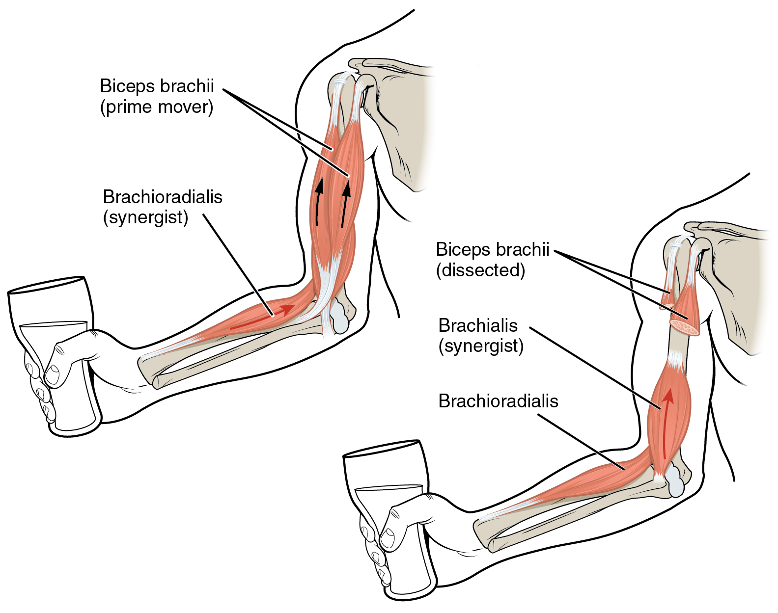
In anatomy, flexor is a muscle that contracts to perform flexion (from the Latin verb ''flectere'', to bend), a movement that decreases the angle between the bones converging at a joint. For example, one's elbow joint flexes when one brings their hand closer to the shoulder, thus decreasing the angle between the upper arm and the forearm. Flexors Upper limb *of the humerus bone (the bone in the upper arm) at the shoulder ** Pectoralis major ** Anterior deltoid ** Coracobrachialis **Biceps brachii * of the forearm at the elbow ** Brachialis **Brachioradialis **Biceps brachii *of carpus (the carpal bones) at the wrist **flexor carpi radialis **flexor carpi ulnaris ** palmaris longus *of the hand ** flexor pollicis longus muscle ** flexor pollicis brevis muscle **flexor digitorum profundus muscle ** flexor digitorum superficialis muscle Lower limb Hip The hip flexors are (in descending order of importance to the action of flexing the hip joint):Platzer (2004), p 246 *Collecti ... [...More Info...] [...Related Items...] OR: [Wikipedia] [Google] [Baidu] |
Sports Injury
Sports injuries occur during participation in sports or exercise in general. Globally, around 40% of individuals engage in some form of regular exercise or organized sports, with upwards of 60% of US high school students participating in one or more sports. Sports injuries account for 15 - 20% of annual acute care visits with an incidence of 1.79 - 6.36 injuries per 1,000 hours of participation. Sports injuries can be broken down into the types of injuries, risk factors and prevention and the overall impact that injuries have on athletes. Types of sport injury The type of sports injury suffered varies greatly based on gender, age and sport. Nonetheless, those with the highest prevalence remain contusions, fractures and sprains, followed closely by wounds and overuse injuries. Also common, the possible severity of head and neck injuries are important to consider. It is also paramount to place emphasis on the specific injuries that are most commonly encountered by sports medicine ... [...More Info...] [...Related Items...] OR: [Wikipedia] [Google] [Baidu] |
Avulsion Fracture
An avulsion fracture is a bone fracture which occurs when a fragment of bone tears away from the main mass of bone as a result of physical trauma. This can occur at the ligament by the application of forces external to the body (such as a fall or pull) or at the tendon by a muscular contraction that is stronger than the forces holding the bone together. Generally muscular avulsion is prevented by the neurological limitations placed on muscle contractions. Highly trained Athlete, athletes can overcome this neurological inhibition of strength and produce a much greater force output capable of breaking or avulsing a bone. Types Dental avulsion dental trauma, Traumatic complete displacement of a tooth from its socket in alveolar bone. It is a serious dental emergency in which Treatment of knocked-out (avulsed) teeth, prompt management (within 20–40 minutes of injury) affects the prognosis of the tooth. Tuberosity avulsion of the 5th metatarsal file:Proximal fractures of 5th me ... [...More Info...] [...Related Items...] OR: [Wikipedia] [Google] [Baidu] |
Hamstring
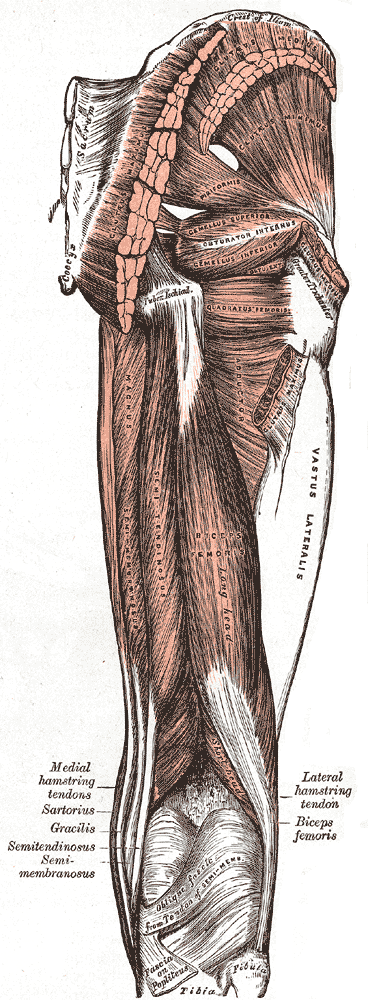
A hamstring () is any one of the three posterior thigh muscles in human anatomy between the hip and the knee: from medial to lateral, the semimembranosus, semitendinosus and biceps femoris. Etymology The word " ham" is derived from the Old English “ham” or “hom” meaning the hollow or bend of the knee, from a Germanic base where it meant "crooked". It gained the meaning of the leg of an animal around the 15th century. ''String'' refers to tendons, and thus the hamstrings' string-like tendons felt on either side of the back of the knee. Criteria The common criteria of any hamstring muscles are: # Muscles should originate from ischial tuberosity. # Muscles should be inserted over the knee joint, in the tibia or in the fibula. # Muscles will be innervated by the tibial branch of the sciatic nerve. # Muscle will participate in flexion of the knee joint and extension of the hip joint. Those muscles which fulfill all of the four criteria are called true hamstrings. ... [...More Info...] [...Related Items...] OR: [Wikipedia] [Google] [Baidu] |
List Of Flexors Of The Human Body
In anatomy, flexor is a muscle that contracts to perform flexion (from the Latin verb ''flectere'', to bend), a movement that decreases the angle between the bones converging at a joint. For example, one's elbow joint flexes when one brings their hand closer to the shoulder, thus decreasing the angle between the upper arm and the forearm. Flexors Upper limb *of the humerus bone (the bone in the upper arm) at the shoulder ** Pectoralis major **Anterior deltoid ** Coracobrachialis **Biceps brachii * of the forearm at the elbow ** Brachialis **Brachioradialis **Biceps brachii *of carpus (the carpal bones) at the wrist **flexor carpi radialis **flexor carpi ulnaris ** palmaris longus *of the hand ** flexor pollicis longus muscle ** flexor pollicis brevis muscle **flexor digitorum profundus muscle **flexor digitorum superficialis muscle Lower limb Hip The hip flexors are (in descending order of importance to the action of flexing the hip joint):Platzer (2004), p 246 *Collectively ... [...More Info...] [...Related Items...] OR: [Wikipedia] [Google] [Baidu] |
Tensor Fasciae Latae Muscle
The tensor fasciae latae (or tensor fasciæ latæ or, formerly, tensor vaginae femoris) is a muscle of the thigh. Together with the gluteus maximus, it acts on and is continuous with the iliotibial band, which attaches to the tibia. The muscle assists in keeping the balance of the pelvis while standing, walking, or running. Structure The tensor fasciae latae arises from the anterior part of the outer lip of the iliac crest; from the outer surface of the anterior superior iliac spine, and part of the outer border of the notch below it, between the gluteus medius and sartorius; and from the deep surface of the fascia lata. The tensor fasciae latae is inserted between the two layers of the iliotibial tract of the fascia lata about the junction of the middle and upper thirds of the thigh. It tautens the iliotibial tract and braces the knee, especially when the opposite foot is lifted.Saladin, Kenneth. Anatomy and Physiology. 6th ed. Mc-Graw Hill. 2010. The terminal insertion poin ... [...More Info...] [...Related Items...] OR: [Wikipedia] [Google] [Baidu] |
Psoas Major Muscle
The psoas major ( or ; from ) is a long fusiform muscle located in the lateral lumbar region between the vertebral column and the brim of the lesser pelvis. It joins the iliacus muscle to form the iliopsoas. In other animals, this muscle is equivalent to the tenderloin. Structure The psoas major is divided into a superficial and a deep part. The deep part originates from the transverse processes of lumbar vertebrae L1–L5. The superficial part originates from the lateral surfaces of the last thoracic vertebra, lumbar vertebrae L1–L4, and the neighboring intervertebral discs. The lumbar plexus lies between the two layers. Together, the iliacus muscle and the psoas major form the iliopsoas, which is surrounded by the iliac fascia. The iliopsoas runs across the iliopubic eminence through the muscular lacuna to its insertion on the lesser trochanter of the femur. The iliopectineal bursa separates the tendon of the iliopsoas muscle from the external surface of the hip-joi ... [...More Info...] [...Related Items...] OR: [Wikipedia] [Google] [Baidu] |
Iliacus Muscle
The iliacus is a flat, triangular muscle which fills the iliac fossa. It forms the lateral portion of iliopsoas, providing flexion of the thigh and lower limb at the acetabulofemoral joint. Structure The iliacus arises from the iliac fossa on the interior side of the hip bone, and also from the region of the anterior inferior iliac spine (AIIS). It joins the psoas major to form the iliopsoas. It proceeds across the iliopubic eminence through the muscular lacuna to its insertion on the lesser trochanter of the femur. Its fibers are often inserted in front of those of the psoas major and extend distally over the lesser trochanter.Platzer (2004), p 234 Nerve supply The iliopsoas is innervated by the femoral nerve and direct branches from the lumbar plexus.''Thieme Atlas of Anatomy'' (2006), p 422 Function In open-chain exercises, as part of the iliopsoas, the iliacus is important for lifting (flexing) the femur forward (e.g. front scale). In closed-chain exercises, ... [...More Info...] [...Related Items...] OR: [Wikipedia] [Google] [Baidu] |
Iliopsoas
The iliopsoas muscle (; ) refers to the joined psoas major and the iliacus muscles. The two muscles are separate in the abdomen, but usually merge in the thigh. They are usually given the common name ''iliopsoas''. The iliopsoas muscle joins to the femur at the lesser trochanter. It acts as the strongest flexor of the hip. The iliopsoas muscle is supplied by the lumbar spinal nerves L1– L3 (psoas) and parts of the femoral nerve (iliacus). Structure The iliopsoas muscle is a composite muscle formed from the psoas major muscle, and the iliacus muscle. The psoas major originates along the outer surfaces of the vertebral bodies of T12 and L1– L3 and their associated intervertebral discs. The iliacus originates in the iliac fossa of the pelvis. The psoas major unites with the iliacus at the level of the inguinal ligament. It crosses the hip joint to insert on the lesser trochanter of the femur. The iliopsoas is classified as an "anterior hip muscle" or "inner ... [...More Info...] [...Related Items...] OR: [Wikipedia] [Google] [Baidu] |
Sartorius Muscle
The sartorius muscle () is the longest muscle in the human body. It is a long, thin, superficial muscle that runs down the length of the thigh in the anterior compartment. Structure The sartorius muscle originates from the anterior superior iliac spine, and part of the notch between the anterior superior iliac spine and anterior inferior iliac spine. It runs obliquely across the upper and anterior part of the thigh in an inferomedial direction. It passes behind the medial condyle of the femur to end in a tendon. This tendon curves anteriorly to join the tendons of the gracilis and semitendinosus muscles in the pes anserinus, where it inserts into the superomedial surface of the tibia. Its upper portion forms the lateral border of the femoral triangle, and the point where it crosses adductor longus marks the apex of the triangle. Deep to sartorius and its fascia is the adductor canal, through which the saphenous nerve, femoral artery and vein, and nerve to vastus medialis pa ... [...More Info...] [...Related Items...] OR: [Wikipedia] [Google] [Baidu] |

