|
Pterygopalatine Ganglion
The pterygopalatine ganglion (aka Meckel's ganglion, nasal ganglion, or sphenopalatine ganglion) is a parasympathetic ganglion in the pterygopalatine fossa. It is one of four parasympathetic ganglia of the head and neck, (the others being the submandibular, otic, and ciliary ganglion). It is innervated by the Vidian nerve (formed by the greater superficial petrosal nerve branch of the facial nerve and deep petrosal nerve) and maxillary division of the trigeminal nerve. Its postsynaptic axons project to the lacrimal glands and nasal mucosa. The flow of blood to the nasal mucosa, in particular the venous plexus of the conchae, is regulated by the pterygopalatine ganglion and heats or cools the air in the nose. Structure The pterygopalatine ganglion (of Meckel), the largest of the parasympathetic ganglia associated with the branches of the maxillary nerve, is deeply placed in the pterygopalatine fossa, close to the sphenopalatine foramen. It is triangular or heart-shaped ... [...More Info...] [...Related Items...] OR: [Wikipedia] [Google] [Baidu] |
Dental Alveolus
Dental alveoli (singular ''alveolus'') are sockets in the jaws in which the roots of teeth are held in the alveolar process with the periodontal ligament. The lay term for dental alveoli is tooth sockets. A joint that connects the roots of the teeth and the alveolus is called a ''gomphosis'' (plural ''gomphoses''). Alveolar bone is the bone that surrounds the roots of the teeth forming bone sockets. In mammals, tooth sockets are found in the maxilla, the premaxilla, and the mandible. Etymology 1706, "a hollow", especially "the socket of a tooth", from Latin alveolus "a tray, trough, basin; bed of a small river; small hollow or cavity", diminutive of alvus "belly, stomach, paunch, bowels; hold of a ship", from PIE root *aulo- "hole, cavity" (source also of Greek aulos "flute, tube, pipe"; Serbo-Croatian, Polish, Russian ulica "street", originally "narrow opening"; Old Church Slavonic uliji, Lithuanian aulys "beehive" (hollow trunk), Armenian yli "pregnant"). The word was extended ... [...More Info...] [...Related Items...] OR: [Wikipedia] [Google] [Baidu] |
Trigeminal Nerve
In neuroanatomy, the trigeminal nerve (literal translation, lit. ''triplet'' nerve), also known as the fifth cranial nerve, cranial nerve V, or simply CN V, is a cranial nerve responsible for Sense, sensation in the face and motor functions such as biting and chewing; it is the most complex of the cranial nerves. Its name (''trigeminal'', ) derives from each of the two nerves (one on each side of the pons) having three major branches: the ophthalmic nerve (V), the maxillary nerve (V), and the mandibular nerve (V). The ophthalmic and maxillary nerves are purely sensory, whereas the mandibular nerve supplies motor as well as sensory (or "cutaneous") functions. Adding to the complexity of this nerve is that Autonomic nervous system, autonomic nerve fibers as well as special sensory fibers (taste) are contained within it. The motor division of the trigeminal nerve derives from the Basal plate (neural tube), basal plate of the embryonic pons, and the sensory division originates in ... [...More Info...] [...Related Items...] OR: [Wikipedia] [Google] [Baidu] |
Nervus Intermedius
The intermediate nerve, nervus intermedius, nerve of Wrisberg or glossopalatine nerve is the part of the facial nerve (cranial nerve VII) located between the motor component of the facial nerve and the vestibulocochlear nerve (cranial nerve VIII). It contains the sensory and parasympathetic fibers of the facial nerve. Upon reaching the facial canal, it joins with the motor root of the facial nerve at the geniculate ganglion. Alex Alfieri postulates that the intermediate nerve should be considered as a separate cranial nerve and not a part of the facial nerve. Parasympathetic fibers The superior salivatory nucleus contains the cell bodies of parasympathetic axons within the intermediate nerve. These fibers reach the geniculate ganglion but do not synapse. Some of these preganglionic parasympathetic fibers persist within the greater petrosal nerve as they exit the geniculate ganglion and subsequently synapse with neurons in the pterygopalatine ganglion. These postganglionic neuron ... [...More Info...] [...Related Items...] OR: [Wikipedia] [Google] [Baidu] |
Palatine Nerves
The palatine nerves (descending branches) are distributed to the roof of the mouth, soft palate, tonsil, and lining membrane of the nasal cavity. Most of their fibers are derived from the sphenopalatine branches of the maxillary nerve. In older texts, they are usually categorized as three in number: anterior, middle, and posterior. (In newer texts, and in Terminologia anatomica, they are broken down into "greater palatine nerve" and "lesser palatine nerve The lesser palatine nerves (posterior palatine nerve) are branches of the maxillary nerve (CN V2). They descends through the greater palatine canal alongside the greater palatine nerve, and emerge (separately) through the lesser palatine foramen t ...".) References External links * * Diagram at adi-visuals.com Peripheral nervous system Palate {{Portal bar, Anatomy ... [...More Info...] [...Related Items...] OR: [Wikipedia] [Google] [Baidu] |
Sphenopalatine Nerves
The two pterygopalatine nerves (or sphenopalatine branches) descend to the pterygopalatine ganglion. Although it is closely related to the pterygopalatine ganglion, it is still considered a branch of the maxillary nerve and does not synapse in the ganglion. It is found in the pterygopalatine fossa In human anatomy, the pterygopalatine fossa (sphenopalatine fossa) is a fossa in the skull. A human skull contains two pterygopalatine fossae—one on the left side, and another on the right side. Each fossa is a cone-shaped paired depression dee .... Additional images File:Gray778.png, Distribution of the maxillary and mandibular nerves, and the submaxillary ganglion. References Maxillary nerve {{Neuroanatomy-stub ... [...More Info...] [...Related Items...] OR: [Wikipedia] [Google] [Baidu] |
Hard Palate
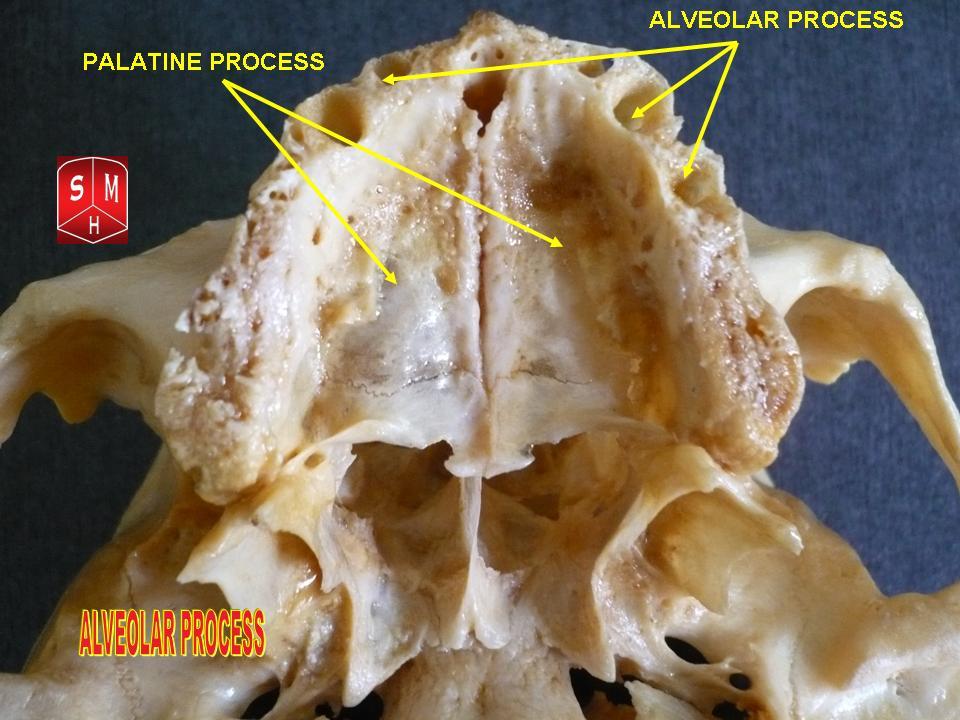
The hard palate is a thin horizontal bony plate made up of two bones of the facial skeleton, located in the roof of the mouth. The bones are the palatine process of the maxilla and the horizontal plate of palatine bone. The hard palate spans the alveolar arch formed by the alveolar process that holds the upper teeth (when these are developed). Structure The hard palate is formed by the palatine process of the maxilla and horizontal plate of palatine bone. It forms a partition between the nasal passages and the mouth. On the anterior portion of the hard palate are the ''plicae'', irregular ridges in the mucous membrane that help hold food while the teeth are biting into it while also facilitating the movement of food backward towards the larynx once pieces have been bitten off. This partition is continued deeper into the mouth by a fleshy extension called the soft palate. On the ventral surface of the hard palate, some projections or transverse ridges are present which ... [...More Info...] [...Related Items...] OR: [Wikipedia] [Google] [Baidu] |
Gland
A gland is a Cell (biology), cell or an Organ (biology), organ in an animal's body that produces and secretes different substances that the organism needs, either into the bloodstream or into a body cavity or outer surface. A gland may also function to remove unwanted substances such as urine from the body. There are two types of gland, each with a different method of secretion. Endocrine glands are ductless and secrete their products, hormones, directly into interstitial spaces to be taken up into the bloodstream. Exocrine glands secrete their products through a duct into a body cavity or outer surface. Glands are mostly composed of epithelium, epithelial tissue, and typically have a supporting framework of connective tissue, and a capsule. Structure Development Every gland is formed by an ingrowth from an epithelium, epithelial surface. This ingrowth may in the beginning possess a tubular structure, but in other instances glands may start as a solid column of cells which ... [...More Info...] [...Related Items...] OR: [Wikipedia] [Google] [Baidu] |
Gingiva
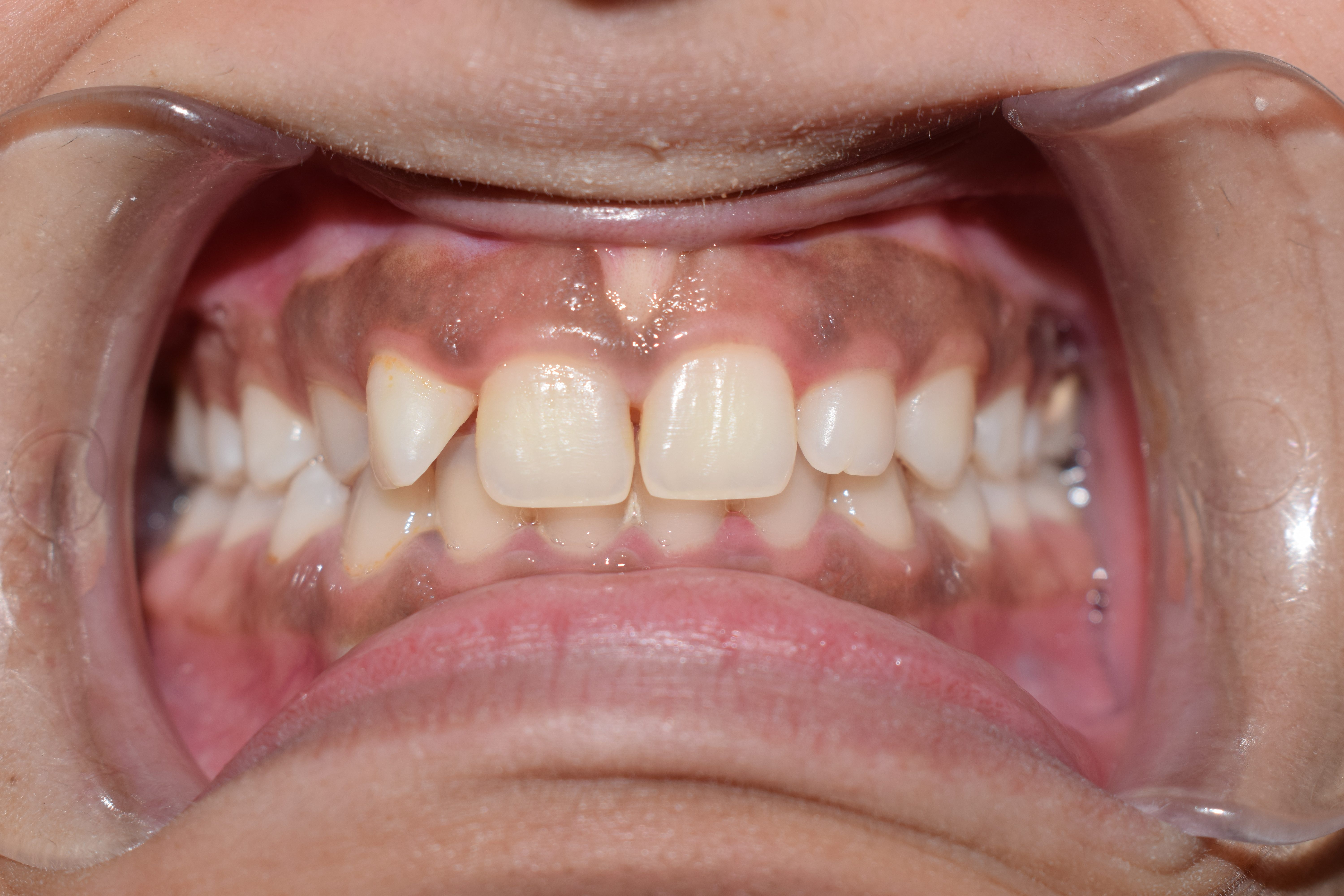
The gums or gingiva (: gingivae) consist of the mucosal tissue that lies over the mandible and maxilla inside the mouth. Gum health and disease can have an effect on general health. Structure The gums are part of the soft tissue lining of the mouth. They surround the teeth and provide a seal around them. Unlike the soft tissue linings of the lips and cheeks, most of the gums are tightly bound to the underlying bone which helps resist the friction of food passing over them. Thus when healthy, it presents an effective barrier to the barrage of periodontal insults to deeper tissue. Healthy gums are usually coral pink in light skinned people, and may be naturally darker with melanin pigmentation. Changes in color, particularly increased redness, together with swelling and an increased tendency to bleed, suggest an inflammation that is possibly due to the accumulation of bacterial plaque. Overall, the clinical appearance of the tissue reflects the underlying histology, both in hea ... [...More Info...] [...Related Items...] OR: [Wikipedia] [Google] [Baidu] |
Pharynx
The pharynx (: pharynges) is the part of the throat behind the human mouth, mouth and nasal cavity, and above the esophagus and trachea (the tubes going down to the stomach and the lungs respectively). It is found in vertebrates and invertebrates, though its structure varies across species. The pharynx carries food to the esophagus and air to the larynx. The flap of cartilage called the epiglottis stops food from entering the larynx. In humans, the pharynx is part of the Digestion, digestive system and the conducting zone of the respiratory system. (The conducting zone—which also includes the nostrils of the Human nose, nose, the larynx, trachea, bronchus, bronchi, and bronchioles—filters, warms, and moistens air and conducts it into the lungs). The human pharynx is conventionally divided into three sections: the nasopharynx, oropharynx, and laryngopharynx (hypopharynx). In humans, two sets of pharyngeal muscles form the pharynx and determine the shape of its lumen (anatomy), ... [...More Info...] [...Related Items...] OR: [Wikipedia] [Google] [Baidu] |
Paranasal Sinuses
Paranasal sinuses are a group of four paired air-filled spaces that surround the nasal cavity. The maxillary sinuses are located under the eyes; the frontal sinuses are above the eyes; the ethmoidal sinuses are between the eyes and the sphenoidal sinuses are behind the eyes. The sinuses are named for the facial bones and sphenoid bone in which they are located. Their role is disputed. Structure Humans possess four pairs of paranasal sinuses, divided into subgroups that are named according to the bones within which the sinuses lie. They are all innervated by branches of the trigeminal nerve (CN V). * The maxillary sinuses, the largest of the paranasal sinuses, are under the eyes, in the maxillary bones (open in the back of the semilunar hiatus of the nose). They are innervated by the maxillary nerve (CN V2). * The frontal sinuses, superior to the eyes, in the frontal bone, which forms the hard part of the forehead. They are innervated by the ophthalmic nerve (CN V1 ... [...More Info...] [...Related Items...] OR: [Wikipedia] [Google] [Baidu] |
Sphenopalatine Foramen
The sphenopalatine foramen is a foramen of the skull that connects the nasal cavity and the pterygopalatine fossa. It gives passage to the sphenopalatine artery, nasopalatine nerve, and the superior nasal nerve (all passing from the pterygopalatine fossa into the nasal cavity). Structure The processes of the superior border of the palatine bone are separated by the ''sphenopalatine notch'', which is converted into the sphenopalatine foramen by the under surface of the body of the sphenoid. The sphenopalatine foramen is situated posterior to the middle nasal meatus orbital process of palatine bone, anterior to the sphenoidal process of palatine bone, inferior to the body and of the sphenoid bone, and superior to the superior margin of the perpendicular plate of palatine bone. Relations The ethmoid crest (a reliable surgical landmark A landmark is a recognizable natural or artificial feature used for navigation, a feature that stands out from its near environment and ... [...More Info...] [...Related Items...] OR: [Wikipedia] [Google] [Baidu] |
Johann Friedrich Meckel, The Elder
Johann Friedrich Meckel the Elder (31 July 1724 – 18 September 1774) was a German anatomist born in Wetzlar. He often has "the Elder" appended to his name to avoid confusion with his famous grandson Johann Friedrich Meckel (1781–1833), who was also an anatomist and often has "the Younger" included with his name. The elder Meckel's son, Philipp Friedrich Theodor Meckel (1755–1803) and another grandson, August Albrecht Meckel (1790–1829) were also anatomists. Meckel earned his medical doctorate from the University of Göttingen in 1748, and in his thesis, "''Tractatus anatomico physiologicus de quinto pare nervorum cerebri''", he documented his discovery of the submandibular ganglion. Subsequently, he moved to Berlin, where he worked as a prosector and taught classes on midwifery. In 1751 he became a professor of anatomy, botany and obstetrics. In 1773, Meckel was elected a foreign member of the Royal Swedish Academy of Sciences. Eponyms Meckel has a number of anatomical ... [...More Info...] [...Related Items...] OR: [Wikipedia] [Google] [Baidu] |


