|
Pterygopalatine Fossa
In human anatomy, the pterygopalatine fossa (sphenopalatine fossa) is a fossa in the skull. A human skull contains two pterygopalatine fossae—one on the left side, and another on the right side. Each fossa is a cone-shaped paired depression deep to the infratemporal fossa and posterior to the maxilla on each side of the skull, located between the pterygoid process and the maxillary tuberosity close to the apex of the orbit. It is the indented area medial to the pterygomaxillary fissure leading into the sphenopalatine foramen. It communicates with the nasal and oral cavities, infratemporal fossa, orbit, pharynx, and middle cranial fossa through eight foramina. Structure Boundaries It has the following boundaries: * ''anterior'': superomedial part of the infratemporal surface of maxilla * ''posterior'': root of the pterygoid process and adjoining anterior surface of the greater wing of sphenoid bone * ''medial'': perpendicular plate of the palatine bone and its orbital an ... [...More Info...] [...Related Items...] OR: [Wikipedia] [Google] [Baidu] |
Fossa (anatomy)
In anatomy, a fossa (; : fossae ( or ); ) is a depression or hollow, usually in a bone, such as the hypophyseal fossa (the depression in the sphenoid bone).Venieratos D, Anagnostopoulou S, Garidou A., A new morphometric method for the sella turcica and the hypophyseal fossa and its clinical relevance.;Folia Morphol (Warsz). 2005 Nov;64(4):240-7. Some examples include: In the skull: * Cranial fossa ** Anterior cranial fossa ** Middle cranial fossa *** Interpeduncular fossa ** Posterior cranial fossa * Hypophyseal fossa * Temporal bone fossa ** Mandibular fossa ** Jugular fossa * Infratemporal fossa * Pterygopalatine fossa * Pterygoid fossa * Lacrimal fossa ** Fossa for lacrimal gland ** Fossa for lacrimal sac * Scaphoid fossa * Condyloid fossa * Rhomboid fossa In the mandible: * Retromolar fossa In the torso: * Fossa ovalis (heart) * Infraclavicular fossa * Pyriform fossa * Substernal fossa * Iliac fossa * Ovarian fossa * Paravesical fossa * Coccygeal f ... [...More Info...] [...Related Items...] OR: [Wikipedia] [Google] [Baidu] |
Middle Cranial Fossa
The middle cranial fossa is formed by the sphenoid bones, and the temporal bones. It lodges the temporal lobes, and the pituitary gland. It is deeper than the anterior cranial fossa, is narrow medially and widens laterally to the sides of the skull. It is separated from the posterior cranial fossa by the clivus and the petrous crest. It is bounded in front by the posterior margins of the lesser wings of the sphenoid bone, the anterior clinoid processes, and the ridge forming the anterior margin of the chiasmatic groove; behind, by the superior angles of the petrous portions of the temporal bones and the dorsum sellae; laterally by the temporal squamae, sphenoidal angles of the parietals, and greater wings of the sphenoid. It is traversed by the squamosal, sphenoparietal, sphenosquamosal, and sphenopetrosal sutures. Anatomy Features Middle part The middle part of the fossa presents, in front, the chiasmatic groove and tuberculum sellae; the chiasmatic groove ends o ... [...More Info...] [...Related Items...] OR: [Wikipedia] [Google] [Baidu] |
Maxillary Artery
The maxillary artery (eg, internal maxillary artery) supplies deep structures of the face. It branches from the external carotid artery just deep to the neck of the mandible. Structure The maxillary artery, the larger of the two terminal branches of the external carotid artery, arises behind the neck of the Human mandible, mandible, and is at first imbedded in the substance of the parotid gland; it passes forward between the ramus of the mandible and the sphenomandibular ligament, and then runs, either superficial or deep to the lateral pterygoid muscle, to the pterygopalatine fossa. It supplies the deep structures of the face, and may be divided into Human mandible, mandibular, Pterygoid processes of the sphenoid, pterygoid, and pterygopalatine ganglion, pterygopalatine portions. First portion The ''first'' or ''mandibular '' or ''bony'' portion passes horizontally forward, between the neck of the mandible and the sphenomandibular ligament, where it lies parallel to and a little ... [...More Info...] [...Related Items...] OR: [Wikipedia] [Google] [Baidu] |
Maxillary Nerve
In neuroanatomy, the maxillary nerve (V) is one of the three branches or divisions of the trigeminal nerve, the fifth (CN V) cranial nerve. It comprises the principal functions of Sense, sensation from the maxilla, nasal cavity, Sinus (anatomy), sinuses, the palate and subsequently that of the mid-face, and is intermediate, both in position and size, between the ophthalmic nerve and the mandibular nerve.Illustrated Anatomy of the Head and Neck, Fehrenbach and Herring, Elsevier, 2012, page 180 Structure It begins at the middle of the trigeminal ganglion as a flattened plexiform band then it passes through the lateral wall of the cavernous sinus. It leaves the skull through the foramen rotundum, where it becomes more cylindrical in form, and firmer in texture. After leaving foramen rotundum it gives two branches to the pterygopalatine ganglion. It then crosses the pterygopalatine fossa, inclines lateralward on the back of the maxilla, and enters the orbit through the inferior orb ... [...More Info...] [...Related Items...] OR: [Wikipedia] [Google] [Baidu] |
Pterygopalatine Ganglion
The pterygopalatine ganglion (aka Meckel's ganglion, nasal ganglion, or sphenopalatine ganglion) is a parasympathetic ganglion in the pterygopalatine fossa. It is one of four parasympathetic ganglia of the head and neck, (the others being the submandibular, otic, and ciliary ganglion). It is innervated by the Vidian nerve (formed by the greater superficial petrosal nerve branch of the facial nerve and deep petrosal nerve) and maxillary division of the trigeminal nerve. Its postsynaptic axons project to the lacrimal glands and nasal mucosa. The flow of blood to the nasal mucosa, in particular the venous plexus of the conchae, is regulated by the pterygopalatine ganglion and heats or cools the air in the nose. Structure The pterygopalatine ganglion (of Meckel), the largest of the parasympathetic ganglia associated with the branches of the maxillary nerve, is deeply placed in the pterygopalatine fossa, close to the sphenopalatine foramen. It is triangular or heart-shaped ... [...More Info...] [...Related Items...] OR: [Wikipedia] [Google] [Baidu] |
Lesser Palatine Canals
The lesser palatine canals (also accessory palatine canals) are passages in the palatine bone that carry the lesser and middle palatine nerves and vessels. Structure The lesser palatine canals start from the greater palatine canal, and run with them, also opening into the roof of the oral cavity. Their openings are known as the lesser palatine foramina, and they transmit the lesser palatine artery, vein, and nerve, as well as the middle palatine vessels and nerve. See also * Pterygopalatine fossa In human anatomy, the pterygopalatine fossa (sphenopalatine fossa) is a fossa in the skull. A human skull contains two pterygopalatine fossae—one on the left side, and another on the right side. Each fossa is a cone-shaped paired depression dee ... References Bones of the head and neck {{musculoskeletal-stub ro:Canale palatine mici ... [...More Info...] [...Related Items...] OR: [Wikipedia] [Google] [Baidu] |
Oral Cavity
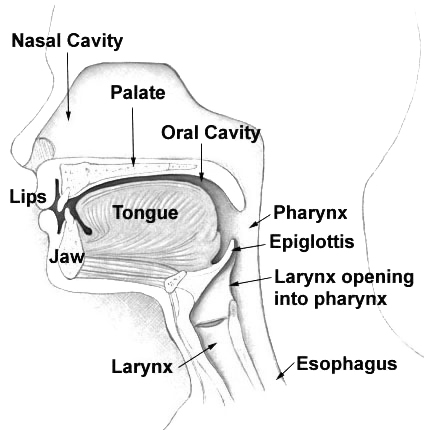
A mouth also referred to as the oral is the body orifice through which many animals ingest food and vocalize. The body cavity immediately behind the mouth opening, known as the oral cavity (or in Latin), is also the first part of the alimentary canal, which leads to the pharynx and the gullet. In tetrapod vertebrates, the mouth is bounded on the outside by the lips and cheeks — thus the oral cavity is also known as the buccal cavity (from Latin ', meaning "cheek") — and contains the tongue on the inside. Except for some groups like birds and lissamphibians, vertebrates usually have teeth in their mouths, although some fish species have pharyngeal teeth instead of oral teeth. Most bilaterian phyla, including arthropods, molluscs and chordates, have a two-opening gut tube with a mouth at one end and an anus at the other. Which end forms first in ontogeny is a criterion used to classify bilaterian animals into protostomes and deuterostomes. Development In t ... [...More Info...] [...Related Items...] OR: [Wikipedia] [Google] [Baidu] |
Greater Palatine Canal
The greater palatine canal (or pterygopalatine canal) is a passage in the skull that transmits the descending palatine artery, vein, and greater and lesser palatine nerves between the pterygopalatine fossa and the oral cavity. Structure The greater palatine canal starts on the inferior aspect of the pterygopalatine fossa. It goes through the maxilla and palatine bones to reach the palate, ending at the greater palatine foramen. From this canal, accessory canals branch off; these are known as the lesser palatine canals. The canal is formed by a vertical groove on the posterior part of the maxillary surface of the palatine bone; it is converted into a canal by articulation with the maxilla. The canal transmits the descending palatine vessels, the greater palatine nerve, and the lesser palatine nerve. See also * Greater palatine foramen * Pterygopalatine fossa In human anatomy, the pterygopalatine fossa (sphenopalatine fossa) is a fossa in the skull. A human skull conta ... [...More Info...] [...Related Items...] OR: [Wikipedia] [Google] [Baidu] |
Orbit (anatomy)
In anatomy Anatomy () is the branch of morphology concerned with the study of the internal structure of organisms and their parts. Anatomy is a branch of natural science that deals with the structural organization of living things. It is an old scien ..., the orbit is the Body cavity, cavity or socket/hole of the skull in which the eye and Accessory visual structures, its appendages are situated. "Orbit" can refer to the bony socket, or it can also be used to imply the contents. In the adult human, the volume of the orbit is about , of which the eye occupies . The orbital contents comprise the eye, the Orbital fascia, orbital and retrobulbar fascia, extraocular muscles, cranial nerves optic nerve, II, oculomotor nerve, III, trochlear nerve, IV, trigeminal nerve, V, and abducens nerve, VI, blood vessels, fat, the lacrimal gland with its Lacrimal sac, sac and nasolacrimal duct, duct, the eyelids, Medial palpebral ligament, medial and Lateral palpebral raphe, lateral palpebr ... [...More Info...] [...Related Items...] OR: [Wikipedia] [Google] [Baidu] |
Inferior Orbital Fissure
The inferior orbital fissure is a gap between the Greater wing of sphenoid bone, greater wing of sphenoid bone, and the maxilla. It connects the Orbit (anatomy), orbit (anteriorly) with the infratemporal fossa and pterygopalatine fossa (posteriorly). Anatomy The medial end of the inferior orbital fissure diverges laterally from the medial end of the superior orbital fissure. It is situated between the lateral wall of the orbit and the floor of the orbit. Contents The fissure gives passage to multiple structures, including: * Infraorbital nerve, Infraorbital artery, artery and Infraorbital vein, vein * Inferior ophthalmic vein * Zygomatic nerve * Orbital branches of the pharyngeal nerve * Maxillary nerve Additional images File:Gray189.png, Left infratemporal fossa. File:Gray191.png, Horizontal section of nasal and orbital cavities. File:Gray787.png, Dissection showing origins of right ocular muscles, and nerves entering by the superior orbital fissure. File:Slide2rome. ... [...More Info...] [...Related Items...] OR: [Wikipedia] [Google] [Baidu] |
Nasopharynx
The pharynx (: pharynges) is the part of the throat behind the mouth and nasal cavity, and above the esophagus and trachea (the tubes going down to the stomach and the lungs respectively). It is found in vertebrates and invertebrates, though its structure varies across species. The pharynx carries food to the esophagus and air to the larynx. The flap of cartilage called the epiglottis stops food from entering the larynx. In humans, the pharynx is part of the digestive system and the conducting zone of the respiratory system. (The conducting zone—which also includes the nostrils of the nose, the larynx, trachea, bronchi, and bronchioles—filters, warms, and moistens air and conducts it into the lungs). The human pharynx is conventionally divided into three sections: the nasopharynx, oropharynx, and laryngopharynx (hypopharynx). In humans, two sets of pharyngeal muscles form the pharynx and determine the shape of its lumen. They are arranged as an inner layer of longitudina ... [...More Info...] [...Related Items...] OR: [Wikipedia] [Google] [Baidu] |
Nasal Cavity
The nasal cavity is a large, air-filled space above and behind the nose in the middle of the face. The nasal septum divides the cavity into two cavities, also known as fossae. Each cavity is the continuation of one of the two nostrils. The nasal cavity is the uppermost part of the respiratory system and provides the nasal passage for inhaled air from the nostrils to the nasopharynx and rest of the respiratory tract. The paranasal sinuses surround and drain into the nasal cavity. Structure The term "nasal cavity" can refer to each of the two cavities of the nose, or to the two sides combined. The lateral wall of each nasal cavity mainly consists of the maxilla. However, there is a deficiency that is compensated for by the perpendicular plate of the palatine bone, the medial pterygoid plate, the labyrinth of ethmoid and the inferior concha. The paranasal sinuses are connected to the nasal cavity through small orifices called ostia. Most of these ostia communicat ... [...More Info...] [...Related Items...] OR: [Wikipedia] [Google] [Baidu] |


