|
Posterior Auricular Vein
The posterior auricular vein is a vein of the head. It begins from a plexus with the occipital vein and the superficial temporal vein, descends behind the auricle, and drains into the external jugular vein. Structure The posterior auricular vein begins upon the side of the head, in a plexus which communicates with the tributaries of the occipital vein and the superficial temporal vein. It descends behind the auricle. It joins the posterior division of the retromandibular vein. It drains into the external jugular vein. It receive the stylomastoid vein, and some tributaries from the cranial surface of the auricle. Variation The posterior auricular vein may drain into the internal jugular vein or a posterior jugular vein if there are variations in the external jugular vein The external jugular vein receives the greater part of the blood from the exterior of the cranium and the deep parts of the face, being formed by the junction of the posterior division of the retroman ... [...More Info...] [...Related Items...] OR: [Wikipedia] [Google] [Baidu] |
Scalp
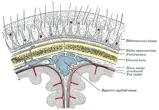
The scalp is the anatomical area bordered by the human face at the front, and by the neck at the sides and back. Structure The scalp is usually described as having five layers, which can conveniently be remembered as a mnemonic: * S: The skin on the head from which head hair grows. It contains numerous sebaceous glands and hair follicles. * C: Connective tissue. A dense subcutaneous layer of fat and fibrous tissue that lies beneath the skin, containing the nerves and vessels of the scalp. * A: The aponeurosis called epicranial aponeurosis (or galea aponeurotica) is the next layer. It is a tough layer of dense fibrous tissue which runs from the frontalis muscle anteriorly to the occipitalis posteriorly. * L: The loose areolar connective tissue layer provides an easy plane of separation between the upper three layers and the pericranium. In scalping the scalp is torn off through this layer. It also provides a plane of access in craniofacial surgery and neurosurgery. Th ... [...More Info...] [...Related Items...] OR: [Wikipedia] [Google] [Baidu] |
External Jugular Vein
The external jugular vein receives the greater part of the blood from the exterior of the cranium and the deep parts of the face, being formed by the junction of the posterior division of the retromandibular vein with the posterior auricular vein. Structure It commences in the substance of the parotid gland, on a level with the angle of the mandible, and runs perpendicularly down the neck, in the direction of a line drawn from the angle of the mandible to the middle of the clavicle superficial to the sternocleidomastoideus. In its course it crosses the sternocleidomastoideus obliquely, and in the subclavian triangle perforates the deep fascia, and ends in the subclavian vein lateral to or in front of the scalenus anterior, piercing the roof of the posterior triangle. It is separated from the sternocleidomastoideus by the investing layer of the deep cervical fascia, and is covered by the platysma, the superficial fascia, and the integument; it crosses the cutaneous cervica ... [...More Info...] [...Related Items...] OR: [Wikipedia] [Google] [Baidu] |
Posterior Auricular Artery
The posterior auricular artery is a small artery that arises from the external carotid artery, above the digastric muscle and stylohyoid muscle, opposite the apex of the styloid process. It ascends posteriorly beneath the parotid gland, along the styloid process of the temporal bone, between the cartilage of the ear and the mastoid process of the temporal bone along the lateral side of the head. The posterior auricular artery gives off the stylomastoid artery, small branches to the auricle, and supplies blood to the scalp posterior to the auricle. A person may be able to "hear" their own heart rate via this artery, under certain conditions. See also * Anterior auricular branches of superficial temporal artery * Posterior auricular nerve The posterior auricular nerve is a nerve of the head. It is a branch of the facial nerve (CN VII). It communicates with branches from the vagus nerve, the great auricular nerve, and the lesser occipital nerve. Its auricular branch suppl ... [...More Info...] [...Related Items...] OR: [Wikipedia] [Google] [Baidu] |
Vein
Veins are blood vessels in humans and most other animals that carry blood towards the heart. Most veins carry deoxygenated blood from the tissues back to the heart; exceptions are the pulmonary and umbilical veins, both of which carry oxygenated blood to the heart. In contrast to veins, arteries carry blood away from the heart. Veins are less muscular than arteries and are often closer to the skin. There are valves (called ''pocket valves'') in most veins to prevent backflow. Structure Veins are present throughout the body as tubes that carry blood back to the heart. Veins are classified in a number of ways, including superficial vs. deep, pulmonary vs. systemic, and large vs. small. * Superficial veins are those closer to the surface of the body, and have no corresponding arteries. * Deep veins are deeper in the body and have corresponding arteries. * Perforator veins drain from the superficial to the deep veins. These are usually referred to in the lower limbs and feet. * ... [...More Info...] [...Related Items...] OR: [Wikipedia] [Google] [Baidu] |
Head
A head is the part of an organism which usually includes the ears, brain, forehead, cheeks, chin, eyes, nose, and mouth, each of which aid in various sensory functions such as sight, hearing, smell, and taste. Some very simple animals may not have a head, but many bilaterally symmetric forms do, regardless of size. Heads develop in animals by an evolutionary trend known as cephalization. In bilaterally symmetrical animals, nervous tissue concentrate at the anterior region, forming structures responsible for information processing. Through biological evolution, sense organs and feeding structures also concentrate into the anterior region; these collectively form the head. Human head The human head is an anatomical unit that consists of the skull, hyoid bone and cervical vertebrae. The term "skull" collectively denotes the mandible (lower jaw bone) and the cranium (upper portion of the skull that houses the brain). Sculptures of human heads are generally based on ... [...More Info...] [...Related Items...] OR: [Wikipedia] [Google] [Baidu] |
Occipital Vein
The occipital vein is a vein of the scalp. It originates from a plexus around the external occipital protuberance and superior nuchal line to the back part of the vertex of the skull. It usually drains into the internal jugular vein, but may also drain into the posterior auricular vein (which joins the external jugular vein). It drains part of the scalp. Structure The occipital vein is part of the scalp. It begins as a plexus at the posterior aspect of the scalp from the external occipital protuberance and superior nuchal line to the back part of the vertex of the skull. It pierces the cranial attachment of the trapezius and, dipping into the venous plexus of the suboccipital triangle, joins the deep cervical vein and the vertebral vein. Occasionally it follows the course of the occipital artery, and ends in the internal jugular vein. Alternatively, it joins the posterior auricular vein, and ends in the external jugular vein. The parietal emissary vein connects it with th ... [...More Info...] [...Related Items...] OR: [Wikipedia] [Google] [Baidu] |
Superficial Temporal Vein
The superficial temporal vein is a vein of the side of the head. It begins on the side and vertex of the skull in a network of veins which communicates with the frontal vein and supraorbital vein, with the corresponding vein of the opposite side, and with the posterior auricular vein and occipital vein. It ultimately crosses the posterior root of the zygomatic arch, enters the parotid gland, and unites with the internal maxillary vein to form the posterior facial vein. Structure It begins on the side and vertex of the skull in a network () which communicates with the frontal vein and supraorbital vein, with the corresponding vein of the opposite side, and with the posterior auricular vein and occipital vein. From this network frontal and parietal branches arise, and join above the zygomatic arch to form the trunk of the vein, which is joined by the middle temporal vein emerging from the temporalis muscle. It then crosses the posterior root of the zygomatic arch, enters the subs ... [...More Info...] [...Related Items...] OR: [Wikipedia] [Google] [Baidu] |
Auricle (anatomy)
The auricle or auricula is the visible part of the ear that is outside the head. It is also called the pinna ( Latin for " wing" or " fin", plural pinnae), a term that is used more in zoology. Structure The diagram shows the shape and location of most of these components: * '' antihelix'' forms a 'Y' shape where the upper parts are: ** ''Superior crus'' (to the left of the ''fossa triangularis'' in the diagram) ** ''Inferior crus'' (to the right of the ''fossa triangularis'' in the diagram) * '' Antitragus'' is below the ''tragus'' * ''Aperture'' is the entrance to the ear canal * ''Auricular sulcus'' is the depression behind the ear next to the head * ''Concha'' is the hollow next to the ear canal * Conchal angle is the angle that the back of the ''concha'' makes with the side of the head * ''Crus'' of the helix is just above the ''tragus'' * ''Cymba conchae'' is the narrowest end of the ''concha'' * External auditory meatus is the ear canal * ''Fossa triangularis'' is the ... [...More Info...] [...Related Items...] OR: [Wikipedia] [Google] [Baidu] |
External Jugular Vein
The external jugular vein receives the greater part of the blood from the exterior of the cranium and the deep parts of the face, being formed by the junction of the posterior division of the retromandibular vein with the posterior auricular vein. Structure It commences in the substance of the parotid gland, on a level with the angle of the mandible, and runs perpendicularly down the neck, in the direction of a line drawn from the angle of the mandible to the middle of the clavicle superficial to the sternocleidomastoideus. In its course it crosses the sternocleidomastoideus obliquely, and in the subclavian triangle perforates the deep fascia, and ends in the subclavian vein lateral to or in front of the scalenus anterior, piercing the roof of the posterior triangle. It is separated from the sternocleidomastoideus by the investing layer of the deep cervical fascia, and is covered by the platysma, the superficial fascia, and the integument; it crosses the cutaneous cervica ... [...More Info...] [...Related Items...] OR: [Wikipedia] [Google] [Baidu] |
Plexus
In neuroanatomy, a plexus (from the Latin term for "braid") is a branching network of vessels or nerves. The vessels may be blood vessels (veins, capillaries) or lymphatic vessels. The nerves are typically axons outside the central nervous system. The standard plural form in English is plexuses. Alternatively, the Latin plural plexūs may be used. Types Nerve plexuses The four primary nerve plexuses are the cervical plexus, brachial plexus, lumbar plexus, and the sacral plexus. Cardiac plexus Celiac plexus Renal plexus Venous plexus Choroid plexus The choroid plexus is a part of the central nervous system in the brain and consists of capillaries, brain ventricles, and ependymal cells. Invertebrates The plexus is the characteristic form of nervous system in the coelenterates and persists with modifications in the flatworms. The nerves of the radially symmetric echinoderm An echinoderm () is any member of the phylum Echinodermata (). The adults are re ... [...More Info...] [...Related Items...] OR: [Wikipedia] [Google] [Baidu] |
British Journal Of Plastic Surgery
The ''Journal of Plastic, Reconstructive & Aesthetic Surgery'', formerly the ''British Journal of Plastic Surgery'', is the journal of plastic surgery Plastic surgery is a surgical specialty involving the restoration, reconstruction or alteration of the human body. It can be divided into two main categories: reconstructive surgery and cosmetic surgery. Reconstructive surgery includes cranio ... of the British Association of Plastic, Reconstructive and Aesthetic Surgeons. It is open access and abstracted and indexed in Scopus and other databases. References Surgery journals Plastic surgery Open access journals English-language journals {{surgery-journal-stub ... [...More Info...] [...Related Items...] OR: [Wikipedia] [Google] [Baidu] |
Retromandibular Vein
The retromandibular vein (temporomaxillary vein, posterior facial vein) is a major vein of the face. Anatomy Origin The retromandibular vein is formed by the union of the superficial temporal and maxillary veins. Course It descends in the substance of the parotid gland, superficial to the external carotid artery (but beneath the facial nerve), between the ramus of the mandible and the sternocleidomastoideus muscle. It terminates by dividing into two branches: * an ''anterior'', which passes forward and joins anterior facial vein, to form the common facial vein, which then drains into the internal jugular vein. * a ''posterior'', which is joined by the posterior auricular vein and becomes the external jugular vein. Function The retromandibular vein provides venous drainage to the superior cranium The skull is a bone protective cavity for the brain. The skull is composed of four types of bone i.e., cranial bones, facial bones, ear ossicles and hyoid bone. Ho ... [...More Info...] [...Related Items...] OR: [Wikipedia] [Google] [Baidu] |


