|
Superficial Temporal Vein
The superficial temporal vein is a vein of the side of the head. It begins on the side and vertex of the skull in a network of veins which communicates with the frontal vein and supraorbital vein, with the corresponding vein of the opposite side, and with the posterior auricular vein and occipital vein. It ultimately crosses the posterior root of the zygomatic arch, enters the parotid gland, and unites with the internal maxillary vein to form the posterior facial vein. Structure It begins on the side and vertex of the skull in a network () which communicates with the frontal vein and supraorbital vein, with the corresponding vein of the opposite side, and with the posterior auricular vein and occipital vein. From this network frontal and parietal branches arise, and join above the zygomatic arch to form the trunk of the vein, which is joined by the middle temporal vein emerging from the temporalis muscle. It then crosses the posterior root of the zygomatic arch, enters the subs ... [...More Info...] [...Related Items...] OR: [Wikipedia] [Google] [Baidu] |
Eyelids
An eyelid is a thin fold of skin that covers and protects an eye. The levator palpebrae superioris muscle retracts the eyelid, exposing the cornea to the outside, giving vision. This can be either voluntarily or involuntarily. The human eyelid features a row of eyelashes along the eyelid margin, which serve to heighten the protection of the eye from dust and foreign debris, as well as from perspiration. "Palpebral" (and "blepharal") means relating to the eyelids. Its key function is to regularly spread the tears and other secretions on the eye surface to keep it moist, since the cornea must be continuously moist. They keep the eyes from drying out when asleep. Moreover, the blink reflex protects the eye from foreign bodies. The appearance of the human upper eyelid often varies between different populations. The prevalence of an epicanthic fold covering the inner corner of the eye account for the majority of East Asian and Southeast Asian populations, and is also found ... [...More Info...] [...Related Items...] OR: [Wikipedia] [Google] [Baidu] |
Parotid Gland
The parotid gland is a major salivary gland in many animals. In humans, the two parotid glands are present on either side of the mouth and in front of both ears. They are the largest of the salivary glands. Each parotid is wrapped around the mandibular ramus, and secretes serous saliva through the parotid duct into the mouth, to facilitate mastication and swallowing and to begin the digestion of starches. There are also two other types of salivary glands; they are submandibular and sublingual glands. Sometimes accessory parotid glands are found close to the main parotid glands. Etymology The word ''parotid'' literally means "beside the ear". From Greek παρωτίς (stem παρωτιδ-) : (gland) behind the ear < παρά - pará : in front, and οὖς - ous (stem ὠτ-, ōt-) : ear. Structure The parotid glands are a pair of mainly serous salivary gland ...[...More Info...] [...Related Items...] OR: [Wikipedia] [Google] [Baidu] |
Anterior Auricular Veins
The anterior auricular veins are veins which drain the anterior aspect of the external ear The outer ear, external ear, or auris externa is the external part of the ear, which consists of the auricle (also pinna) and the ear canal. It gathers sound energy and focuses it on the eardrum (tympanic membrane). Structure Auricle Th .... The veins drains to the superficial temporal vein. See also * Posterior auricular vein References Veins of the head and neck {{circulatory-stub ... [...More Info...] [...Related Items...] OR: [Wikipedia] [Google] [Baidu] |
Temporomandibular Joint
In anatomy, the temporomandibular joints (TMJ) are the two joints connecting the jawbone to the skull. It is a bilateral synovial articulation between the temporal bone of the skull above and the mandible below; it is from these bones that its name is derived. This joint is unique in that it is a bilateral joint that functions as one unit. Since the TMJ is connected to the mandible, the right and left joints must function together and therefore are not independent of each other. Structure The main components are the joint capsule, articular disc, mandibular condyles, articular surface of the temporal bone, temporomandibular ligament, stylomandibular ligament, sphenomandibular ligament, and lateral pterygoid muscle. Capsule The articular capsule (capsular ligament) is a thin, loose envelope, attached above to the circumference of the mandibular fossa and the articular tubercle immediately in front; below, to the neck of the condyle of the mandible. Its loose attachment t ... [...More Info...] [...Related Items...] OR: [Wikipedia] [Google] [Baidu] |
Articular Veins
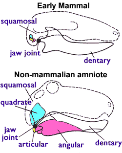
The articular bone is part of the lower jaw of most vertebrates, including most jawed fish, amphibians, birds and various kinds of reptiles, as well as ancestral mammals. Anatomy In most vertebrates, the articular bone is connected to two other lower jaw bones, the suprangular and the angular. Developmentally, it originates from the embryonic mandibular cartilage. The most caudal portion of the mandibular cartilage ossifies to form the articular bone, while the remainder of the mandibular cartilage either remains cartilaginous or disappears. In snakes In snakes, the articular, surangular, and prearticular bones have fused to form the compound bone. The mandible is suspended from the quadrate bone and articulates at this compound bone. Function In amphibians and reptiles In most tetrapods, the articular bone forms the lower portion of the jaw joint. The upper jaw articulates at the quadrate bone. In mammals In mammals, the articular bone evolves to form the malleus, ... [...More Info...] [...Related Items...] OR: [Wikipedia] [Google] [Baidu] |
Parotid Veins
The parotid gland is a major salivary gland in many animals. In humans, the two parotid glands are present on either side of the mouth and in front of both ears. They are the largest of the salivary glands. Each parotid is wrapped around the mandibular ramus, and secretes serous saliva through the parotid duct into the mouth, to facilitate mastication and swallowing and to begin the digestion of starches. There are also two other types of salivary glands; they are submandibular and sublingual glands. Sometimes accessory parotid glands are found close to the main parotid glands. Etymology The word ''parotid'' literally means "beside the ear". From Greek παρωτίς (stem παρωτιδ-) : (gland) behind the ear < παρά - pará : in front, and οὖς - ous (stem ὠτ-, ōt-) : ear. Structure The parotid glands are a pair of mainly |
Temporalis Muscle
In anatomy, the temporalis muscle, also known as the temporal muscle, is one of the muscles of mastication (chewing). It is a broad, fan-shaped convergent muscle on each side of the head that fills the temporal fossa, superior to the zygomatic arch so it covers much of the temporal bone.Illustrated Anatomy of the Head and Neck, Fehrenbach and Herring, Elsevier, 2012, page 98''Temporal'' refers to the head's temples. Structure In humans, the temporalis muscle arises from the temporal fossa and the deep part of temporal fascia. This is a very broad area of attachment. It passes medial to the zygomatic arch. It forms a tendon which inserts onto the coronoid process of the mandible, with its insertion extending into the retromolar fossa posterior to the most distal mandibular molar.Human Anatomy, Jacobs, Elsevier, 2008, page 194 In other mammals, the muscle usually spans the dorsal part of the skull all the way up to the medial line. There, it may be attached to a sagittal cre ... [...More Info...] [...Related Items...] OR: [Wikipedia] [Google] [Baidu] |
Middle Temporal Vein
Middle or The Middle may refer to: * Centre (geometry), the point equally distant from the outer limits. Places * Middle (sheading), a subdivision of the Isle of Man * Middle Bay (other) * Middle Brook (other) * Middle Creek (other) * Middle Island (other) * Middle Lake (other) * Middle Mountain, California * Middle Peninsula, Chesapeake Bay, Virginia * Middle Range, a former name of the Xueshan Range on Taiwan Island * Middle River (other) * Middle Rocks, two rocks at the eastern opening of the Straits of Singapore * Middle Sound, a bay in North Carolina * Middle Township (other) * Middle East Music * "Middle" (song), 2015 * "The Middle" (Jimmy Eat World song), 2001 * "The Middle" (Zedd, Maren Morris and Grey song), 2018 *"Middle", a song by Rocket from the Crypt from their 1995 album ''Scream, Dracula, Scream!'' *"The Middle", a song by Demi Lovato from their debut album ''Don't Forget'' *"The Middle", a song by Th ... [...More Info...] [...Related Items...] OR: [Wikipedia] [Google] [Baidu] |
Posterior Facial Vein
The retromandibular vein (temporomaxillary vein, posterior facial vein) is a major vein of the face. Anatomy Origin The retromandibular vein is formed by the union of the superficial temporal and maxillary veins. Course It descends in the substance of the parotid gland, superficial to the external carotid artery (but beneath the facial nerve), between the ramus of the mandible and the sternocleidomastoideus muscle. It terminates by dividing into two branches: * an ''anterior'', which passes forward and joins anterior facial vein, to form the common facial vein, which then drains into the internal jugular vein. * a ''posterior'', which is joined by the posterior auricular vein and becomes the external jugular vein. Function The retromandibular vein provides venous drainage to the superior cranium, and significant drainage to the ear. Clinical significance Parrot's sign is a sensation of pain when pressure is applied to the retromandibular region. Additional images ... [...More Info...] [...Related Items...] OR: [Wikipedia] [Google] [Baidu] |
Internal Maxillary Vein
The maxillary vein, or internal maxillary vein, is a vein of the head. It is a short trunk which accompanies the first part of the maxillary artery. It is formed by a confluence of the veins of the pterygoid plexus and the interpterygoid emissary vein, and passes posteriorly between the sphenomandibular ligament and the neck of the mandible. It unites with the superficial temporal vein to form the retromandibular vein. Structure The maxillary vein is a short trunk which accompanies the first part of the maxillary artery. It is formed from the merging of the veins of the pterygoid plexus, and the interpterygoid emissary vein. It passes posteriorly between the sphenomandibular ligament and the neck of the mandible. It unites with the superficial temporal vein. It drains into the retromandibular vein (posterior facial vein). The maxillary vein anastomoses with the retroglenoid vein. Development The maxillary vein may be the embryological origin of the central retinal vein ... [...More Info...] [...Related Items...] OR: [Wikipedia] [Google] [Baidu] |
Zygomatic Arch
In anatomy, the zygomatic arch, or cheek bone, is a part of the skull formed by the zygomatic process of the temporal bone (a bone extending forward from the side of the skull, over the opening of the ear) and the temporal process of the zygomatic bone (the side of the cheekbone), the two being united by an oblique suture (the zygomaticotemporal suture); the tendon of the temporal muscle passes medial to (i.e. through the middle of) the arch, to gain insertion into the coronoid process of the mandible (jawbone). The jugal point is the point at the anterior (towards face) end of the upper border of the zygomatic arch where the masseteric and maxillary edges meet at an angle, and where it meets the process of the zygomatic bone. The arch is typical of '' Synapsida'' (“fused arch”), a clade of amniotes that includes mammals and their extinct relatives, such as '' Moschops'' and ''Dimetrodon''. Structure The zygomatic process of the temporal arises by two roots: * an ... [...More Info...] [...Related Items...] OR: [Wikipedia] [Google] [Baidu] |
Temple (anatomy)
The temple is a latch where four skull bones fuse: the frontal, parietal, temporal, and sphenoid. It is located on the side of the head behind the eye between the forehead and the ear. The temporal muscle covers this area and is used during mastication. Cladists classify land vertebrates based on the presence of an upper hole, a lower hole, both, or neither in the cover of dermal bone that formerly covered the temporalis muscle, whose origin is the temple and whose insertion is the jaw. The brain has a lobe called the temporal lobe. Etymology The word "templar" as used in anatomy has a separate etymology from the other meaning of word ''temple'', meaning "place of worship". Both come from Latin, but the word for the place of worship comes from ', whereas the word for the part of the head comes from Vulgar Latin *', modified from ', plural form ("both temples") of ', a word that meant both "time" and the part of the head. Due to the common source with the word for ti ... [...More Info...] [...Related Items...] OR: [Wikipedia] [Google] [Baidu] |

.jpg)


