|
Posterior Atlantoaxial Ligament
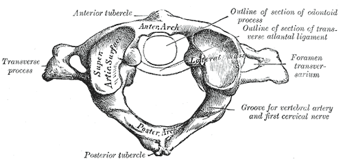
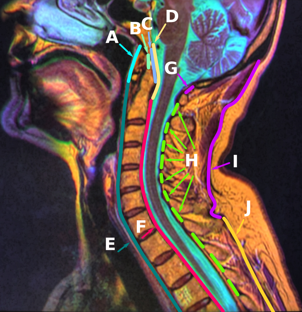
The posterior atlantoaxial ligament is a broad, thin membrane attached, above, to the lower border of the posterior arch of the atlas; below, to the upper edges of the laminæ of the axis. It supplies the place of the ligamenta flava, and is in relation, behind, with the obliqui capitis inferiores. See also * Atlanto-axial joint The atlanto-axial joint is a joint in the upper part of the neck between the atlas bone and the axis bone, which are the first and second cervical vertebrae. It is a pivot joint, that can start from C2 To C7. Structure The atlanto-axial joint ... References External links Description at spineuniverse.com Ligaments of the head and neck Bones of the vertebral column {{Portal bar, Anatomy ... [...More Info...] [...Related Items...] OR: [Wikipedia] [Google] [Baidu] |
Atlas (anatomy)
In anatomy, the atlas (C1) is the most superior (first) cervical vertebra of the spine and is located in the neck. The bone is named for Atlas of Greek mythology, just as Atlas bore the weight of the heavens, the first cervical vertebra supports the head. However, the term atlas was first used by the ancient Romans for the seventh cervical vertebra (C7) due to its suitability for supporting burdens. In Greek mythology, Atlas was condemned to bear the weight of the heavens as punishment for rebelling against Zeus. Ancient depictions of Atlas show the globe of the heavens resting at the base of his neck, on C7. Sometime around 1522, anatomists decided to call the first cervical vertebra the atlas. Scholars believe that by switching the designation atlas from the seventh to the first cervical vertebra Renaissance anatomists were commenting that the point of man's burden had shifted from his shoulders to his head—that man's true burden was not a physical load, but rather, his m ... [...More Info...] [...Related Items...] OR: [Wikipedia] [Google] [Baidu] |
Axis (anatomy)
In anatomy, the axis (from Latin ''axis'', "axle") is the second cervical vertebra (C2) of the spine, immediately inferior to the atlas, upon which the head rests. The spinal cord passes through the axis. The defining feature of the axis is its strong bony protrusion known as the dens, which rises from the superior aspect of the bone. Structure The body is deeper in front or in the back and is prolonged downward anteriorly to overlap the upper and front part of the third vertebra. It presents a median longitudinal ridge in front, separating two lateral depressions for the attachment of the longus colli muscles. Dens The dens, also called the odontoid process, or the peg, is the most pronounced projecting feature of the axis. The dens exhibits a slight constriction where it joins the main body of the vertebra. The condition where the dens is separated from the body of the axis is called ''os odontoideum'' and may cause nerve and circulation compression syndrome. On its an ... [...More Info...] [...Related Items...] OR: [Wikipedia] [Google] [Baidu] |
Ligamenta Flava
The ligamenta flava (: ligamentum flavum, Latin for ''yellow ligament'') are a series of ligaments that connect the ventral parts of the laminae of adjacent vertebrae. They help to preserve upright posture, preventing hyperflexion, and ensuring that the vertebral column straightens after flexion. Hypertrophy can cause spinal stenosis. They appear yellowish in colour due to their high elastic fibre content. Anatomy Each ligamentum flavum connects the laminae of two adjacent vertebrae. They attach to the anterior portion of the upper lamina above, and the posterior portion of the lower lamina below. They begin with the junction of the axis and third cervical vertebra, continuing down to the junction of the 5th lumbar vertebra and the sacrum. In the neck region the ligaments are thin, but broad and long; they are thicker in the thoracic region, and thickest in the lumbar region. They are thinnest between the atlas bone (C1) and the axis bone (C2), and may be absent in some ... [...More Info...] [...Related Items...] OR: [Wikipedia] [Google] [Baidu] |
Obliqui Capitis Inferiores
The obliquus capitis inferior muscle () is a muscle in the upper back of the neck. It is one of the suboccipital muscles. Its inferior attachment is at the spinous process of the axis; its superior attachment is at the transverse process of the atlas. It is innervated by the suboccipital nerve (the posterior ramus of first cervical spinal nerve). The muscle rotates the head to its side. Despite what its name suggest, it is the only capitis (Latin: "head") muscle that does not actually attach to the skull. Anatomy The obliquus capitis inferior is one of the suboccipital muscles (and the only one of these to have no attachment to the skull). It is larger than the obliquus capitis superior muscle. It forms the inferolateral boundary of the suboccipital triangle. The muscle extends laterally and somewhat superiorly from its inferior attachment to its superior attachment. Attachments its inferior attachment is at the lateral external aspect of the bifid spinous process of the ... [...More Info...] [...Related Items...] OR: [Wikipedia] [Google] [Baidu] |
Atlanto-axial Joint
The atlanto-axial joint is a joint in the upper part of the neck between the atlas bone and the axis bone, which are the first and second cervical vertebrae. It is a pivot joint, that can start from C2 To C7. Structure The atlanto-axial joint is a joint between the atlas bone and the axis bone, which are the first and second cervical vertebrae. It is a pivot joint that provides 40 to 70% of axial rotation of the head. There is a pivot articulation between the odontoid process of the axis and the ring formed by the anterior arch and the transverse ligament of the atlas. Lateral and median joints There are three atlanto-axial joints: one median and two lateral: * The median atlanto-axial joint is sometimes considered a triple joint: ** one between the posterior surface of the anterior arch of atlas and the front of the odontoid process ** one between the anterior surface of the ligament and the back of the odontoid process * The lateral atlantoaxial joint involves the ... [...More Info...] [...Related Items...] OR: [Wikipedia] [Google] [Baidu] |
Ligaments Of The Head And Neck
A ligament is a type of Connective tissue#Types, fibrous connective tissue in the body that connects bones to other bones. It also connects Flight_feather#Remiges, flight feathers to bones, in dinosaurs and birds. All 30,000 species of amniotes (land animals with internal bones) have ligaments. It is also known as ''articular ligament'', ''articular larua'', ''fibrous ligament'', or ''true ligament''. Comparative anatomy Ligaments are similar to tendons and fasciae as they are all made of connective tissue. The differences among them are in the connections that they make: ligaments connect one bone to another bone, tendons connect muscle to bone, and fasciae connect muscles to other muscles. These are all found in the skeletal system of the human body. Ligaments cannot usually be regenerated naturally; however, there are periodontal ligament stem cells located near the periodontal ligament which are involved in the adult regeneration of periodontist ligament. The study of li ... [...More Info...] [...Related Items...] OR: [Wikipedia] [Google] [Baidu] |


