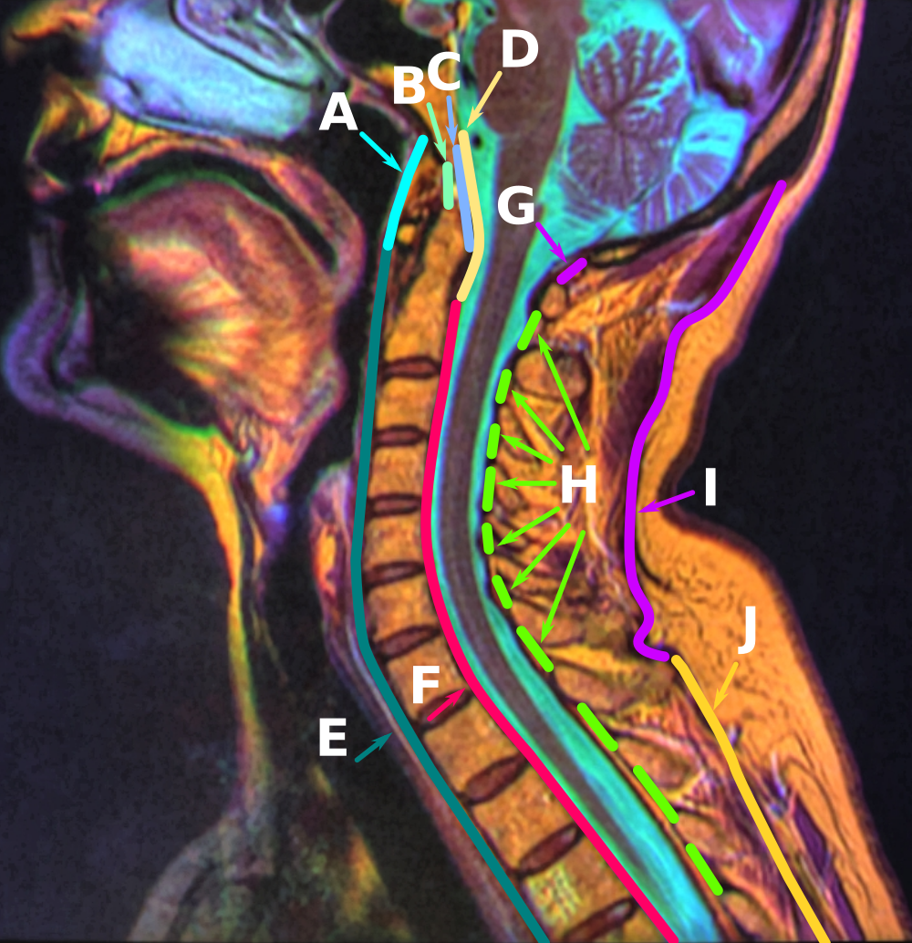
Ligamenta Flava on:
[Wikipedia]
[Google]
[Amazon]
The ligamenta flava (: ligamentum flavum, Latin for ''yellow ligament'') are a series of
 Ligamenta flava undergo slight fibrotic and chondrometaplastic changes with aging. In spinal stenosis, the density of the ligaments is reduced possibly causing a bulge into the spinal canal in the standing position.
ligament
A ligament is a type of fibrous connective tissue in the body that connects bones to other bones. It also connects flight feathers to bones, in dinosaurs and birds. All 30,000 species of amniotes (land animals with internal bones) have liga ...
s that connect the ventral parts of the lamina
Lamina may refer to:
People
* Saa Emerson Lamina, Sierra Leonean politician
* Tamba Lamina, Sierra Leonean politician and diplomat
Science and technology
* Planar lamina, a two-dimensional planar closed surface with mass and density, in mathem ...
e of adjacent vertebra
Each vertebra (: vertebrae) is an irregular bone with a complex structure composed of bone and some hyaline cartilage, that make up the vertebral column or spine, of vertebrates. The proportions of the vertebrae differ according to their spina ...
e. They help to preserve upright posture, preventing hyperflexion, and ensuring that the vertebral column
The spinal column, also known as the vertebral column, spine or backbone, is the core part of the axial skeleton in vertebrates. The vertebral column is the defining and eponymous characteristic of the vertebrate. The spinal column is a segmente ...
straightens after flexion. Hypertrophy
Hypertrophy is the increase in the volume of an organ or tissue due to the enlargement of its component cells. It is distinguished from hyperplasia, in which the cells remain approximately the same size but increase in number. Although hypertro ...
can cause spinal stenosis
Spinal stenosis is an abnormal narrowing of the spinal canal or neural foramen that results in pressure on the spinal cord or nerve roots. Symptoms may include pain, numbness, or weakness in the arms or legs. Symptoms are typically gradual in ...
.
They appear yellowish in colour due to their high elastic fibre content.
Anatomy
Each ligamentum flavum connects the laminae of two adjacent vertebrae. They attach to the anterior portion of the upper lamina above, and the posterior portion of the lower lamina below. They begin with the junction of theaxis
An axis (: axes) may refer to:
Mathematics
*A specific line (often a directed line) that plays an important role in some contexts. In particular:
** Coordinate axis of a coordinate system
*** ''x''-axis, ''y''-axis, ''z''-axis, common names ...
and third cervical vertebra, continuing down to the junction of the 5th lumbar vertebra
The lumbar vertebrae are located between the thoracic vertebrae and pelvis. They form the lower part of the back in humans, and the tail end of the back in quadrupeds. In humans, there are five lumbar vertebrae. The term is used to describe the ...
and the sacrum
The sacrum (: sacra or sacrums), in human anatomy, is a triangular bone at the base of the spine that forms by the fusing of the sacral vertebrae (S1S5) between ages 18 and 30.
The sacrum situates at the upper, back part of the pelvic cavity, ...
.
In the neck
The neck is the part of the body in many vertebrates that connects the head to the torso. It supports the weight of the head and protects the nerves that transmit sensory and motor information between the brain and the rest of the body. Addition ...
region the ligaments are thin, but broad and long; they are thicker in the thoracic
The thorax (: thoraces or thoraxes) or chest is a part of the anatomy of mammals and other tetrapod animals located between the neck and the abdomen.
In insects, crustaceans, and the extinct trilobites, the thorax is one of the three main ...
region, and thickest in the lumbar
In tetrapod anatomy, lumbar is an adjective that means of or pertaining to the abdominal segment of the torso, between the diaphragm (anatomy), diaphragm and the sacrum.
Naming and location
The lumbar region is sometimes referred to as the lowe ...
region. They are thinnest between the atlas bone (C1) and the axis bone (C2), and may be absent in some people. They become longer inferiorly in the cervical spine
In tetrapods, cervical vertebrae (: vertebra) are the vertebrae of the neck, immediately below the skull. Truncal vertebrae (divided into thoracic and lumbar vertebrae in mammals) lie caudal (toward the tail) of cervical vertebrae. In sauro ...
, as the distance between adjacent laminae increases.
They are best seen from the interior of the vertebral canal
In human anatomy, the spinal canal, vertebral canal or spinal cavity is an elongated body cavity enclosed within the dorsal bony arches of the vertebral column, which contains the spinal cord, spinal roots and dorsal root ganglia. It is a proc ...
. when looked at from the outer surface they appear short, being overlapped by the lamina of the vertebral arch
Each vertebra (: vertebrae) is an irregular bone with a complex structure composed of bone and some hyaline cartilage, that make up the vertebral column or spine, of vertebrates. The proportions of the vertebrae differ according to their spina ...
.
Structure
Each ligament consists of twolateral
Lateral is a geometric term of location which may also refer to:
Biology and healthcare
* Lateral (anatomy), a term of location meaning "towards the side"
* Lateral cricoarytenoid muscle, an intrinsic muscle of the larynx
* Lateral release ( ...
portions which commence one on either side of the roots of the articular processes
The articular process or zygapophysis ( + apophysis) of a vertebra is a projection of the vertebra that serves the purpose of fitting with an adjacent vertebra. The actual region of contact is called the ''articular facet''.Moore, Keith L. et al. ...
, and extend backward to the point where the laminae meet to form the spinous process
Each vertebra (: vertebrae) is an irregular bone with a complex structure composed of bone and some hyaline cartilage, that make up the vertebral column or spine, of vertebrates. The proportions of the vertebrae differ according to their spina ...
; the posterior margins of the two portions are in contact and to a certain extent united, slight intervals being left for the passage of small vessels. Small veins that form anastomotic connections between the internal
Internal may refer to:
*Internality as a concept in behavioural economics
*Neijia, internal styles of Chinese martial arts
*Neigong or "internal skills", a type of exercise in meditation associated with Daoism
* ''Internal'' (album) by Safia, 2016 ...
and external vertebral venous plexuses may pass between a pair of the ligaments.
 Ligamenta flava undergo slight fibrotic and chondrometaplastic changes with aging. In spinal stenosis, the density of the ligaments is reduced possibly causing a bulge into the spinal canal in the standing position.
Function
The ligamenta flava become stretched with flexion of the spine. The marked elasticity of the ligamenta flava serves to preserve upright posture, and to assist thevertebral column
The spinal column, also known as the vertebral column, spine or backbone, is the core part of the axial skeleton in vertebrates. The vertebral column is the defining and eponymous characteristic of the vertebrate. The spinal column is a segmente ...
in resuming it after flexion
Motion, the process of movement, is described using specific anatomical terminology, anatomical terms. Motion includes movement of Organ (anatomy), organs, joints, Limb (anatomy), limbs, and specific sections of the body. The terminology used de ...
. The elastin
Elastin is a protein encoded by the ''ELN'' gene in humans and several other animals. Elastin is a key component in the extracellular matrix of gnathostomes (jawed vertebrates). It is highly Elasticity (physics), elastic and present in connective ...
, fairly unique to the ligamenta flava among other ligaments
A ligament is a type of fibrous connective tissue in the body that connects bones to other bones. It also connects flight feathers to bones, in dinosaurs and birds. All 30,000 species of amniotes (land animals with internal bones) have ligam ...
, prevents buckling of the ligament into the spinal canal during extension, which would cause spinal cord compression
Spinal cord compression is a form of myelopathy in which the spinal cord is compressed. Causes can be bone fragments from a vertebral fracture, a tumor, abscess, ruptured intervertebral disc or other lesion.
When acute it can cause a medical eme ...
.
Clinical significance
Because these ligaments lie in the posterior part of thevertebral canal
In human anatomy, the spinal canal, vertebral canal or spinal cavity is an elongated body cavity enclosed within the dorsal bony arches of the vertebral column, which contains the spinal cord, spinal roots and dorsal root ganglia. It is a proc ...
, their hypertrophy
Hypertrophy is the increase in the volume of an organ or tissue due to the enlargement of its component cells. It is distinguished from hyperplasia, in which the cells remain approximately the same size but increase in number. Although hypertro ...
can cause spinal stenosis
Spinal stenosis is an abnormal narrowing of the spinal canal or neural foramen that results in pressure on the spinal cord or nerve roots. Symptoms may include pain, numbness, or weakness in the arms or legs. Symptoms are typically gradual in ...
, particularly in patients with diffuse idiopathic skeletal hyperostosis. The ligamenta flava may also become fatty or calcify during ageing. These cause degeneration of elastin
Elastin is a protein encoded by the ''ELN'' gene in humans and several other animals. Elastin is a key component in the extracellular matrix of gnathostomes (jawed vertebrates). It is highly Elasticity (physics), elastic and present in connective ...
. Some studies indicate that the hypertrophy of these ligaments may be linked to a fibrotic process associated with increased collagen VI, which could represent an adaptive and reparative process in response to the rupture of elastic fibers.
Epidural
During anepidural
Epidural administration (from Ancient Greek ἐπί, "upon" + '' dura mater'') is a method of medication administration in which a medicine is injected into the epidural space around the spinal cord. The epidural route is used by physicians ...
, the needle has to be inserted into the spinal space through a ligamentum flavum. Once it passes through, this is felt as a decrease in the pressure requited to further advance the needle. This makes the ligamentum flavum an important landmark to overcome to ensure proper needle placement.
Removal
During a microdiscectomy, a procedure to remove part of anintervertebral disc
An intervertebral disc (British English), also spelled intervertebral disk (American English), lies between adjacent vertebrae in the vertebral column. Each disc forms a fibrocartilaginous joint (a symphysis), to allow slight movement of the ver ...
that is pressing on the spinal nerves
A spinal nerve is a mixed nerve, which carries motor, sensory, and autonomic signals between the spinal cord and the body. In the human body there are 31 pairs of spinal nerves, one on each side of the vertebral column. These are grouped into ...
, the ligamenta flava may need to be removed or reshaped. A hook can be placed underneath a ligamentum flavum to ensure it is separated from the dura mater.
References
External links
* {{Authority control Ligaments of the torso Bones of the vertebral column