|
Plethysmograph
A plethysmograph is an instrument for measuring changes in volume within an organ or whole body (usually resulting from fluctuations in the amount of blood or air it contains). The word is derived from the Greek "plethysmos" (increasing, enlarging, becoming full), and "graphein" (to write). Organs studied Lungs Pulmonary plethysmographs are commonly used to measure the functional residual capacity (FRC) of the lungs—the volume in the lungs when the muscles of respiration are relaxed—and total lung capacity. In a traditional plethysmograph (or "body box"), the test subject, or patient, is placed inside a sealed chamber the size of a small telephone booth with a single mouthpiece. At the end of normal expiration, the mouthpiece is closed. The patient is then asked to make an inspiratory effort. As the patient tries to inhale (a maneuver which looks and feels like panting), the lungs expand, decreasing pressure within the lungs and increasing lung volume. This, in turn ... [...More Info...] [...Related Items...] OR: [Wikipedia] [Google] [Baidu] |
Penile Plethysmograph
Penile plethysmography (PPG) or phallometry is measurement of blood flow to the penis, typically used as a proxy for measurement of sexual arousal. The most commonly reported methods of conducting penile plethysmography involve the measurement of the circumference of the penis with a mercury-in-rubber or electromechanical strain gauge, or the volume of the penis with an airtight cylinder and inflatable cuff at the base of the penis. Corpora cavernosa nerve penile plethysmographs measure changes in response to inter-operative electric stimulation during surgery. The volumetric procedure was invented by Kurt Freund and is considered to be particularly sensitive at low arousal levels. The easier to use circumferential measures are more widely used, however, and more common in studies using erotic film stimuli. A corresponding device in women is the vaginal photoplethysmograph. For sexual offenders it is typically used to determine the level of sexual arousal as the subject is ex ... [...More Info...] [...Related Items...] OR: [Wikipedia] [Google] [Baidu] |
Photoplethysmograph
A photoplethysmogram (PPG) is an optically obtained plethysmogram that can be used to detect blood volume changes in the microvascular bed of tissue. A PPG is often obtained by using a pulse oximeter which illuminates the skin and measures changes in light absorption. A conventional pulse oximeter monitors the perfusion of blood to the dermis and subcutaneous tissue of the skin. With each cardiac cycle the heart pumps blood to the periphery. Even though this pressure pulse is somewhat damped by the time it reaches the skin, it is enough to distend the arteries and arterioles in the subcutaneous tissue. If the pulse oximeter is attached without compressing the skin, a pressure pulse can also be seen from the venous plexus, as a small secondary peak. The change in volume caused by the pressure pulse is detected by illuminating the skin with the light from a light-emitting diode (LED) and then measuring the amount of light either transmitted or reflected to a photodiode. Each c ... [...More Info...] [...Related Items...] OR: [Wikipedia] [Google] [Baidu] |
Optoelectronic Plethysmography
Optoelectronic plethysmography is a method to evaluate ventilation through an external measurement of the chest wall surface motion. A number of small reflective markers are placed on the thoraco-abdominal surface by hypoallergenic adhesive tape. A system for human motion analysis measures the three-dimensional coordinates of these markers and the enclosed volume is computed by connecting the points to form triangles. From optoelectronic plethysmography it is thus possible to obtain volume variations of the entire chest wall and its different compartments. The chest wall can be modeled as being composed of three different compartments: pulmonary rib cage, abdominal rib cage, and the abdomen. This model is the most appropriate for the study of chest wall kinematics in the majority of conditions, including exercise. It takes into consideration the fact that the lung- and diaphragm-apposed parts of the rib cage are exposed to substantially different pressures on their inner surface du ... [...More Info...] [...Related Items...] OR: [Wikipedia] [Google] [Baidu] |
Vaginal Photoplethysmography
Vaginal photoplethysmography (VPG, VPP) is a technique using light to measure the amount of blood in the walls of the vagina. The device that is used is called a vaginal photometer. Use The device is used to try to obtain an objective measure of a woman's sexual arousal. There is an overall poor correlation (r = 0.26) between women's self-reported levels of desire and their VPG readings. Instrument The instrument used in the procedure is called vaginal photometer. The device has a clear shell, inside of which is a light source and a photocell, which senses reflected light. The use of the device is done with the assumption that the more light that is scattered back, and that the photocell senses, the more blood is in the walls of the vagina. The output of the VPG can be filtered into two types of signals, which have different properties. The direct current signal is a measure of vaginal blood volume (VBV) and reflects the total blood volume in the vaginal tissues.Hatch, J. P. ... [...More Info...] [...Related Items...] OR: [Wikipedia] [Google] [Baidu] |
Sexual Arousal
Sexual arousal (also known as sexual excitement) describes the Physiology, physiological and psychological responses in preparation for sexual intercourse or when exposed to Sexual stimulation, sexual stimuli. A number of physiological responses occur in the body and mind as preparation for sexual intercourse, and continue during intercourse. #Male physiological response, Male arousal will lead to an erection, and in #Female physiological response, female arousal the body's response is engorged sexual tissues such as Erection of nipples, nipples, vulva, Clitoral erection, clitoris, Vagina#Sexual activity, vaginal walls, and vaginal lubrication. Stimulus (psychology), Mental stimuli and Stimulus (physiology), physical stimuli such as touch, and the internal fluctuation of hormones, can influence sexual arousal. Sexual arousal has several stages and may not lead to any actual sexual activity beyond a mental arousal and the physiological changes that accompany it. Given sufficient ... [...More Info...] [...Related Items...] OR: [Wikipedia] [Google] [Baidu] |
Respiratory Inductance Plethysmography
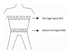
Respiratory inductance plethysmography (RIP) is a method of evaluating pulmonary ventilation by measuring the movement of the chest and abdominal wall. Accurate measurement of pulmonary ventilation or breathing often requires the use of devices such as masks or mouthpieces coupled to the airway opening. These devices are often both encumbering and invasive, and thus ill suited for continuous or ambulatory measurements. As an alternative RIP devices that sense respiratory excursions at the body surface can be used to measure pulmonary ventilation. According to a paper by Konno and Mead "the chest can be looked upon as a system of two compartments with only one degree of freedom each". Therefore, any volume change of the abdomen must be equal and opposite to that of the rib cage. The paper suggests that the volume change is close to being linearly related to changes in antero-posterior (front to back of body) diameter. When a known air volume is inhaled and measured with a spirome ... [...More Info...] [...Related Items...] OR: [Wikipedia] [Google] [Baidu] |
Pulmonary Function Testing
Pulmonary function testing (PFT) is a complete evaluation of the respiratory system including patient history, physical examinations, and tests of pulmonary function. The primary purpose of pulmonary function testing is to identify the severity of pulmonary impairment. Pulmonary function testing has diagnostic and therapeutic roles and helps clinicians answer some general questions about patients with lung disease. PFTs are normally performed by a pulmonary function technician, respiratory therapist, respiratory physiologist, physiotherapist, pulmonologist, or general practitioner. Indications Pulmonary function testing is a diagnostic and management tool used for a variety of reasons, such as: * Diagnose lung disease. * Monitor the effect of chronic diseases like asthma, chronic obstructive lung disease, or cystic fibrosis. * Detect early changes in lung function. * Identify narrowing in the airways. * Evaluate airway bronchodilator reactivity. * Show if environmental f ... [...More Info...] [...Related Items...] OR: [Wikipedia] [Google] [Baidu] |
Functional Residual Capacity
Functional residual capacity (FRC) is the volume of air present in the lungs at the end of passive expiration. At FRC, the opposing elastic recoil forces of the lungs and chest wall are in equilibrium and there is no exertion by the diaphragm or other respiratory muscles. FRC is the sum of expiratory reserve volume (ERV) and residual volume (RV) and measures approximately 2500 mL in a 70 kg, average-sized male (or approximately 30ml/kg). It cannot be estimated through spirometry, since it includes the residual volume. In order to measure RV precisely, one would need to perform a test such as nitrogen washout, helium dilution or body plethysmography. A lowered or elevated FRC is often an indication of some form of respiratory disease. For instance, in emphysema Emphysema, or pulmonary emphysema, is a lower respiratory tract disease, characterised by air-filled spaces ( pneumatoses) in the lungs, that can vary in size and may be very large. The spaces are caused ... [...More Info...] [...Related Items...] OR: [Wikipedia] [Google] [Baidu] |
Spirometry
Spirometry (meaning ''the measuring of breath'') is the most common of the pulmonary function tests (PFTs). It measures lung function, specifically the amount (volume) and/or speed (flow) of air that can be inhaled and exhaled. Spirometry is helpful in assessing breathing patterns that identify conditions such as asthma, pulmonary fibrosis, cystic fibrosis, and COPD. It is also helpful as part of a system of health surveillance, in which breathing patterns are measured over time. Spirometry generates pneumotachographs, which are charts that plot the volume and flow of air coming in and out of the lungs from one inhalation and one exhalation. Indications Spirometry is indicated for the following reasons: * to diagnose or manage asthma * to detect respiratory disease in patients presenting with symptoms of breathlessness, and to distinguish respiratory from cardiac disease as the cause * to measure bronchial responsiveness in patients suspected of having asthma * to diagnose ... [...More Info...] [...Related Items...] OR: [Wikipedia] [Google] [Baidu] |
Sexual Orientation
Sexual orientation is an enduring pattern of romantic or sexual attraction (or a combination of these) to persons of the opposite sex or gender, the same sex or gender, or to both sexes or more than one gender. These attractions are generally subsumed under heterosexuality, homosexuality, and bisexuality, while asexuality (the lack of sexual attraction to others) is sometimes identified as the fourth category. These categories are aspects of the more nuanced nature of sexual identity and terminology. For example, people may use other Label (sociology), labels, such as '' pansexual'' or ''polysexual'', or none at all. According to the American Psychological Association, sexual orientation "also refers to a person's sense of identity based on those attractions, related behaviors, and membership in a community of others who share those attractions". ''Androphilia'' and ''gynephilia'' are terms used in behavioral science to describe sexual orientation as an alternative to a ... [...More Info...] [...Related Items...] OR: [Wikipedia] [Google] [Baidu] |
Lung
The lungs are the primary organs of the respiratory system in humans and most other animals, including some snails and a small number of fish. In mammals and most other vertebrates, two lungs are located near the backbone on either side of the heart. Their function in the respiratory system is to extract oxygen from the air and transfer it into the bloodstream, and to release carbon dioxide from the bloodstream into the atmosphere, in a process of gas exchange. Respiration is driven by different muscular systems in different species. Mammals, reptiles and birds use their different muscles to support and foster breathing. In earlier tetrapods, air was driven into the lungs by the pharyngeal muscles via buccal pumping, a mechanism still seen in amphibians. In humans, the main muscle of respiration that drives breathing is the diaphragm. The lungs also provide airflow that makes vocal sounds including human speech possible. Humans have two lungs, one on the left ... [...More Info...] [...Related Items...] OR: [Wikipedia] [Google] [Baidu] |
Impedance Plethysmography
Impedance cardiography (ICG) is a non-invasive technology measuring total electrical conductivity of the thorax and its changes in time to process continuously a number of cardiodynamic parameters, such as stroke volume (SV), heart rate (HR), cardiac output (CO), ventricular ejection time (VET), pre-ejection period and used to detect the impedance changes caused by a high-frequency, low magnitude current flowing through the thorax between additional two pairs of electrodes located outside of the measured segment. The sensing electrodes also detect the ECG signal, which is used as a timing clock of the system. Introduction Impedance cardiography (ICG), also referred to as electrical impedance plethysmography (EIP) or Thoracic Electrical Bioimpedance (TEB) has been researched since the 1940s. NASA helped develop the technology in the 1960s. The use of impedance cardiography in psychophysiological research was pioneered by the publication of an article by Miller and Horvath in 1978 ... [...More Info...] [...Related Items...] OR: [Wikipedia] [Google] [Baidu] |


