|
Palatoglossal Arch
The palatoglossal arch (glossopalatine arch, anterior pillar of fauces) on either side runs downward, lateral (to the side), and forward to the side of the base of the tongue, and is formed by the projection of the glossopalatine muscle with its covering mucous membrane. It is the anterior border of the isthmus of the fauces and marks the border between the mouth and the palatopharyngeal arch. The latter marks the beginning of the pharynx The pharynx (: pharynges) is the part of the throat behind the human mouth, mouth and nasal cavity, and above the esophagus and trachea (the tubes going down to the stomach and the lungs respectively). It is found in vertebrates and invertebrates .... References External links * * Palate {{anatomy-stub ... [...More Info...] [...Related Items...] OR: [Wikipedia] [Google] [Baidu] |
Tongue
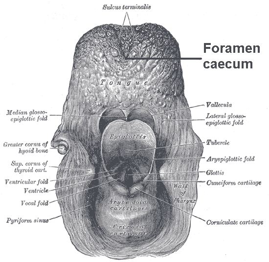
The tongue is a Muscle, muscular organ (anatomy), organ in the mouth of a typical tetrapod. It manipulates food for chewing and swallowing as part of the digestive system, digestive process, and is the primary organ of taste. The tongue's upper surface (dorsum) is covered by taste buds housed in numerous lingual papillae. It is sensitive and kept moist by saliva and is richly supplied with nerves and blood vessels. The tongue also serves as a natural means of cleaning the teeth. A major function of the tongue is to enable speech in humans and animal communication, vocalization in other animals. The human tongue is divided into two parts, an oral cavity, oral part at the front and a pharynx, pharyngeal part at the back. The left and right sides are also separated along most of its length by a vertical section of connective tissue, fibrous tissue (the lingual septum) that results in a groove, the median sulcus, on the tongue's surface. There are two groups of glossal muscles. The f ... [...More Info...] [...Related Items...] OR: [Wikipedia] [Google] [Baidu] |
Lateral (anatomy)
Standard anatomical terms of location are used to describe unambiguously the anatomy of humans and other animals. The terms, typically derived from Latin or Greek roots, describe something in its standard anatomical position. This position provides a definition of what is at the front ("anterior"), behind ("posterior") and so on. As part of defining and describing terms, the body is described through the use of anatomical planes and axes. The meaning of terms that are used can change depending on whether a vertebrate is a biped or a quadruped, due to the difference in the neuraxis, or if an invertebrate is a non-bilaterian. A non-bilaterian has no anterior or posterior surface for example but can still have a descriptor used such as proximal or distal in relation to a body part that is nearest to, or furthest from its middle. International organisations have determined vocabularies that are often used as standards for subdisciplines of anatomy. For example, '' Terminolo ... [...More Info...] [...Related Items...] OR: [Wikipedia] [Google] [Baidu] |
Posterior Tongue
The tongue is a muscular organ in the mouth of a typical tetrapod. It manipulates food for chewing and swallowing as part of the digestive process, and is the primary organ of taste. The tongue's upper surface (dorsum) is covered by taste buds housed in numerous lingual papillae. It is sensitive and kept moist by saliva and is richly supplied with nerves and blood vessels. The tongue also serves as a natural means of cleaning the teeth. A major function of the tongue is to enable speech in humans and vocalization in other animals. The human tongue is divided into two parts, an oral part at the front and a pharyngeal part at the back. The left and right sides are also separated along most of its length by a vertical section of fibrous tissue (the lingual septum) that results in a groove, the median sulcus, on the tongue's surface. There are two groups of glossal muscles. The four intrinsic muscles alter the shape of the tongue and are not attached to bone. The four paired extri ... [...More Info...] [...Related Items...] OR: [Wikipedia] [Google] [Baidu] |
Glossopalatine Muscle
The palatoglossal muscle is a muscle of the soft palate and an extrinsic muscle of the tongue. Its surface is covered by oral mucosa and forms the visible palatoglossal arch. Structure From its origin, it passes anteroinferiorly and laterally. It passes anterior to the palatine tonsil. Origin The palatoglossus arises (the oral aspect of) the palatine aponeurosis of the soft palate, where it is continuous with its contralateral partner (i.e. the same muscle of the opposite side). Insertion It inserts onto the side of the tongue; some of its fibers extend over the dorsum of the tongue, and some pass into the substance of the tongue to intermingle with the transverse muscle of tongue. Innervation The palatoglossus muscle receives motor innervation from the pharyngeal plexus of vagus nerve. It is the only muscle of the tongue ''not'' innervated by the hypoglossal nerve (CN XII). Controversy Some sources state that the palatoglossus is innervated by fibers from the cranial pa ... [...More Info...] [...Related Items...] OR: [Wikipedia] [Google] [Baidu] |
Mucous Membrane
A mucous membrane or mucosa is a membrane that lines various cavities in the body of an organism and covers the surface of internal organs. It consists of one or more layers of epithelial cells overlying a layer of loose connective tissue. It is mostly of endodermal origin and is continuous with the skin at body openings such as the eyes, eyelids, ears, inside the nose, inside the mouth, lips, the genital areas, the urethral opening and the anus. Some mucous membranes secrete mucus, a thick protective fluid. The function of the membrane is to stop pathogens and dirt from entering the body and to prevent bodily tissues from becoming dehydrated. Structure The mucosa is composed of one or more layers of epithelial cells that secrete mucus, and an underlying lamina propria of loose connective tissue. The type of cells and type of mucus secreted vary from organ to organ and each can differ along a given tract. Mucous membranes line the digestive, respiratory and rep ... [...More Info...] [...Related Items...] OR: [Wikipedia] [Google] [Baidu] |
Isthmus Of The Fauces
The fauces (also termed the isthmus of fauces or oropharyngeal isthmus) is the opening at the back of the mouth into the throat. It is a narrow passage between the velum and the base of the tongue. The fauces is a part of the oropharynx directly behind the oral cavity as a subdivision, bounded superiorly by the soft palate, laterally by the palatoglossal arch, palatoglossal and palatopharyngeal arches, and inferiorly by the tongue The tongue is a Muscle, muscular organ (anatomy), organ in the mouth of a typical tetrapod. It manipulates food for chewing and swallowing as part of the digestive system, digestive process, and is the primary organ of taste. The tongue's upper s .... The arches form the pillars of the fauces (faucial pillars, tonsillar pillars, palatine arches). The anterior (front) pillar is the palatoglossal arch formed of the palatoglossus muscle. The posterior (back) pillar is the palatopharyngeal arch formed of the palatopharyngeus muscle. Between these two arch ... [...More Info...] [...Related Items...] OR: [Wikipedia] [Google] [Baidu] |
Mouth
A mouth also referred to as the oral is the body orifice through which many animals ingest food and animal communication#Auditory, vocalize. The body cavity immediately behind the mouth opening, known as the oral cavity (or in Latin), is also the first part of the alimentary canal, which leads to the pharynx and the gullet. In tetrapod vertebrates, the mouth is bounded on the outside by the lips and cheeks — thus the oral cavity is also known as the buccal cavity (from Latin ', meaning "cheek") — and contains the tongue on the inside. Except for some groups like birds and lissamphibians, vertebrates usually have teeth in their mouths, although some fish species have pharyngeal teeth instead of oral teeth. Most bilaterian phylum, phyla, including arthropods, molluscs and chordates, have a two-opening gut tube with a mouth at one end and an anus at the other. Which end forms first in ontogeny is a criterion used to classify bilaterian animals into protostomes and deuterostomes ... [...More Info...] [...Related Items...] OR: [Wikipedia] [Google] [Baidu] |
Palatopharyngeal Arch
The palatopharyngeal arch (pharyngopalatine arch, posterior pillar of fauces) is larger and projects further toward the middle line than the palatoglossal arch; it runs downward, lateralward, and backward to the side of the pharynx, and is formed by the projection of the palatopharyngeal muscle, covered by mucous membrane A mucous membrane or mucosa is a membrane that lines various cavities in the body of an organism and covers the surface of internal organs. It consists of one or more layers of epithelial cells overlying a layer of loose connective tissue. It .... References External links * * * Palate Pharynx {{anatomy-stub ... [...More Info...] [...Related Items...] OR: [Wikipedia] [Google] [Baidu] |
Pharynx
The pharynx (: pharynges) is the part of the throat behind the human mouth, mouth and nasal cavity, and above the esophagus and trachea (the tubes going down to the stomach and the lungs respectively). It is found in vertebrates and invertebrates, though its structure varies across species. The pharynx carries food to the esophagus and air to the larynx. The flap of cartilage called the epiglottis stops food from entering the larynx. In humans, the pharynx is part of the Digestion, digestive system and the conducting zone of the respiratory system. (The conducting zone—which also includes the nostrils of the Human nose, nose, the larynx, trachea, bronchus, bronchi, and bronchioles—filters, warms, and moistens air and conducts it into the lungs). The human pharynx is conventionally divided into three sections: the nasopharynx, oropharynx, and laryngopharynx (hypopharynx). In humans, two sets of pharyngeal muscles form the pharynx and determine the shape of its lumen (anatomy), ... [...More Info...] [...Related Items...] OR: [Wikipedia] [Google] [Baidu] |

