|
Ovarian Artery
The ovarian artery is an artery that supplies oxygenated blood to the ovary in females. It arises from the abdominal aorta below the renal artery. It can be found within the suspensory ligament of the ovary, anterior to the ovarian vein and ureter. Structure The ovarian arteries are paired structures that arise from the abdominal aorta, usually at the level of L2. After emerging from the aorta, the artery travels within the suspensory ligament of the ovary and enters the mesovarium. The ovarian arteries are the corresponding arteries in the female to the testicular artery in the male. They are shorter than the testicular arteries, as the testicular arteries courses through the abdominal wall to the external scrotum. The origin and course of the first part of each artery are the same as those of the testicular artery, but on arriving at the upper opening of the lesser pelvis the ovarian artery passes inward, between the two layers of the ovariopelvic ligament and of the bro ... [...More Info...] [...Related Items...] OR: [Wikipedia] [Google] [Baidu] |
Uterine Artery
The uterine artery is an artery that supplies blood to the uterus in females. Structure The uterine artery usually arises from the anterior division of the internal iliac artery. It travels to the uterus, crossing the ureter anteriorly, to the uterus by traveling in the cardinal ligament. It travels through the parametrium of the inferior broad ligament of the uterus. It commonly anastomoses (connects with) the ovarian artery. The uterine artery is the major blood supply to the uterus and enlarges significantly during pregnancy. Branches and organs supplied * round ligament of the uterus * ovary ("ovarian branches") * uterus ( arcuate vessels) * vagina ( Vaginal branches of uterine artery) * uterine tube ("tubal branch") Anatomical variants Uterine artery can arise from the first branch of inferior gluteal artery. It can also arise as the 2nd or 3rd branch from the inferior gluteal artery. On the other hand, uterine artery can be first branch from internal iliac artery bef ... [...More Info...] [...Related Items...] OR: [Wikipedia] [Google] [Baidu] |
Ureter
The ureters are tubes composed of smooth muscle that transport urine from the kidneys to the urinary bladder. In an adult human, the ureters typically measure 20 to 30 centimeters in length and about 3 to 4 millimeters in diameter. They are lined with urothelial cells, a form of transitional epithelium, and feature an extra layer of smooth muscle in the lower third to aid in peristalsis. The ureters can be affected by a number of diseases, including urinary tract infections and kidney stone. is when a ureter is narrowed, due to for example chronic inflammation. Congenital abnormalities that affect the ureters can include the development of two ureters on the same side or abnormally placed ureters. Additionally, reflux of urine from the bladder back up the ureters is a condition commonly seen in children. The ureters have been identified for at least two thousand years, with the word "ureter" stemming from the stem relating to urinating and seen in written records since at ... [...More Info...] [...Related Items...] OR: [Wikipedia] [Google] [Baidu] |
Inguinal Canal
The inguinal canal is a passage in the anterior abdominal wall on each side of the body (one on each side of the midline), which in males, convey the spermatic cords and in females, the round ligament of the uterus. The inguinal canals are larger and more prominent in males. Structure The inguinal canals are situated just above the medial half of the inguinal ligament. The canals are approximately 4 to 6 cm long, angled anteroinferiorly and medially. In males, its diameter is normally 2 cm (±1 cm in standard deviation) at the deep inguinal ring.The diameter has been estimated to be ±2.2cm ±1.08cm in Africans, and 2.1 cm ±0.41cm in Europeans. A first-order approximation is to visualize each canal as a cylinder. Walls To help define the boundaries, these canals are often further approximated as boxes with six sides. Not including the two rings, the remaining four sides are usually called the "anterior wall", "inferior wall ("floor")", "superior wall ("roof")", and "po ... [...More Info...] [...Related Items...] OR: [Wikipedia] [Google] [Baidu] |
Round Ligament Of The Uterus
The round ligament of the uterus is a ligament that connects the uterus to the labia majora. It originates at the junction of the uterus and uterine tube. It passes through the inguinal canal to insert at the labium majus. The two round ligaments of uterus develop from the gubernaculum; they are the female homologue of the male gubernaculum testis. Structure The round ligament of the uterus originates at the uterine horns, in the parametrium. The round ligament exits the pelvis via the deep inguinal ring. It passes through the inguinal canal to reach the labium majus, inserting into the fibro-fatty substance of the labium majus. Blood supply The round ligament is supplied by the artery of the round ligament of uterus, also known as ''Sampson's artery''. Development The round ligament develops from the gubernaculum which attaches the gonad to the labioscrotal swellings in the embryo. Function The round ligament of uterus acts to hold the uterus anterior-ward to in antef ... [...More Info...] [...Related Items...] OR: [Wikipedia] [Google] [Baidu] |
Uterine Tube
The fallopian tubes, also known as uterine tubes, oviducts or salpinges (: salpinx), are paired tubular sex organs in the human female body that stretch from the ovaries to the uterus. The fallopian tubes are part of the female reproductive system. In other vertebrates, they are only called oviducts. Each tube is a muscular hollow organ that is on average between in length, with an external diameter of . It has four described parts: the intramural part, isthmus, ampulla, and infundibulum with associated fimbriae. Each tube has two openings: a proximal opening nearest to the uterus, and a distal opening nearest to the ovary. The fallopian tubes are held in place by the mesosalpinx, a part of the broad ligament mesentery that wraps around the tubes. Another part of the broad ligament, the mesovarium suspends the ovaries in place. An egg cell is transported from an ovary to a fallopian tube where it may be fertilized in the ampulla of the tube. The fallopian tubes are lined with ... [...More Info...] [...Related Items...] OR: [Wikipedia] [Google] [Baidu] |
Broad Ligament
The broad ligament of the uterus is the wide fold of peritoneum that connects the sides of the uterus to the walls and floor of the pelvis. Structure Subdivisions Contents The contents of the broad ligament include the following: * Reproductive ** uterine tubes (or fallopian tube) ** ovary (some sources consider the ovary to be on the broad ligament, but not in it.) * vessels ** ovarian artery (in the suspensory ligament) ** uterine artery (in reality, travels in the cardinal ligament) * ligaments ** ovarian ligament ** round ligament of uterus ** suspensory ligament of the ovary (Some sources consider it a part of the broad ligament, while other sources just consider it a "termination" of the ligament.) Relations The peritoneum surrounds the uterus like a flat sheet that folds over its fundus, covering it anteriorly and posteriorly; on the sides of the uterus, this sheet of peritoneum comes in direct contact with itself, forming the double layer of peritoneum known as t ... [...More Info...] [...Related Items...] OR: [Wikipedia] [Google] [Baidu] |
Lesser Pelvis
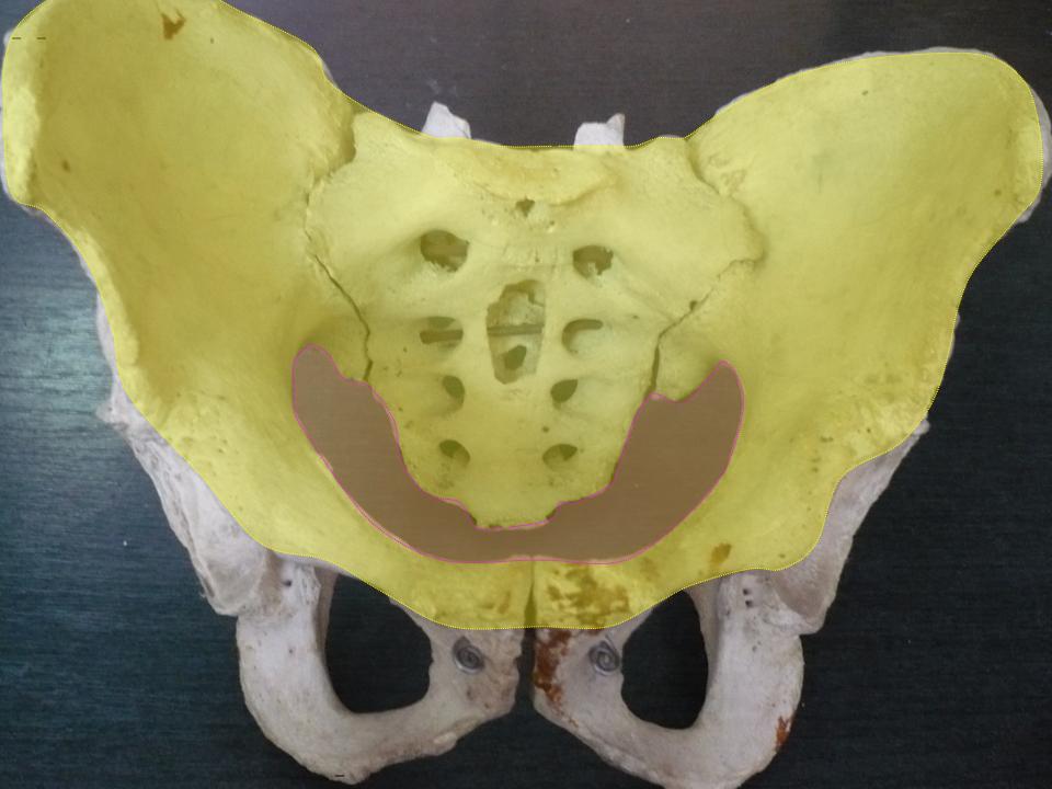
The pelvic cavity is a body cavity that is bounded by the bones of the pelvis. Its oblique roof is the pelvic inlet (the superior opening of the pelvis). Its lower boundary is the pelvic floor. The pelvic cavity primarily contains the reproductive organs, urinary bladder, distal ureters, proximal urethra, terminal sigmoid colon, rectum, and anal canal. In females, the uterus, fallopian tubes, ovaries and upper vagina occupy the area between the other viscera. The rectum is located at the back of the pelvis, in the curve of the sacrum and coccyx; the bladder is in front, behind the pubic symphysis. The pelvic cavity also contains major arteries, veins, muscles, and nerves. These structures coexist in a crowded space, and disorders of one pelvic component may impact upon another; for example, constipation may overload the rectum and compress the urinary bladder, or childbirth might damage the pudendal nerves and later lead to anal weakness. Structure The pelvis has ... [...More Info...] [...Related Items...] OR: [Wikipedia] [Google] [Baidu] |
Scrotum
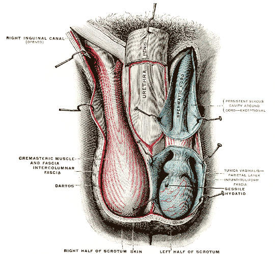
In most terrestrial mammals, the scrotum (: scrotums or scrota; possibly from Latin ''scortum'', meaning "hide" or "skin") or scrotal sac is a part of the external male genitalia located at the base of the penis. It consists of a sac of skin containing the external spermatic fascia, testicles, epididymides, and vasa deferentia. The scrotum will usually tighten when exposed to cold temperatures. The scrotum is homologous to the labia majora in females. Structure In regards to humans, the scrotum is a suspended two-chambered sac of skin and muscular tissue containing the testicles and the lower part of the spermatic cords. It is located behind the penis and above the perineum. The perineal raphe is a small, vertical ridge of skin that expands from the anus and runs through the middle of the scrotum front to back. The scrotum is also a distention of the perineum and carries some abdominal tissues into its cavity including the testicular artery, testicular vein, and ... [...More Info...] [...Related Items...] OR: [Wikipedia] [Google] [Baidu] |
Male
Male (Planet symbols, symbol: ♂) is the sex of an organism that produces the gamete (sex cell) known as sperm, which fuses with the larger female gamete, or Egg cell, ovum, in the process of fertilisation. A male organism cannot sexual reproduction, reproduce sexually without access to at least one ovum from a female, but some organisms can reproduce both sexually and Asexual reproduction, asexually. Most male mammals, including male humans, have a Y chromosome, which codes for the production of larger amounts of testosterone to develop male reproductive organs. In humans, the word ''male'' can also be used to refer to gender, in the social sense of gender role or gender identity. Overview The existence of separate sexes has evolved independently at different times and in different lineage (evolution), lineages, an example of convergent evolution. The repeated pattern is sexual reproduction in isogamy, isogamous species with two or more mating types with gametes of identic ... [...More Info...] [...Related Items...] OR: [Wikipedia] [Google] [Baidu] |
Testicular Artery
The testicular artery (the male gonadal artery, also called the internal spermatic arteries in older texts) is a branch of the abdominal aorta that supplies blood to the testicle. It is a paired artery, with one for each of the testicles. It is the male equivalent of the ovarian artery. Because the testis is found in a different location than that of its female equivalent, it has a different course than the ovarian artery. They are two slender vessels of considerable length, and arise from the front of the aorta a little below the renal arteries. Each passes obliquely downward and lateralward behind the peritoneum, resting on the psoas major, the right lying in front of the inferior vena cava and behind the middle colic and ileocolic arteries and the terminal part of the ileum, the left behind the left colic and sigmoid arteries and the iliac colon. Each crosses obliquely over the ureter and the lower part of the external iliac artery to reach the abdominal inguinal rin ... [...More Info...] [...Related Items...] OR: [Wikipedia] [Google] [Baidu] |


