|
Opponens Digiti Minimi Muscle
The opponens digiti minimi (opponens digiti quinti in older texts) is a muscle in the hand. It is of a triangular form, and placed immediately beneath the palmaris brevis, abductor digiti minimi and flexor digiti minimi brevis. It is one of the three hypothenar muscles that control the little finger. It arises from the convexity of the hamulus of the hamate bone and the contiguous portion of the transverse carpal ligament; it is inserted into the whole length of the metacarpal bone of the little finger, along its ulnar margin. The opponens digiti minimi muscle serves to flex and laterally rotate the 5th metacarpal about the 5th carpometacarpal joint, as when bringing the little finger and thumb into opposition. It is innervated by the deep branch of the ulnar nerve. See also * Hypothenar * Opponens pollicis muscle Additional images Image:Gray426.png, The muscles of the thumb The thumb is the first digit of the hand, next to the index finger. When a person is st ... [...More Info...] [...Related Items...] OR: [Wikipedia] [Google] [Baidu] [Amazon] |
Hamate
The hamate bone (from Latin language, Latin wiktionary:hamatus, hamatus, "hooked"), or unciform bone (from Latin language, Latin ''wikt:uncus, uncus'', "hook"), Latin os hamatum and occasionally abbreviated as just hamatum, is a bone in the human wrist readily distinguishable by its wedge shape and a hook-like process ("hamulus") projecting from its Radioulnar, palmar surface. Structure The hamate is an irregularly shaped carpal bone found within the hand. The hamate is found within the distal row of carpal bones, and abuts the metacarpals of the little finger and ring finger. Adjacent to the hamate on the ulnar side, and slightly proximal and ulnar to it, is the pisiform bone. Adjacent on the radial side is the capitate, and proximal is the lunate bone. Surfaces The hamate bone has six surfaces: * The ''superior'', the apex of the wedge, is narrow, convex, smooth, and articulates with the lunate bone, lunate. * The ''inferior'' articulates with the fourth and fifth metacarpal ... [...More Info...] [...Related Items...] OR: [Wikipedia] [Google] [Baidu] [Amazon] |
Little Finger
The little finger or pinkie, also known as the baby finger, fifth digit, or pinky finger, is the most ulnar and smallest digit of the human hand, and next to the ring finger. Etymology The word "pinkie" is derived from the Dutch word ''pink'', meaning "little finger". The earliest recorded use of the term "pinkie" is from Scotland in 1808. The term (sometimes spelled "pinky") is common in Scottish English and American English, and is also used extensively in other Commonwealth countries such as New Zealand, Canada, and Australia. Nerves and muscles There are nine muscles that control the fifth digit: Three in the hypothenar eminence, two extrinsic flexors, two extrinsic extensors, and two more intrinsic muscles: * Hypothenar eminence: ** Opponens digiti minimi muscle ** Abductor minimi digiti muscle (adduction from third palmar interossei) ** Flexor digiti minimi brevis (the "longus" is absent in most humans) * Two extrinsic flexors: ** Flexor digitorum superficialis ** ... [...More Info...] [...Related Items...] OR: [Wikipedia] [Google] [Baidu] [Amazon] |
Thumb
The thumb is the first digit of the hand, next to the index finger. When a person is standing in the medical anatomical position (where the palm is facing to the front), the thumb is the outermost digit. The Medical Latin English noun for thumb is ''pollex'' (compare ''hallux'' for big toe), and the corresponding adjective for thumb is ''pollical''. Definition Thumb and fingers The English word ''finger'' has two senses, even in the context of appendages of a single typical human hand: 1) Any of the five terminal members of the hand. 2) Any of the four terminal members of the hand, other than the thumb. Linguistically, it appears that the original sense was the first of these two: (also rendered as ) was, in the inferred Proto-Indo-European language, a suffixed form of (or ), which has given rise to many Indo-European-family words (tens of them defined in English dictionaries) that involve, or stem from, concepts of fiveness. The thumb shares the following with each of ... [...More Info...] [...Related Items...] OR: [Wikipedia] [Google] [Baidu] [Amazon] |
Opponens Pollicis Muscle
The opponens pollicis is a small, triangular muscle in the hand, which functions to oppose the thumb. It is one of the three thenar muscles. It lies deep to the abductor pollicis brevis and lateral to the flexor pollicis brevis. Structure The opponens pollicis muscle is one of the three thenar muscles. It originates from the flexor retinaculum of the hand and the tubercle of the trapezium. It passes downward and laterally, and is inserted into the whole length of the metacarpal bone of the thumb on its radial side. Innervation Like the other thenar muscles, the opponens pollicis is innervated by the recurrent branch of the median nerve. In 20% of the population, opponens pollicis is innervated by the ulnar nerve. Blood supply The opponens pollicis receives its blood supply from the superficial palmar arch. Function ''Opposition of the thumb'' is a combination of actions that allows the tip of the thumb to touch the tips of other fingers. The part of apposition that this m ... [...More Info...] [...Related Items...] OR: [Wikipedia] [Google] [Baidu] [Amazon] |
Hypothenar
The hypothenar muscles are a group of three muscles of the hand, palm that control the motion of the little finger. The three muscles are: * Abductor minimi digiti muscle (hand), Abductor digiti minimi * Flexor digiti minimi brevis (hand), Flexor digiti minimi brevis * Opponens digiti minimi Structure The muscles of hypothenar eminence are from medial to lateral: * Opponens digiti minimi muscle, Opponens digiti minimi * Flexor digiti minimi brevis muscle (hand), Flexor digiti minimi brevis * Abductor digiti minimi muscle of hand, Abductor digiti minimi The intrinsic muscles of hand can be remembered using the mnemonic, "A OF A OF A" for, Abductor pollicis brevis, Opponens pollicis, Flexor pollicis brevis (the three thenar muscles), Adductor pollicis, and the three hypothenar muscles, Opponens digiti minimi, Flexor digiti minimi brevis, Abductor digiti minimi. Clinical significance "Hypothenar atrophy" is associated with the lesion of the ulnar nerve, which supplies the three hyp ... [...More Info...] [...Related Items...] OR: [Wikipedia] [Google] [Baidu] [Amazon] |
Deep Branch Of The Ulnar Nerve
The deep branch of the ulnar nerve is a terminal, primarily motor branch of the ulnar nerve. It is accompanied by the deep palmar branch of ulnar artery. Structure It passes between the abductor digiti minimi and the flexor digiti minimi brevis. It then perforates the opponens digiti minimi and follows the course of the deep palmar arch beneath the flexor tendons. As the deep ulnar nerve passes across the palm, it lies in a fibrous tunnel formed between the hook of the hamate and the pisiform ( Guyon's canal). Function At its origin it innervates the hypothenar muscles. As it crosses the deep part of the hand, it innervates all the interosseous muscles and the third and fourth lumbricals. It ends by innervating the adductor pollicis and the medial (deep) head of the flexor pollicis brevis The flexor pollicis brevis is a muscle in the hand that flexes the thumb. It is one of three thenar muscles. It has both a superficial part and a deep part. Origin and insertion The mus ... [...More Info...] [...Related Items...] OR: [Wikipedia] [Google] [Baidu] [Amazon] |
Carpometacarpal Joint
The carpometacarpal (CMC) joints are five joints in the wrist that articulate the distal row of carpal bones and the proximal bases of the five metacarpal bones. The CMC joint of the thumb or the first CMC joint, also known as the trapeziometacarpal (TMC) joint, differs significantly from the other four CMC joints and is therefore described separately. Thumb The carpometacarpal joint of the thumb (''pollex''), also known as the first carpometacarpal joint, or the trapeziometacarpal joint (TMC) because it connects the trapezium to the first metacarpal bone, plays an irreplaceable role in the normal functioning of the thumb. The most important joint connecting the wrist to the metacarpus, osteoarthritis of the TMC is a severely disabling condition; it is up to twenty times more common among elderly women than in the average. Pronation-supination of the first metacarpal is especially important for the action of opposition. The movements of the first CMC are limited by the sha ... [...More Info...] [...Related Items...] OR: [Wikipedia] [Google] [Baidu] [Amazon] |
Metacarpal Bone
In human anatomy, the metacarpal bones or metacarpus, also known as the "palm bones", are the appendicular bones that form the intermediate part of the hand between the phalanges (fingers) and the carpal bones ( wrist bones), which articulate with the forearm. The metacarpal bones are homologous to the metatarsal bones in the foot. Structure The metacarpals form a transverse arch to which the rigid row of distal carpal bones are fixed. The peripheral metacarpals (those of the thumb and little finger) form the sides of the cup of the palmar gutter and as they are brought together they deepen this concavity. The index metacarpal is the most firmly fixed, while the thumb metacarpal articulates with the trapezium and acts independently from the others. The middle metacarpals are tightly united to the carpus by intrinsic interlocking bone elements at their bases. The ring metacarpal is somewhat more mobile while the fifth metacarpal is semi-independent.Tubiana ''et al'' 1998, p 11 ... [...More Info...] [...Related Items...] OR: [Wikipedia] [Google] [Baidu] [Amazon] |
Transverse Carpal Ligament
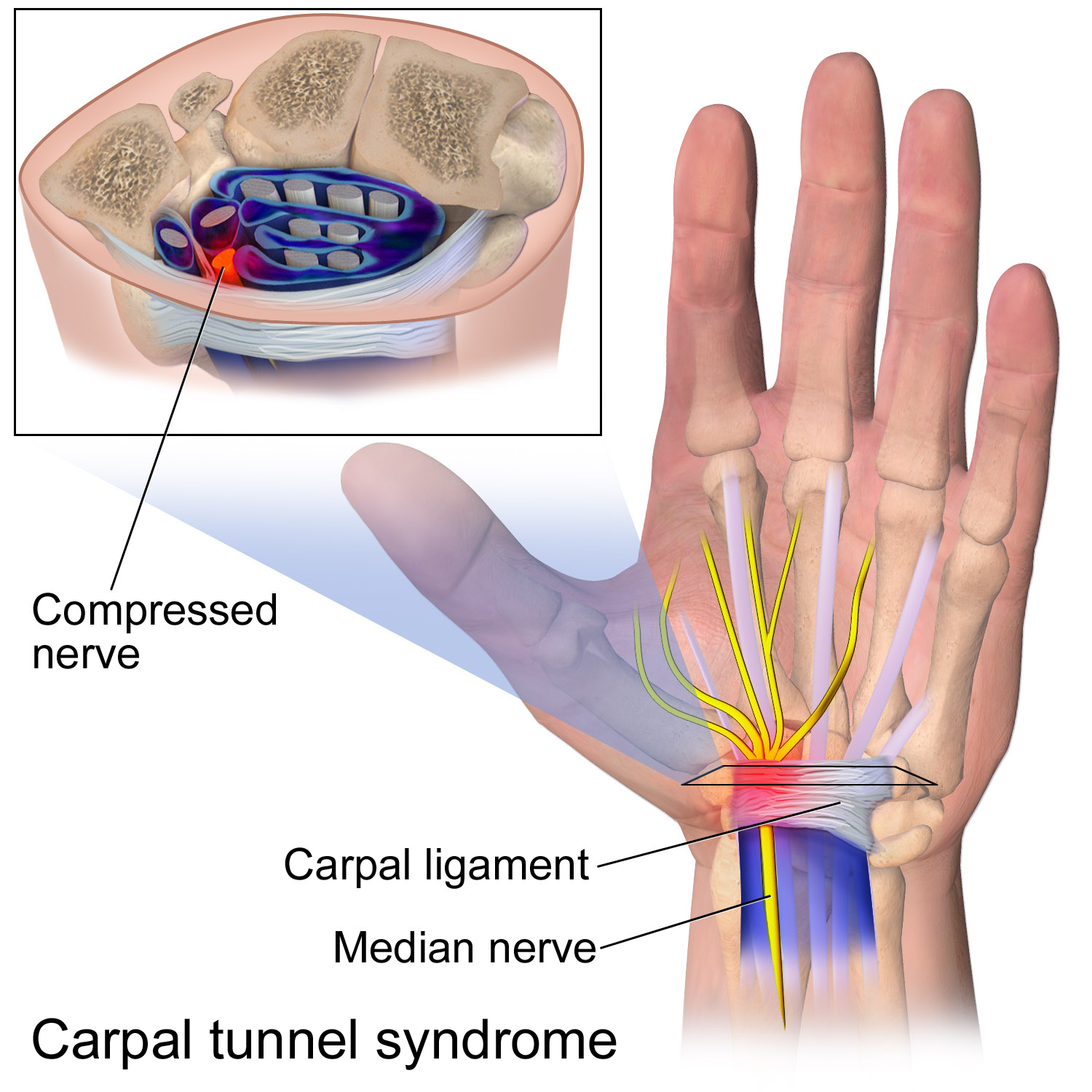
The flexor retinaculum (transverse carpal ligament or anterior annular ligament) is a fibrous band on the palmar side of the hand near the wrist. It arches over the carpal bones of the hands, covering them and forming the carpal tunnel. Structure The flexor retinaculum is a strong, fibrous band that covers the carpal bones on the palmar side of the hand near the wrist. It attaches to the bones near the radius and ulna. On the ulnar side, the flexor retinaculum attaches to the pisiform bone and the hook of the hamate bone. On the radial side, it attaches to the tubercle of the scaphoid bone, and to the medial part of the palmar surface and the ridge of the trapezium bone. The flexor retinaculum is continuous with the palmar carpal ligament, and deeper with the palmar aponeurosis. The ulnar artery and ulnar nerve, and the cutaneous branches of the median and ulnar nerves, pass on top of the flexor retinaculum. On the radial side of the retinaculum is the tendon of the flexor car ... [...More Info...] [...Related Items...] OR: [Wikipedia] [Google] [Baidu] [Amazon] |
Hamate Bone
The hamate bone (from Latin hamatus, "hooked"), or unciform bone (from Latin '' uncus'', "hook"), Latin os hamatum and occasionally abbreviated as just hamatum, is a bone in the human wrist readily distinguishable by its wedge shape and a hook-like process ("hamulus") projecting from its palmar surface. Structure The hamate is an irregularly shaped carpal bone found within the hand. The hamate is found within the distal row of carpal bones, and abuts the metacarpals of the little finger and ring finger. Adjacent to the hamate on the ulnar side, and slightly proximal and ulnar to it, is the pisiform bone. Adjacent on the radial side is the capitate, and proximal is the lunate bone. Surfaces The hamate bone has six surfaces: * The ''superior'', the apex of the wedge, is narrow, convex, smooth, and articulates with the lunate. * The ''inferior'' articulates with the fourth and fifth metacarpal bones, by concave facets which are separated by a ridge. * The ''dorsal'' is triangul ... [...More Info...] [...Related Items...] OR: [Wikipedia] [Google] [Baidu] [Amazon] |
Hamulus
A hamus or hamulus is a structure functioning as, or in the form of, hooks or hooklets. Etymology The terms are directly from Latin, in which ''hamus'' means "hook". The plural is ''hami''. ''Hamulus'' is the diminutive – hooklet or little hook. The plural is ''hamuli''. Adjectives are ''hamate'' and ''hamulate'', as in "a hamulate wing-coupling", in which the wings of certain insects in flight are joined by hooking hamuli on one wing into folds on a matching wing. ''Hamulate'' can also mean "having hamuli". The terms ''hamose'', ''hamular'', ''hamous'' and ''hamiform'' also have been used to mean "hooked", or "hook-shaped". Terms such as ''hamate'' that do not indicate a diminutive usually refer particularly to a hook at the tip, whereas diminutive terms such as ''hamulose'' tend to imply that something is beset with small hooks. Anatomy In vertebrate anatomy, a hamulus is a small, hook-shaped portion of a bone, or possibly of other hard tissue. In human anatomy, example ... [...More Info...] [...Related Items...] OR: [Wikipedia] [Google] [Baidu] [Amazon] |
Hypothenar Muscles
The hypothenar muscles are a group of three muscles of the palm that control the motion of the little finger. The three muscles are: * Abductor digiti minimi * Flexor digiti minimi brevis * Opponens digiti minimi Structure The muscles of hypothenar eminence are from medial to lateral: * Opponens digiti minimi * Flexor digiti minimi brevis * Abductor digiti minimi The intrinsic muscles of hand can be remembered using the mnemonic, "A OF A OF A" for, Abductor pollicis brevis, Opponens pollicis, Flexor pollicis brevis (the three thenar muscles), Adductor pollicis, and the three hypothenar muscles, Opponens digiti minimi, Flexor digiti minimi brevis, Abductor digiti minimi. Clinical significance "Hypothenar atrophy" is associated with the lesion of the ulnar nerve, which supplies the three hypothenar muscles. Hypothenar hammer syndrome is a vascular occlusion of this region. See also * Thenar eminence * Palmaris brevis Palmaris brevis muscle is a thin, quadrilateral muscle, ... [...More Info...] [...Related Items...] OR: [Wikipedia] [Google] [Baidu] [Amazon] |


