|
Hamate Bone
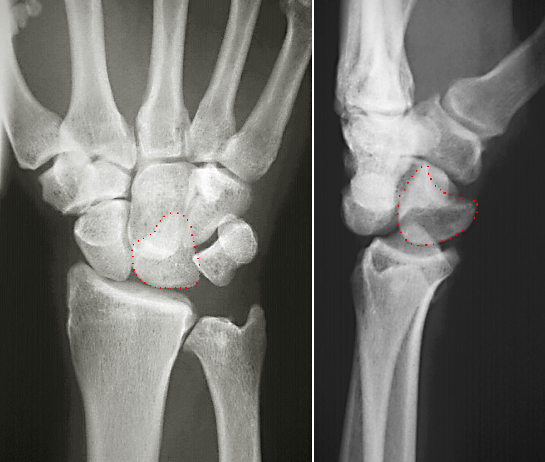
The hamate bone (from Latin hamatus, "hooked"), or unciform bone (from Latin '' uncus'', "hook"), Latin os hamatum and occasionally abbreviated as just hamatum, is a bone in the human wrist readily distinguishable by its wedge shape and a hook-like process ("hamulus") projecting from its palmar surface. Structure The hamate is an irregularly shaped carpal bone found within the hand. The hamate is found within the distal row of carpal bones, and abuts the metacarpals of the little finger and ring finger. Adjacent to the hamate on the ulnar side, and slightly proximal and ulnar to it, is the pisiform bone. Adjacent on the radial side is the capitate, and proximal is the lunate bone. Surfaces The hamate bone has six surfaces: * The ''superior'', the apex of the wedge, is narrow, convex, smooth, and articulates with the lunate. * The ''inferior'' articulates with the fourth and fifth metacarpal bones, by concave facets which are separated by a ridge. * The ''dorsal'' is triangul ... [...More Info...] [...Related Items...] OR: [Wikipedia] [Google] [Baidu] |
Lunate Bone
The lunate bone (semilunar bone) is a carpal bone in the human hand. It is distinguished by its deep concavity and crescentic outline. It is situated in the center of the proximal row carpal bones, which lie between the ulna and radius and the hand. The lunate carpal bone is situated between the lateral scaphoid bone and medial triquetral bone. Structure The lunate is a crescent-shaped carpal bone found within the hand. The lunate is found within the proximal row of carpal bones. Proximally, it abuts the radius. Laterally, it articulates with the scaphoid bone, medially with the triquetral bone, and distally with the capitate bone. The lunate also articulates on its distal and medial surface with the hamate bone. The lunate is stabilised by a medial ligament to the scaphoid bone and a lateral ligament to the triquetral bone. Ligaments between the radius and carpal bone also stabilise the position of the lunate, as does its position in the lunate fossa of the radius. Bone Th ... [...More Info...] [...Related Items...] OR: [Wikipedia] [Google] [Baidu] |
Hamulus Of Hamate Bone
The hamate bone (from Latin language, Latin wiktionary:hamatus, hamatus, "hooked"), or unciform bone (from Latin language, Latin ''wikt:uncus, uncus'', "hook"), Latin os hamatum and occasionally abbreviated as just hamatum, is a bone in the human wrist readily distinguishable by its wedge shape and a hook-like process ("hamulus") projecting from its Radioulnar, palmar surface. Structure The hamate is an irregularly shaped carpal bone found within the hand. The hamate is found within the distal row of carpal bones, and abuts the metacarpals of the little finger and ring finger. Adjacent to the hamate on the ulnar side, and slightly proximal and ulnar to it, is the pisiform bone. Adjacent on the radial side is the capitate, and proximal is the lunate bone. Surfaces The hamate bone has six surfaces: * The ''superior'', the apex of the wedge, is narrow, convex, smooth, and articulates with the lunate bone, lunate. * The ''inferior'' articulates with the fourth and fifth metacarpal ... [...More Info...] [...Related Items...] OR: [Wikipedia] [Google] [Baidu] |
Fracture (bone)
A bone fracture (abbreviated FRX or Fx, Fx, or #) is a medical condition in which there is a partial or complete break in the continuity of any bone in the body. In more severe cases, the bone may be broken into several fragments, known as a ''comminuted fracture''. An open fracture (or compound fracture) is a bone fracture where the broken bone breaks through the skin. A bone fracture may be the result of high force impact or stress, or a minimal trauma injury as a result of certain medical conditions that weaken the bones, such as osteoporosis, osteopenia, bone cancer, or osteogenesis imperfecta, where the fracture is then properly termed a pathologic fracture. Most bone fractures require urgent medical attention to prevent further injury. Signs and symptoms Although bone tissue contains no pain receptors, a bone fracture is painful for several reasons: * Breaking in the continuity of the periosteum, with or without similar discontinuity in endosteum, as both contain mult ... [...More Info...] [...Related Items...] OR: [Wikipedia] [Google] [Baidu] |
Homology (biology)
In biology, homology is similarity in anatomical structures or genes between organisms of different taxa due to shared ancestry, ''regardless'' of current functional differences. Evolutionary biology explains homologous structures as retained heredity from a common descent, common ancestor after having been subjected to adaptation (biology), adaptive modifications for different purposes as the result of natural selection. The term was first applied to biology in a non-evolutionary context by the anatomist Richard Owen in 1843. Homology was later explained by Charles Darwin's theory of evolution in 1859, but had been observed before this from Aristotle's biology onwards, and it was explicitly analysed by Pierre Belon in 1555. A common example of homologous structures is the forelimbs of vertebrates, where the bat wing development, wings of bats and origin of avian flight, birds, the arms of primates, the front flipper (anatomy), flippers of whales, and the forelegs of quadrupedalis ... [...More Info...] [...Related Items...] OR: [Wikipedia] [Google] [Baidu] |
Flexor Tendons
In anatomy, flexor is a muscle that contracts to perform flexion (from the Latin verb ''flectere'', to bend), a movement that decreases the angle between the bones converging at a joint. For example, one's elbow joint flexes when one brings their hand closer to the shoulder, thus decreasing the angle between the upper arm and the forearm. Flexors Upper limb *of the humerus bone (the bone in the upper arm) at the shoulder ** Pectoralis major **Anterior deltoid **Coracobrachialis **Biceps brachii * of the forearm at the elbow **Brachialis **Brachioradialis **Biceps brachii *of carpus (the carpal bones) at the wrist **flexor carpi radialis **flexor carpi ulnaris **palmaris longus *of the hand **flexor pollicis longus muscle **flexor pollicis brevis muscle **flexor digitorum profundus muscle **flexor digitorum superficialis muscle Lower limb Hip The hip flexors are (in descending order of importance to the action of flexing the hip joint):Platzer (2004), p 246 *Collectively know ... [...More Info...] [...Related Items...] OR: [Wikipedia] [Google] [Baidu] |
Opponens Digiti Minimi
The opponens digiti minimi (opponens digiti quinti in older texts) is a muscle in the hand. It is of a triangular form, and placed immediately beneath the palmaris brevis, abductor digiti minimi and flexor digiti minimi brevis. It is one of the three hypothenar muscles that control the little finger. It arises from the convexity of the hamulus of the hamate bone and the contiguous portion of the transverse carpal ligament; it is inserted into the whole length of the metacarpal bone of the little finger, along its ulnar margin. The opponens digiti minimi muscle serves to flex and laterally rotate the 5th metacarpal about the 5th carpometacarpal joint, as when bringing the little finger and thumb into opposition. It is innervated by the deep branch of the ulnar nerve. See also * Hypothenar * Opponens pollicis muscle Additional images Image:Gray426.png, The muscles of the thumb The thumb is the first digit of the hand, next to the index finger. When a person is sta ... [...More Info...] [...Related Items...] OR: [Wikipedia] [Google] [Baidu] |
Flexor Digiti Minimi Brevis (hand)
The flexor digiti minimi brevis is a hypothenar muscle in the hand that flexes the little finger (digit V) at the metacarpophalangeal joint. It lies lateral to the abductor digiti minimi when the hand is in anatomical position. Structure The flexor digiti minimi brevis arises from the hamulus of the hamate bone and the palmar surface of the flexor retinaculum of the hand. It is inserted into the medial side of the base of the proximal phalanx of digit V. It is separated from the abductor digiti minimi, at its origin, by the deep branches of the ulnar artery and the ulnar nerve. The flexor digiti minimi brevis is sometimes not present; in these cases, the abductor digiti minimi is usually larger than normal. The flexor digiti minimi brevis is one of three muscles in the hypothenar muscle group. These three muscles form the fleshy mass at the base of the little finger, and are solely concerned with the movement of digit V. The other two muscles that make up the hypothenar ... [...More Info...] [...Related Items...] OR: [Wikipedia] [Google] [Baidu] |
Flexor Carpi Ulnaris
The flexor carpi ulnaris (FCU) is a skeletal muscle, muscle of the forearm that flexion, flexes and Adduction, adducts at the wrist joint. Structure Origin The flexor carpi ulnaris has two heads; a humeral head and ulnar head. The humeral head originates from the medial epicondyle of the humerus via the common flexor tendon. The ulnar head originates from the medial margin of the olecranon of the ulna and the upper two-thirds of the dorsal border of the ulna by an aponeurosis. Between the two heads passes the ulnar nerve and ulnar artery. Insertion The flexor carpi ulnaris inserts onto the pisiform bone, pisiform, hook of the hamate (via the pisohamate ligament) and the anterior surface of the base of the fifth metacarpal bone, fifth metacarpal (via the pisometacarpal ligament). Action The flexor carpi ulnaris flexes and adducts at the Wrist, wrist joint. Innervation The flexor carpi ulnaris is innervated by the ulnar nerve. The corresponding spinal nerves are Cervical spinal ... [...More Info...] [...Related Items...] OR: [Wikipedia] [Google] [Baidu] |
Pisohamate Ligament
The pisohamate ligament is a ligament in the hand. It connects the pisiform, a sesamoid bone in the wrist, to the hook of the hamate. It is a prolongation of the tendon of the flexor carpi ulnaris. It serves as part of the origin for the abductor digiti minimi. It also forms the floor of the ulnar canal, a canal that allows the ulnar nerve and ulnar artery The ulnar artery is the main blood vessel, with oxygenated blood, of the Human Anatomical Terms#Anatomical directions, medial aspects of the forearm. It arises from the brachial artery and terminates in the superficial palmar arch, which joins ... into the hand. References Ligaments of the upper limb {{ligament-stub ... [...More Info...] [...Related Items...] OR: [Wikipedia] [Google] [Baidu] |
Guyon's Canal
The ulnar canal or ulnar tunnel (also known as Guyon's canal or tunnel) is a semi-rigid longitudinal canal in the wrist that allows passage of the ulnar artery and ulnar nerve into the hand. (These are named after the ulna, the long bone on the little finger side of the arm.) The roof of the canal is made up of the superficial palmar carpal ligament, while the deeper flexor retinaculum and hypothenar muscles comprise the floor. The space is medially bounded by the pisiform and pisohamate ligament more proximally, and laterally bounded by the hook of the hamate more distally. It is approximately 4 cm long, beginning proximally at the transverse carpal ligament and ending at the aponeurotic arch of the hypothenar muscles. Eponym The ulnar tunnel is named after the French surgeon Jean Casimir Félix Guyon, who originally described the canal in 1861. Clinical significance Entrapment of the ulnar nerve at the ulnar canal can result in symptoms of ulnar neuropathy, including num ... [...More Info...] [...Related Items...] OR: [Wikipedia] [Google] [Baidu] |
Carpal Tunnel
In the human body, the carpal tunnel or carpal canal is a flattened body cavity on the flexor ( palmar/volar) side of the wrist, bounded by the carpal bones and flexor retinaculum. It forms the passageway that transmits the median nerve and the tendons of the extrinsic flexor muscles of the hand from the forearm to the hand. The median artery is an anatomical variant (increasingly found). When present it lies between the radial artery, and the ulnar artery and runs with the median nerve supplying the same structures innervated. When swelling or degeneration occurs in the tendons and sheaths of any of the nine flexor muscles ( flexor pollicis longus, four flexor digitorum profundus and four flexor digitorum superficialis) passing through the carpal tunnel, the canal can narrow and compress/entrap the median nerve, resulting in a compression neuropathy known as carpal tunnel syndrome (CTS). If untreated, neuropraxia, parasthesia and muscle atrophy (especially of the ... [...More Info...] [...Related Items...] OR: [Wikipedia] [Google] [Baidu] |
Ulnar Nerve
The ulnar nerve is a nerve that runs near the ulna, one of the two long bones in the forearm. The ulnar collateral ligament of elbow joint is in relation with the ulnar nerve. The nerve is the largest in the human body unprotected by muscle or bone, so injury is common. This nerve is directly connected to the little finger, and the adjacent half of the ring finger, innervating the palmar aspect of these fingers, including both front and back of the tips, perhaps as far back as the fingernail beds. This nerve can cause an electric shock-like sensation by striking the medial epicondyle of the humerus posteriorly, or inferiorly with the elbow flexed. The ulnar nerve is trapped between the bone and the overlying skin at this point. This is commonly referred to as bumping one's "funny bone". This name is thought to be a pun, based on the sound resemblance between the name of the bone of the upper arm, the humerus, and the word " humorous". Alternatively, according to the Oxfor ... [...More Info...] [...Related Items...] OR: [Wikipedia] [Google] [Baidu] |


