|
Obturator Nerve
The obturator nerve in human anatomy arises from the ventral divisions of the second, third, and fourth lumbar nerves in the lumbar plexus; the branch from the third is the largest, while that from the second is often very small. Structure The obturator nerve originates from the anterior divisions of the L2, L3, and L4 spinal nerve roots. It descends through the fibers of the psoas major, and emerges from its medial border near the brim of the pelvis. It then passes behind the common iliac arteries, and on the lateral side of the internal iliac artery and vein, and runs along the lateral wall of the lesser pelvis, above and in front of the obturator vessels, to the upper part of the obturator foramen. Here it enters the thigh, through the obturator canal, and divides into an anterior and a posterior branch, which are separated at first by some of the fibers of the obturator externus, and lower down by the adductor brevis. An accessory obturator nerve may be present in ap ... [...More Info...] [...Related Items...] OR: [Wikipedia] [Google] [Baidu] |
Hip-joint
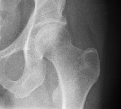
In vertebrate anatomy, the hip, or coxaLatin ''coxa'' was used by Celsus in the sense "hip", but by Pliny the Elder in the sense "hip bone" (Diab, p 77) (: ''coxae'') in medical terminology, refers to either an list of human anatomical regions, anatomical region or a joint on the outer (lateral) side of the pelvis. The hip region is located lateral (anatomy), lateral and anterior (anatomy), anterior to the Buttocks, gluteal region, inferior (anatomy), inferior to the iliac crest, and lateral to the obturator foramen, with muscle tendons and soft tissues overlying the greater trochanter of the femur. In adults, the three pelvic bones (ilium (bone), ilium, ischium and pubis (bone), pubis) have fused into one hip bone, which forms the superomedial/deep wall of the hip region. The hip joint, scientifically referred to as the acetabulofemoral joint (''art. coxae''), is the ball-and-socket joint between the pelvic acetabulum and the femoral head. Its primary function is to weight-bear ... [...More Info...] [...Related Items...] OR: [Wikipedia] [Google] [Baidu] |
Obturator Foramen
The obturator foramen is the large, Bilateral symmetry, bilaterally paired opening of the bony pelvis. It is formed by the pubis and ischium. It is mostly closed by the obturator membrane except for a small opening, the obturator canal, through which the obturator nerve and vessels pass. Structure The obturator foramen is situated inferior and somewhat anterior to the acetabulum. It is bounded by the pubis bone and the ischium: superiorly by the (grooved obturator surface) of the Superior rami of the pubes, superior ramus of pubis, inferiorly by the Ischium#Structure, ramus of ischium, and laterally by (the anterior edge of) the body of ischium (including by the margin of the acetabulum). The margin of the foramen is thin and uneven, and gives attachment to the obturator membrane. Superiorly, it presents a deep groove - the obturator groove - which passes obliquely inferomedially from the pelvis. The foramen is largely closed by the obturator membrane save for a small opening at ... [...More Info...] [...Related Items...] OR: [Wikipedia] [Google] [Baidu] |
Obturator Internus
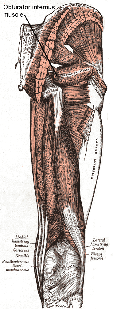
The internal obturator muscle or obturator internus muscle originates on the medial surface of the obturator membrane, the ischium near the membrane, and the rim of the pubis (bone), pubis. It exits the pelvis, pelvic cavity through the lesser sciatic foramen. The internal obturator is situated partly within the lesser pelvis, and partly at the back of the hip-joint. It functions to help laterally rotate femur with hip extension and abduct femur with hip flexion, as well as to steady the femoral head in the acetabulum. Structure Origin The internal obturator muscle arises from the inner surface of the antero-lateral wall of the pelvis. It surrounds the obturator foramen. It is attached to the inferior pubic ramus and ischium, and at the side to the inner surface of the hip bone below and behind the pelvic brim. It reaches from the upper part of the greater sciatic foramen above and behind to the obturator foramen below and in front. It also arises from the pelvic surface of ... [...More Info...] [...Related Items...] OR: [Wikipedia] [Google] [Baidu] |
Pectineus
The pectineus muscle (, from the Latin word ''pecten'', meaning comb) is a flat, quadrangular muscle, situated at the anterior (front) part of the upper and medial (inner) aspect of the thigh. The pectineus muscle is the most anterior adductor of the hip. The muscle's primary action is hip flexion; it also produces adduction and external rotation of the hip. It can be classified in the medial compartment of thigh (when the function is emphasized) or the anterior compartment of thigh (when the nerve is emphasized). Structure The pectineus muscle arises from the pectineal line of the pubis and to a slight extent from the surface of bone in front of it, between the iliopectineal eminence and pubic tubercle, and from the fascia covering the anterior surface of the muscle; the fibers pass downward, backward, and lateral, to be inserted into the pectineal line of the femur which leads from the lesser trochanter to the linea aspera. Relations The pectineus is in relation by it ... [...More Info...] [...Related Items...] OR: [Wikipedia] [Google] [Baidu] |
Gracilis Muscle
The gracilis muscle (; Latin for "slender") is the most superficial muscle on the medial side of the thigh. It is thin and flattened, broad above, narrow and tapering below. Structure It arises by a thin aponeurosis from the anterior margins of the lower half of the symphysis pubis and the upper half of the pubic arch. The muscle's fibers run vertically downward, ending in a rounded tendon. This tendon passes behind the medial condyle of the femur, curves around the medial condyle of the tibia where it becomes flattened, and inserts into the upper part of the medial surface of the body of the tibia, below the condyle. For this reason, the muscle is a lower limb adductor. At its insertion the tendon is situated immediately above that of the semitendinosus muscle, and its upper edge is overlapped by the tendon of the sartorius muscle, which it joins to form the pes anserinus. The pes anserinus is separated from the medial collateral ligament of the knee-joint In huma ... [...More Info...] [...Related Items...] OR: [Wikipedia] [Google] [Baidu] |
Adductor Magnus
The adductor magnus is a large triangular muscle, situated on the medial side of the thigh. It consists of two parts. The portion which arises from the ischiopubic ramus (a small part of the inferior ramus of the pubis, and the inferior ramus of the ischium) is called the pubofemoral portion, adductor portion, or adductor minimus, and the portion arising from the tuberosity of the ischium is called the ischiocondylar portion, extensor portion, or "hamstring portion". Due to its common embryonic origin, innervation, and action the ischiocondylar portion (or hamstring portion) is often considered part of the hamstring group of muscles. The ischiocondylar portion of the adductor magnus is considered a muscle of the posterior compartment of the thigh while the pubofemoral portion of the adductor magnus is considered a muscle of the medial compartment. Structure Pubofemoral (adductor) portion Those fibers which arise from the ramus of the pubis are short, horizontal in direc ... [...More Info...] [...Related Items...] OR: [Wikipedia] [Google] [Baidu] |
Adductor Longus
In the human body, the adductor longus is a skeletal muscle located in the thigh. One of the adductor muscles of the hip, its main function is to Adduction, adduct the thigh and it is innervated by the obturator nerve. It forms the medial wall of the femoral triangle. Structure The adductor longus arises from the body of pubis inferior to pubic crest and lateral to pubic symphysis. It lies ventrally on the adductor magnus, and near the femur, the adductor brevis is interposed between these two muscles. Distally, the fibers of the adductor longus extend into the adductor canal. It is inserted into the middle third of the medial lip of the ''linea aspera''. Innervation As part of the medial compartment of the thigh, the adductor longus is innervated by the anterior division (sometimes the posterior division) of the obturator nerve. The obturator nerve exits via the anterior rami of the spinal cord from L2, L3, and L4.Saladin, Kenneth S. Anatomy & Physiology: The Unity of Form ... [...More Info...] [...Related Items...] OR: [Wikipedia] [Google] [Baidu] |
External Obturator
The external obturator muscle or obturator externus muscle (; OE) is a flat, triangular muscle, which covers the outer surface of the anterior wall of the pelvis. It is sometimes considered part of the medial compartment of thigh, and sometimes considered part of the gluteal region. It is also considered to be part of the short external rotators of the hip, along with the Gemelli muscles, gemellus superior and inferior, Piriformis muscle, piriformis, and Quadratus femoris muscle, quadratus femoris. Structure It arises from the margin of bone immediately around the medial side of the obturator membrane and surrounding bone, viz., from the inferior pubic ramus, and the ramus of the ischium; it also arises from the medial two-thirds of the outer surface of the obturator membrane, and from the tendinous arch which completes the canal for the passage of the obturator vessels and nerves. The fibers springing from the pubic arch extend on to the inner surface of the bone, where they ... [...More Info...] [...Related Items...] OR: [Wikipedia] [Google] [Baidu] |
Lower Limb
Lower may refer to: *Lower (album), ''Lower'' (album), 2025 album by Benjamin Booker *Lower (surname) *Lower Township, New Jersey *Lower Receiver (firearms) *Lower Wick Gloucestershire, England See also *Nizhny {{Disambiguation ... [...More Info...] [...Related Items...] OR: [Wikipedia] [Google] [Baidu] |
Thigh
In anatomy, the thigh is the area between the hip (pelvis) and the knee. Anatomically, it is part of the lower limb. The single bone in the thigh is called the femur. This bone is very thick and strong (due to the high proportion of bone tissue), and forms a ball and socket joint at the hip, and a modified hinge joint at the knee. Structure Bones The femur is the only bone in the thigh and serves as an attachment site for all thigh muscles. The head of the femur articulates with the acetabulum in the pelvic bone forming the hip joint, while the distal part of the femur articulates with the tibia and patella forming the knee. By most measures, the femur is the strongest and longest bone in the body. The femur is categorised as a long bone and comprises a diaphysis, the shaft (or body) and two epiphyses, the lower extremity and the upper extremity of femur, that articulate with adjacent bones in the hip and knee. Muscular compartments In cross-section, the thigh is d ... [...More Info...] [...Related Items...] OR: [Wikipedia] [Google] [Baidu] |
Cutaneous Branch Of The Obturator Nerve
The cutaneous branch of the obturator nerve is an occasional continuation of the communicating branch to the femoral medial cutaneous branches and saphenous branches of the femoral to the thigh and leg. When present it emerges from beneath the distal/inferior border of the adductor longus muscle and descends along the posterior margin of the sartorius muscle to the medial side of the knee where it pierces the deep fascia and communicates with the saphenous nerve The saphenous nerve (long or internal saphenous nerve) is the largest cutaneous branch of the femoral nerve. It is derived from the lumbar plexus (L3-L4). It is a strictly sensory nerve, and has no motor function. It commences in the proximal (u .... When present, it provides sensory innervation to the skin of proximal/superior half of the medial side of the leg. See also * Cutaneous innervation of the lower limbs#Thigh References External links * - "Superficial Anatomy of the Lower Extremity: Cutaneous Nerves of ... [...More Info...] [...Related Items...] OR: [Wikipedia] [Google] [Baidu] |
Anterior Branch Of Obturator Nerve
The anterior branch of the obturator nerve is a branch of the obturator nerve found in the pelvis and leg. It leaves the pelvis in front of the obturator externus and descends anterior to the adductor brevis, and posterior to the pectineus and adductor longus; at the lower border of the latter muscle it communicates with the anterior cutaneous and saphenous branches of the femoral nerve, forming a kind of plexus. It then descends upon the femoral artery, to which it is finally distributed. Near the obturator foramen the nerve gives off an articular branch to the hip joint. Behind the pectineus, it distributes branches to the adductor longus and gracilis, and usually to the adductor brevis The adductor brevis is a muscle in the thigh situated immediately deep to the pectineus and adductor longus. It belongs to the adductor muscle group. The main function of the adductor brevis is to pull the thigh medially. The adductor brevi ..., and in rare cases to the pectineus; it ... [...More Info...] [...Related Items...] OR: [Wikipedia] [Google] [Baidu] |


