|
Nerve To The Subclavius
The subclavian nerve, also known as the nerve to the subclavius, is a small branch of the upper trunk of the brachial plexus. It contains axons from C5 and C6. It innervates the subclavius muscle. Anatomy Origin The subclavian nerve is a branch of the upper trunk of the brachial plexus. It contains axons derived from the ventral rami of the C5 and C6 cervical spinal nerves. The origin is situated within the posterior triangle of the neck. Course Descending, it passes anterior to (the 3rd part of) the subclavian artery and vein. Variation Accessory phrenic nerve The subclavian nerve may issue a branch called the accessory phrenic nerve which innervates the diaphragm. The accessory phrenic nerve may rather branch from the C4 or C6 segments or ansa cervicalis. This nerve usually joins with the phrenic nerve before innervating the diaphragm, ventral to the subclavian vein. Function The subclavian nerve innervates the subclavius muscle The subclavius is a small ... [...More Info...] [...Related Items...] OR: [Wikipedia] [Google] [Baidu] |
Brachial Plexus
The brachial plexus is a network of nerves (nerve plexus) formed by the anterior rami of the lower four Spinal nerve#Cervical nerves, cervical nerves and first Spinal nerve#Thoracic nerves, thoracic nerve (cervical spinal nerve 5, C5, Cervical spinal nerve 6, C6, cervical spinal nerve 7, C7, cervical spinal nerve 8, C8, and thoracic spinal nerve 1, T1). This plexus extends from the spinal cord, through the cervicoaxillary canal in the neck, over the first rib, and into the axilla, armpit, it supplies Afferent nerve fiber, afferent and efferent nerve fibers to the chest, shoulder, arm, forearm, and hand. Structure The brachial plexus is divided into five ''roots'', three ''trunks'', six ''divisions'' (three anterior and three posterior), three ''cords'', and five ''branches''. There are five "terminal" branches and numerous other "pre-terminal" or "collateral" branches, such as the subscapular nerve, the thoracodorsal nerve, and the long thoracic nerve, that leave the plexus at vari ... [...More Info...] [...Related Items...] OR: [Wikipedia] [Google] [Baidu] |
Posterior Triangle Of The Neck
The posterior triangle (or lateral cervical region) is a region of the neck. Boundaries The posterior triangle has the following boundaries: Apex: Union of the sternocleidomastoid and the trapezius muscles at the superior nuchal line of the occipital bone Anteriorly: Posterior border of the sternocleidomastoideus Posteriorly: Anterior border of the trapezius Inferiorly: Middle one third of the clavicle Roof: Investing layer of the deep cervical fascia Floor: (From superior to inferior) 1) M. semispinalis capitis 2) M. splenius capitis 3) M. levator scapulae 4) M. scalenus posterior 5) M. scalenus medius Divisions The posterior triangle is crossed, about 2.5 cm above the clavicle, by the inferior belly of the omohyoid muscle, which divides the space into two triangles: * an upper or occipital triangle * a lower or subclavian triangle (or supraclavicular triangle) Contents A) Nerves and plexuses: * Spinal accessory nerve (Cranial Nerve XI) * Branches of cervic ... [...More Info...] [...Related Items...] OR: [Wikipedia] [Google] [Baidu] |
Elsevier
Elsevier ( ) is a Dutch academic publishing company specializing in scientific, technical, and medical content. Its products include journals such as ''The Lancet'', ''Cell (journal), Cell'', the ScienceDirect collection of electronic journals, ''Trends (journals), Trends'', the ''Current Opinion (Elsevier), Current Opinion'' series, the online citation database Scopus, the SciVal tool for measuring research performance, the ClinicalKey search engine for clinicians, and the ClinicalPath evidence-based cancer care service. Elsevier's products and services include digital tools for Data management platform, data management, instruction, research analytics, and assessment. Elsevier is part of the RELX Group, known until 2015 as Reed Elsevier, a publicly traded company. According to RELX reports, in 2022 Elsevier published more than 600,000 articles annually in over 2,800 journals. As of 2018, its archives contained over 17 million documents and 40,000 Ebook, e-books, with over one b ... [...More Info...] [...Related Items...] OR: [Wikipedia] [Google] [Baidu] |
Subclavian Vein
The subclavian vein is a paired large vein, one on either side of the body, that is responsible for draining blood from the upper extremities, allowing this blood to return to the heart. The left subclavian vein plays a key role in the absorption of lipids, by allowing products that have been carried by lymph in the thoracic duct to enter the bloodstream. The diameter of the subclavian veins is approximately 1–2 cm, depending on the individual. Structure Each subclavian vein is a continuation of the axillary vein and runs from the outer border of the first rib to the medial border of anterior scalene muscle. From here it joins with the internal jugular vein to form the brachiocephalic vein (also known as "innominate vein"). The angle of union is termed the venous angle. The subclavian vein follows the subclavian artery and is separated from the subclavian artery by the insertion of anterior scalene. Thus, the subclavian vein lies anterior to the anterior scalene while ... [...More Info...] [...Related Items...] OR: [Wikipedia] [Google] [Baidu] |
Phrenic Nerve
The phrenic nerve is a mixed nerve that originates from the C3–C5 spinal nerves in the neck. The nerve is important for breathing because it provides exclusive motor control of the diaphragm, the primary muscle of respiration. In humans, the right and left phrenic nerves are primarily supplied by the C4 spinal nerve, but there is also a contribution from the C3 and C5 spinal nerves. From its origin in the neck, the nerve travels downward into the chest to pass between the heart and lungs towards the diaphragm. In addition to motor fibers, the phrenic nerve contains sensory fibers, which receive input from the central tendon of the diaphragm and the mediastinal pleura, as well as some sympathetic nerve fibers. Although the nerve receives contributions from nerve roots of the cervical plexus and the brachial plexus, it is usually considered separate from either plexus. The name of the nerve comes from Ancient Greek ''phren'' 'diaphragm'. Structure The phrenic nerve or ... [...More Info...] [...Related Items...] OR: [Wikipedia] [Google] [Baidu] |
Ansa Cervicalis
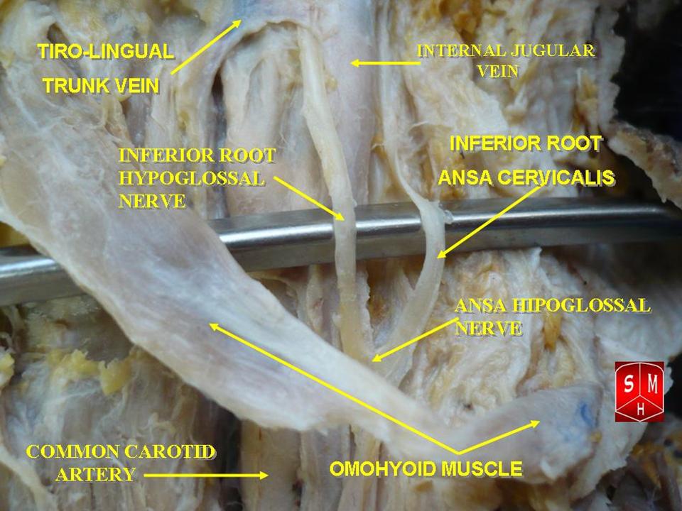
The ansa cervicalis (or ansa hypoglossi in older literature) is a loop formed by muscular branches of the cervical plexus formed by branches of cervical spinal nerves C1-C3. The ansa cervicalis has two roots - a superior root (formed by branch of C1) and an inferior root (formed by union of branches of C2 and C3) - that unite distally, forming a loop. It is situated anterior to the carotid sheath. Branches of the ansa cervicalis innervate three of the four infrahyoid muscles: the sternothyroid, sternohyoid, and omohyoid muscles (note that the thyrohyoid muscle is the one infrahyoid muscle not innervated by the ansa cervicalis - it is instead innervated by cervical spinal nerve 1 via a separate thyrohyoid branch). Its name means "handle of the neck" in Latin. Anatomy The ansa cervicalis is typically embedded within the anterior wall of the carotid sheath anterior to the internal jugular vein. Superior root The superior root of the ansa cervicalis (formerly known as d ... [...More Info...] [...Related Items...] OR: [Wikipedia] [Google] [Baidu] |
Thoracic Diaphragm
The thoracic diaphragm, or simply the diaphragm (; ), is a sheet of internal Skeletal striated muscle, skeletal muscle in humans and other mammals that extends across the bottom of the thoracic cavity. The diaphragm is the most important Muscles of respiration, muscle of respiration, and separates the thoracic cavity, containing the heart and lungs, from the abdominal cavity: as the diaphragm contracts, the volume of the thoracic cavity increases, creating a negative pressure there, which draws air into the lungs. Its high oxygen consumption is noted by the many mitochondria and capillaries present; more than in any other skeletal muscle. The term ''diaphragm'' in anatomy, created by Gerard of Cremona, can refer to other flat structures such as the urogenital diaphragm or Pelvic floor, pelvic diaphragm, but "the diaphragm" generally refers to the thoracic diaphragm. In humans, the diaphragm is slightly asymmetric—its right half is higher up (superior) to the left half, since th ... [...More Info...] [...Related Items...] OR: [Wikipedia] [Google] [Baidu] |
Subclavian Vein
The subclavian vein is a paired large vein, one on either side of the body, that is responsible for draining blood from the upper extremities, allowing this blood to return to the heart. The left subclavian vein plays a key role in the absorption of lipids, by allowing products that have been carried by lymph in the thoracic duct to enter the bloodstream. The diameter of the subclavian veins is approximately 1–2 cm, depending on the individual. Structure Each subclavian vein is a continuation of the axillary vein and runs from the outer border of the first rib to the medial border of anterior scalene muscle. From here it joins with the internal jugular vein to form the brachiocephalic vein (also known as "innominate vein"). The angle of union is termed the venous angle. The subclavian vein follows the subclavian artery and is separated from the subclavian artery by the insertion of anterior scalene. Thus, the subclavian vein lies anterior to the anterior scalene while ... [...More Info...] [...Related Items...] OR: [Wikipedia] [Google] [Baidu] |
Subclavian Artery
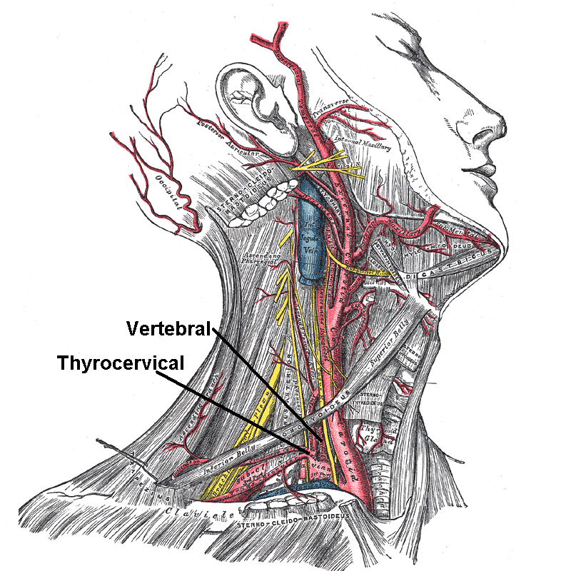
In human anatomy, the subclavian arteries are paired major arteries of the upper thorax, below the clavicle. They receive blood from the aortic arch. The left subclavian artery supplies blood to the left arm and the right subclavian artery supplies blood to the right arm, with some branches supplying the head and thorax. On the left side of the body, the subclavian comes directly off the aortic arch, while on the right side it arises from the relatively short brachiocephalic artery when it bifurcates into the subclavian and the right common carotid artery. The usual branches of the subclavian on both sides of the body are the vertebral artery, the internal thoracic artery, the thyrocervical trunk, the costocervical trunk and the dorsal scapular artery, which may branch off the transverse cervical artery, which is a branch of the thyrocervical trunk. The subclavian becomes the axillary artery at the lateral border of the first rib. Structure From its origin, the subclavian art ... [...More Info...] [...Related Items...] OR: [Wikipedia] [Google] [Baidu] |
Spinal Nerve
A spinal nerve is a mixed nerve, which carries Motor neuron, motor, Sensory neuron, sensory, and Autonomic nervous system, autonomic signals between the spinal cord and the body. In the human body there are 31 pairs of spinal nerves, one on each side of the vertebral column. These are grouped into the corresponding cervical vertebrae, cervical, thoracic vertebrae, thoracic, lumbar vertebrae, lumbar, sacral vertebrae, sacral and coccygeal vertebrae, coccygeal regions of the spine. There are eight pairs of cervical nerves, twelve pairs of thoracic nerves, five pairs of lumbar nerves, five pairs of sacral nerves, and one pair of coccygeal nerves. The spinal nerves are part of the peripheral nervous system. Structure Each spinal nerve is a mixed nerve, formed from the combination of nerve root axon, fibers from its Dorsal root of spinal nerve, dorsal and Ventral root of spinal nerve, ventral roots. The dorsal root is the afferent nerve fiber, afferent sensory root and carries sen ... [...More Info...] [...Related Items...] OR: [Wikipedia] [Google] [Baidu] |
Subclavius Muscle
The subclavius is a small triangular muscle, placed between the clavicle and the first rib. Along with the pectoralis major and pectoralis minor muscles, the subclavius muscle makes up the anterior axioappendicular muscles, also known as anterior wall of the axilla. Structure It arises by a short, thick tendon from the first rib and its cartilage at their junction, in front of the costoclavicular ligament. The fleshy fibers proceed obliquely superolaterally, to be inserted into the groove on the under surface of the clavicle. Innervation The nerve to subclavius (or subclavian nerve) innervates the muscle. This arises from the junction of the fifth and sixth cervical nerves, from the superior/upper trunk of the brachial plexus. Variation Insertion into coracoid process instead of clavicle or into both clavicle and coracoid process. Sternoscapular fasciculus to the upper border of scapula. Sternoclavicularis from manubrium to clavicle between pectoralis major and coracoclav ... [...More Info...] [...Related Items...] OR: [Wikipedia] [Google] [Baidu] |
Ventral Ramus Of Spinal Nerve
The ventral ramus (: rami) (Latin for 'branch') is the anterior division of a spinal nerve. The ventral rami supply the antero-lateral parts of the trunk and the limbs. They are mainly larger than the dorsal rami. Shortly after a spinal nerve exits the intervertebral foramen, it branches into the dorsal ramus, the ventral ramus, and the ramus communicans. Each of these three structures carries both sensory and motor information. Each spinal nerve is a mixed nerve that carries both sensory and motor information, via efferent and afferent nerve fibers—ultimately via the motor cortex in the frontal lobe and to somatosensory cortex in the parietal lobe—but also through the phenomenon of reflex. In the thoracic region they remain distinct from each other and each innervates a narrow strip of muscle and skin along the sides, chest, ribs, and abdominal wall. These rami are called the intercostal nerves. In regions other than the thoracic, ventral rami converge with ea ... [...More Info...] [...Related Items...] OR: [Wikipedia] [Google] [Baidu] |


