|
Nerve Ending
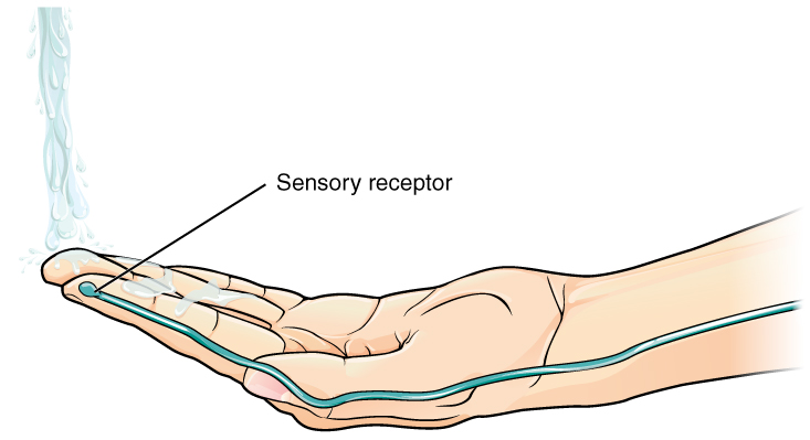
A free nerve ending (FNE) or bare nerve ending, is an unspecialized, afferent nerve fiber sending its signal to a sensory neuron. ''Afferent'' in this case means bringing information from the body's periphery toward the brain. They function as cutaneous nociceptors and are essentially used by vertebrates to detect noxious stimuli that often result in pain. Structure Free nerve endings are unencapsulated and have no complex sensory structures. They are the most common type of nerve ending, and are most frequently found in the skin Skin is the layer of usually soft, flexible outer tissue covering the body of a vertebrate animal, with three main functions: protection, regulation, and sensation. Other animal coverings, such as the arthropod exoskeleton, have different .... They penetrate the dermis and end in the stratum granulosum. FNEs infiltrate the middle layers of the dermis and surround hair follicles. Types Free nerve endings have different rates of adaptation, ... [...More Info...] [...Related Items...] OR: [Wikipedia] [Google] [Baidu] |
Afferent Nerve Fiber
Afferent nerve fibers are axons (nerve fibers) of sensory neurons that carry sensory information from sensory receptors to the central nervous system. Many afferent projections ''arrive'' at a particular brain region. In the peripheral nervous system, afferent nerve fibers are part of the sensory nervous system and arise from outside of the central nervous system. Sensory and mixed nerves contain afferent fibers. Structure Afferent neurons are pseudounipolar neurons that have a single process leaving the cell body dividing into two branches: the long one towards the sensory organ, and the short one toward the central nervous system (e.g. spinal cord). These cells do have sensory afferent dendrites, similar to those typically inherent in neurons. They have a smooth and rounded cell body located in the ganglia of the peripheral nervous system. Just outside the spinal cord, thousands of afferent neuronal cell bodies are aggregated in a swelling in the dorsal root known a ... [...More Info...] [...Related Items...] OR: [Wikipedia] [Google] [Baidu] |
Slowly Adapting
A mechanoreceptor, also called mechanoceptor, is a sensory receptor that responds to mechanical pressure or distortion. Mechanoreceptors are located on sensory neurons that convert mechanical pressure into electrical signals that, in animals, are sent to the central nervous system. Vertebrate mechanoreceptors Cutaneous mechanoreceptors Cutaneous mechanoreceptors respond to mechanical stimuli that result from physical interaction, including pressure and vibration. They are located in the skin, like other cutaneous receptors. They are all innervated by Aβ fibers, except the mechanorecepting free nerve endings, which are innervated by Aδ fibers. Cutaneous mechanoreceptors can be categorized by what kind of sensation they perceive, by the rate of adaptation, and by morphology. Furthermore, each has a different receptive field. By sensation * The Slowly Adapting type 1 (SA1) mechanoreceptor, with the Merkel corpuscle end-organ (also known as Merkel discs) detect sustaine ... [...More Info...] [...Related Items...] OR: [Wikipedia] [Google] [Baidu] |
Group C Nerve Fiber
Group C nerve fibers are one of three classes of nerve fiber in the central nervous system (CNS) and peripheral nervous system (PNS). The Group C fibers are unmyelinated and have a small diameter and low conduction velocity, whereas Groups A and B are myelinated. Group C fibers include postganglionic fibers in the autonomic nervous system (ANS), and nerve fibers at the dorsal roots (IV fiber). These fibers carry sensory information. Damage or injury to nerve fibers causes neuropathic pain. Capsaicin activates C fibre vanilloid receptors, providing the burning sensation associated with chili peppers. Structure and anatomy Location C fibers are one class of nerve fiber found in the nerves of the somatic sensory system. They are afferent fibers, conveying input signals from the periphery to the central nervous system. Structure C fibers are unmyelinated unlike most other fibers in the nervous system. This lack of myelination is the cause of their slow conduction ve ... [...More Info...] [...Related Items...] OR: [Wikipedia] [Google] [Baidu] |
Electron Microscope
An electron microscope is a microscope that uses a beam of electrons as a source of illumination. It uses electron optics that are analogous to the glass lenses of an optical light microscope to control the electron beam, for instance focusing it to produce magnified images or electron diffraction patterns. As the wavelength of an electron can be up to 100,000 times smaller than that of visible light, electron microscopes have a much higher Angular resolution, resolution of about 0.1 nm, which compares to about 200 nm for optical microscope, light microscopes. ''Electron microscope'' may refer to: * Transmission electron microscopy, Transmission electron microscope (TEM) where swift electrons go through a thin sample * Scanning transmission electron microscopy, Scanning transmission electron microscope (STEM) which is similar to TEM with a scanned electron probe * Scanning electron microscope (SEM) which is similar to STEM, but with thick samples * Electron microprobe sim ... [...More Info...] [...Related Items...] OR: [Wikipedia] [Google] [Baidu] |
Optical Microscope
The optical microscope, also referred to as a light microscope, is a type of microscope that commonly uses visible light and a system of lenses to generate magnified images of small objects. Optical microscopes are the oldest design of microscope and were possibly invented in their present compound form in the 17th century. Basic optical microscopes can be very simple, although many complex designs aim to improve resolution and sample contrast. The object is placed on a stage and may be directly viewed through one or two eyepieces on the microscope. In high-power microscopes, both eyepieces typically show the same image, but with a stereo microscope, slightly different images are used to create a 3-D effect. A camera is typically used to capture the image (micrograph). The sample can be lit in a variety of ways. Transparent objects can be lit from below and solid objects can be lit with light coming through ( bright field) or around ( dark field) the objective lens. Polar ... [...More Info...] [...Related Items...] OR: [Wikipedia] [Google] [Baidu] |
C Fiber
Group C nerve fibers are one of three classes of nerve fiber in the central nervous system (CNS) and peripheral nervous system (PNS). The Group C fibers are unmyelinated and have a small diameter and low conduction velocity, whereas Groups A and B are myelinated. Group C fibers include postganglionic fibers in the autonomic nervous system (ANS), and nerve fibers at the dorsal roots (IV fiber). These fibers carry sensory information. Damage or injury to nerve fibers causes neuropathic pain. Capsaicin activates C fibre vanilloid receptors, providing the burning sensation associated with chili peppers. Structure and anatomy Location C fibers are one class of nerve fiber found in the nerves of the somatic sensory system. They are afferent fibers, conveying input signals from the periphery to the central nervous system. Structure C fibers are unmyelinated unlike most other fibers in the nervous system. This lack of myelination is the cause of their slow conduction velocit ... [...More Info...] [...Related Items...] OR: [Wikipedia] [Google] [Baidu] |
Polymodality
Stimulus modality, also called sensory modality, is one aspect of a stimulus or what is perceived after a stimulus. For example, the temperature modality is registered after heat or cold stimulate a receptor. Some sensory modalities include: light, sound, temperature, taste, pressure, and smell. The type and location of the sensory receptor activated by the stimulus plays the primary role in coding the sensation. All sensory modalities work together to heighten stimuli sensation when necessary. Multimodal perception Multimodal perception is the ability of the mammalian nervous system to combine all of the different inputs of the sensory nervous system to result in an enhanced detection or identification of a particular stimulus. Combinations of all sensory modalities are done in cases where a single sensory modality results in an ambiguous and incomplete result. Integration of all sensory modalities occurs when multimodal neurons receive sensory information which overlaps ... [...More Info...] [...Related Items...] OR: [Wikipedia] [Google] [Baidu] |
Nociceptors
A nociceptor (; ) is a sensory neuron that responds to damaging or potentially damaging stimuli by sending "possible threat" signals to the spinal cord and the brain. The brain creates the sensation of pain to direct attention to the body part, so the threat can be mitigated; this process is called nociception. Terminology Nociception and pain are usually evoked only by pressures and temperatures that are potentially damaging to tissues. This barrier or threshold contrasts with the more sensitive visual, auditory, olfactory, taste, and somatosensory responses to stimuli. The experience of pain is individualistic and can be suppressed by stress or exacerbated by anticipation. Simple activation of a nociceptor does not always lead to perceived pain, because the latter also depends on the frequency of the action potentials, integration of pre- and postsynaptic signals, and influences from higher or central processes. Scientific investigation Nociceptors were discovered by Charles ... [...More Info...] [...Related Items...] OR: [Wikipedia] [Google] [Baidu] |
Cutaneous Mechanoreceptors
A mechanoreceptor, also called mechanoceptor, is a sensory receptor that responds to mechanical pressure or distortion. Mechanoreceptors are located on sensory neurons that convert mechanical pressure into electrical signals that, in animals, are sent to the central nervous system. Vertebrate mechanoreceptors Cutaneous mechanoreceptors Cutaneous mechanoreceptors respond to mechanical stimuli that result from physical interaction, including pressure and vibration. They are located in the skin, like other cutaneous receptors. They are all innervated by Aβ fibers, except the mechanorecepting free nerve endings, which are innervated by Aδ fibers. Cutaneous mechanoreceptors can be categorized by what kind of sensation they perceive, by the rate of adaptation, and by morphology. Furthermore, each has a different receptive field. By sensation * The Slowly Adapting type 1 (SA1) mechanoreceptor, with the Merkel corpuscle end-organ (also known as Merkel discs) detect sustained p ... [...More Info...] [...Related Items...] OR: [Wikipedia] [Google] [Baidu] |
Thermoreceptors
A thermoreceptor is a non-specialised sense receptor, or more accurately the receptive portion of a sensory neuron, that codes absolute and relative changes in temperature, primarily within the innocuous range. In the mammalian peripheral nervous system, warmth receptors are thought to be unmyelinated C-fibres (low conduction velocity), while those responding to cold have both C-fibers and thinly myelinated A delta fibers (faster conduction velocity). The adequate stimulus for a warm receptor is warming, which results in an increase in their action potential discharge rate. Cooling results in a decrease in warm receptor discharge rate. For cold receptors their firing rate increases during cooling and decreases during warming. Some cold receptors also respond with a brief action potential discharge to high temperatures, i.e. typically above 45 °C, and this is known as a paradoxical response to heat . The mechanism responsible for this behavior has not been determined. Locatio ... [...More Info...] [...Related Items...] OR: [Wikipedia] [Google] [Baidu] |
A Delta Fiber
Group A nerve fibers are one of the three classes of nerve fiber as ''generally classified'' by Erlanger and Gasser. The other two classes are the group B nerve fibers, and the group C nerve fibers. Group A are heavily myelinated, group B are moderately myelinated, and group C are unmyelinated. The other classification is a sensory grouping that uses the terms '' type Ia and type Ib'', '' type II'', ''type III'', and ''type IV'', sensory fibers. Types There are four subdivisions of group A nerve fibers: alpha (α) Aα; beta (β) Aβ; , gamma (γ) Aγ, and delta (δ) Aδ. These subdivisions have different amounts of myelination and axon thickness and therefore transmit signals at different speeds. Larger diameter axons and more myelin insulation lead to faster signal propagation. Group A nerves are found in both motor and sensory pathways. Different sensory receptors are innervated by different types of nerve fibers. Proprioceptors are innervated by type Ia, Ib and II ... [...More Info...] [...Related Items...] OR: [Wikipedia] [Google] [Baidu] |
Intermediate Adapting
A mechanoreceptor, also called mechanoceptor, is a sensory receptor that responds to mechanical pressure or distortion. Mechanoreceptors are located on sensory neurons that convert mechanical pressure into electrical signals that, in animals, are sent to the central nervous system. Vertebrate mechanoreceptors Cutaneous mechanoreceptors Cutaneous mechanoreceptors respond to mechanical stimuli that result from physical interaction, including pressure and vibration. They are located in the skin, like other cutaneous receptors. They are all innervated by Aβ fibers, except the mechanorecepting free nerve endings, which are innervated by Aδ fibers. Cutaneous mechanoreceptors can be categorized by what kind of sensation they perceive, by the rate of adaptation, and by morphology. Furthermore, each has a different receptive field. By sensation * The Slowly Adapting type 1 (SA1) mechanoreceptor, with the Merkel corpuscle end-organ (also known as Merkel discs) detect sustaine ... [...More Info...] [...Related Items...] OR: [Wikipedia] [Google] [Baidu] |


