|
Metanephros
Kidney development, or nephrogenesis, describes the embryologic origins of the kidney, a major organ in the urinary system. This article covers a 3 part developmental process that is observed in most reptiles, birds and mammals, including humans. Nephrogenesis is often considered in the broader context of the development of the urinary and reproductive organs. Phases The development of the kidney proceeds through a series of successive phases, each marked by the development of a more advanced kidney: the archinephros, pronephros, mesonephros, and metanephros. The pronephros is the most immature form of kidney, while the metanephros is most developed. The metanephros persists as the definitive adult kidney. Archinephros The archinephros is considered as hypothetical or primitive kidney. Pronephros The pronephros develops in the cervical region of the embryo. During approximately day 22 of human gestation, the paired pronephri appears towards the cranial end of the intermediat ... [...More Info...] [...Related Items...] OR: [Wikipedia] [Google] [Baidu] |
Development Of The Urinary And Reproductive Organs
The development of the urinary system begins during prenatal development, and relates to the development of the urogenital system – both the organs of the urinary system and the sex organs of the reproductive system. The development continues as a part of sexual differentiation. The urinary and reproductive organs are developed from the intermediate mesoderm. The permanent organs of the adult are preceded by a set of structures which are purely embryonic, and which with the exception of the ducts disappear almost entirely before birth. These embryonic structures are on either side; the pronephros, the mesonephros and the metanephros of the kidney, and the Wolffian duct, Wolffian and Müllerian ducts of the sex organ. The pronephros disappears very early; the structural elements of the mesonephros mostly degenerate, but the gonad is developed in their place, with which the Wolffian duct remains as the duct in males, and the Müllerian as that of the female. Some of the tubules of ... [...More Info...] [...Related Items...] OR: [Wikipedia] [Google] [Baidu] |
Kidney
In humans, the kidneys are two reddish-brown bean-shaped blood-filtering organ (anatomy), organs that are a multilobar, multipapillary form of mammalian kidneys, usually without signs of external lobulation. They are located on the left and right in the retroperitoneal space, and in adult humans are about in length. They receive blood from the paired renal artery, renal arteries; blood exits into the paired renal veins. Each kidney is attached to a ureter, a tube that carries excreted urine to the urinary bladder, bladder. The kidney participates in the control of the volume of various body fluids, fluid osmolality, Acid-base homeostasis, acid-base balance, various electrolyte concentrations, and removal of toxins. Filtration occurs in the glomerulus (kidney), glomerulus: one-fifth of the blood volume that enters the kidneys is filtered. Examples of substances reabsorbed are solute-free water, sodium, bicarbonate, glucose, and amino acids. Examples of substances secreted are hy ... [...More Info...] [...Related Items...] OR: [Wikipedia] [Google] [Baidu] |
Kidneys
In humans, the kidneys are two reddish-brown bean-shaped blood-filtering organs that are a multilobar, multipapillary form of mammalian kidneys, usually without signs of external lobulation. They are located on the left and right in the retroperitoneal space, and in adult humans are about in length. They receive blood from the paired renal arteries; blood exits into the paired renal veins. Each kidney is attached to a ureter, a tube that carries excreted urine to the bladder. The kidney participates in the control of the volume of various body fluids, fluid osmolality, acid-base balance, various electrolyte concentrations, and removal of toxins. Filtration occurs in the glomerulus: one-fifth of the blood volume that enters the kidneys is filtered. Examples of substances reabsorbed are solute-free water, sodium, bicarbonate, glucose, and amino acids. Examples of substances secreted are hydrogen, ammonium, potassium and uric acid. The nephron is the structural and functi ... [...More Info...] [...Related Items...] OR: [Wikipedia] [Google] [Baidu] |
Intermediate Mesoderm
Intermediate mesoderm or intermediate mesenchyme is a narrow section of the mesoderm (one of the three primary germ layers) located between the paraxial mesoderm and the lateral plate of the developing embryo. The intermediate mesoderm develops into vital parts of the urogenital system (kidneys, gonads and respective tracts). Early formation Factors regulating the formation of the intermediate mesoderm are not fully understood. It is believed that bone morphogenic proteins, or BMPs, specify regions of growth along the dorsal-ventral axis of the mesoderm and plays a central role in formation of the intermediate mesoderm. Vg1/wikt:node, Nodal signalling is an identified regulator of intermediate mesoderm formation acting through BMP signalling. Excess Vg1/Nodal signalling during early gastrulation stages results in expansion of the intermediate mesoderm at the expense of the adjacent paraxial mesoderm, whereas inhibition of Vg1/Nodal signalling represses intermediate mesoderm formati ... [...More Info...] [...Related Items...] OR: [Wikipedia] [Google] [Baidu] |
Mesonephric Tubule
The mesonephros () is one of three excretory organs that develop in vertebrates. It serves as the main excretory organ of aquatic vertebrates and as a temporary kidney in reptiles, birds, and mammals. The mesonephros is included in the Wolffian body after Caspar Friedrich Wolff who described it in 1759. (The Wolffian body is composed of: mesonephros + paramesonephrotic blastema) Structure The mesonephros acts as a structure similar to the kidney that, in humans, functions between the sixth and tenth weeks of embryological life. Despite the similarity in structure, function, and terminology, however, the mesonephric nephrons do not form any part of the mature kidney or nephrons. In humans, the mesonephros consists of units which are similar in structure and function to nephrons of the adult kidney. Each of these consists of a glomerulus, a tuft of capillaries which arises from lateral branches of dorsal aorta and drains into the inferior cardinal vein; a Bowman's capsule, a f ... [...More Info...] [...Related Items...] OR: [Wikipedia] [Google] [Baidu] |
Archinephros
The archinephros, or holonephros, is a primitive kidney that has been retained by the larvae of hagfish and some caecilians. A recent author has referred to this structure as "the hypothetical primitive kidney of ancestral vertebrates". In the earliest vertebrates, this structure potentially extended the entire length of the body and consisted of paired segmental structures which drained via a pair of archinephrenic ducts into the cloaca. The entire structure arises from the nephric ridge, which in higher animal embryos gives rise to nephrotomes and the pronephroi at around 4 weeks gestation in humans. The pronephroi are supplanted by mesonephroi and finally by definitive kidneys, the metanephroi, by around 5 weeks gestation. The archinephros is nonfunctional in humans and other mammals. The three types of mature vertebrate kidneys develop from the archinephros: the pronephros from the front section, the mesonephros from the mid-section and the metanephros Kidney development ... [...More Info...] [...Related Items...] OR: [Wikipedia] [Google] [Baidu] |
Ureters
The ureters are tubes composed of smooth muscle that transport urine from the kidneys to the urinary bladder. In an adult human, the ureters typically measure 20 to 30 centimeters in length and about 3 to 4 millimeters in diameter. They are lined with urothelial cells, a form of transitional epithelium, and feature an extra layer of smooth muscle in the lower third to aid in peristalsis. The ureters can be affected by a number of diseases, including urinary tract infections and kidney stone. is when a ureter is narrowed, due to for example chronic inflammation. Congenital abnormalities that affect the ureters can include the development of two ureters on the same side or abnormally placed ureters. Additionally, reflux of urine from the bladder back up the ureters is a condition commonly seen in children. The ureters have been identified for at least two thousand years, with the word "ureter" stemming from the stem relating to urinating and seen in written records since at ... [...More Info...] [...Related Items...] OR: [Wikipedia] [Google] [Baidu] |
Embryology
Embryology (from Ancient Greek, Greek ἔμβρυον, ''embryon'', "the unborn, embryo"; and -λογία, ''-logy, -logia'') is the branch of animal biology that studies the Prenatal development (biology), prenatal development of gametes (sex cells), fertilization, and development of embryos and fetuses. Additionally, embryology encompasses the study of congenital disorders that occur before birth, known as teratology. Early embryology was proposed by Marcello Malpighi, and known as preformationism, the theory that organisms develop from pre-existing miniature versions of themselves. Aristotle proposed the theory that is now accepted, Epigenesis (biology), epigenesis. Epigenesis (biology), Epigenesis is the idea that organisms develop from seed or egg in a sequence of steps. Modern embryology developed from the work of Karl Ernst von Baer, though accurate observations had been made in Italy by anatomists such as Aldrovandi and Leonardo da Vinci in the Renaissance. Comparative ... [...More Info...] [...Related Items...] OR: [Wikipedia] [Google] [Baidu] |
Collecting Duct System
The collecting duct system of the kidney consists of a series of tubules and ducts that physically connect nephrons to a minor calyx or directly to the renal pelvis. The collecting duct participates in electrolyte and fluid balance through reabsorption and excretion, processes regulated by the hormones aldosterone and vasopressin (antidiuretic hormone). There are several components of the collecting duct system, including the connecting tubules, cortical collecting ducts, and medullary collecting ducts. Structure Segments The segments of the system are as follows: Connecting tubule With respect to the renal corpuscle, the connecting tubule (CNT, or junctional tubule, or arcuate renal tubule) is the most proximal part of the collecting duct system. It is adjacent to the distal convoluted tubule, the most distal segment of the renal tubule. Connecting tubules from several adjacent nephrons merge to form cortical collecting tubules, and these may join to form cortical col ... [...More Info...] [...Related Items...] OR: [Wikipedia] [Google] [Baidu] |
Renal Calyx
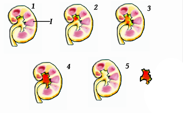
The renal calyces ( calyx) are conduits in the kidney through which urine passes. The minor calyces form a cup-shaped drain around the apex of the renal pyramids. Urine formed in the kidney passes through a renal papilla at the apex into the minor calyx; four or five minor calyces converge to form a major calyx through which urine passes into the renal pelvis (which in turn drains urine out of the kidney through the ureter). Function Peristalsis of the smooth muscle originating in pace-maker cells originating in the walls of the calyces propels urine through the renal pelvis and ureters to the bladder. The initiation is caused by the increase in volume that stretches the walls of the calyces. This causes them to fire impulses which stimulate rhythmical contraction and relaxation, called peristalsis. Parasympathetic innervation enhances the peristalsis while sympathetic innervation inhibits it. Clinical significance A " staghorn calculus" is a kidney stone that may exten ... [...More Info...] [...Related Items...] OR: [Wikipedia] [Google] [Baidu] |
Connecting Tubule
The collecting duct system of the kidney consists of a series of tubules and ducts that physically connect nephrons to a minor calyx or directly to the renal pelvis. The collecting duct participates in electrolyte and fluid balance through reabsorption and excretion, processes regulated by the hormones aldosterone and vasopressin (antidiuretic hormone). There are several components of the collecting duct system, including the connecting tubules, cortical collecting ducts, and medullary collecting ducts. Structure Segments The segments of the system are as follows: Connecting tubule With respect to the renal corpuscle, the connecting tubule (CNT, or junctional tubule, or arcuate renal tubule) is the most proximal part of the collecting duct system. It is adjacent to the distal convoluted tubule, the most distal segment of the renal tubule. Connecting tubules from several adjacent nephrons merge to form cortical collecting tubules, and these may join to form cortical col ... [...More Info...] [...Related Items...] OR: [Wikipedia] [Google] [Baidu] |


