Renal Calyx on:
[Wikipedia]
[Google]
[Amazon]
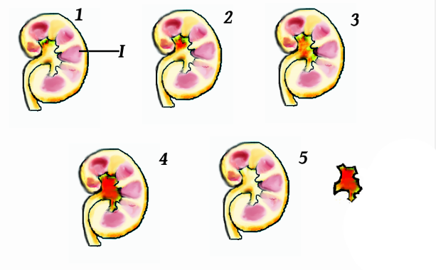
The renal calyces ( calyx) are conduits in the
 A " staghorn calculus" is a kidney stone that may extend into the renal calyces.
A renal diverticulum is diverticulum of renal calyces.
Diagram at bway.net
{{kidney Kidney anatomy
kidney
In humans, the kidneys are two reddish-brown bean-shaped blood-filtering organ (anatomy), organs that are a multilobar, multipapillary form of mammalian kidneys, usually without signs of external lobulation. They are located on the left and rig ...
through which urine passes. The minor calyces form a cup-shaped drain around the apex of the renal pyramids. Urine formed in the kidney passes through a renal papilla at the apex into the minor calyx; four or five minor calyces converge to form a major calyx through which urine passes into the renal pelvis (which in turn drains urine out of the kidney through the ureter
The ureters are tubes composed of smooth muscle that transport urine from the kidneys to the urinary bladder. In an adult human, the ureters typically measure 20 to 30 centimeters in length and about 3 to 4 millimeters in diameter. They are lin ...
).
Function
Peristalsis
Peristalsis ( , ) is a type of intestinal motility, characterized by symmetry in biology#Radial symmetry, radially symmetrical contraction and relaxation of muscles that propagate in a wave down a tube, in an wikt:anterograde, anterograde dir ...
of the smooth muscle originating in pace-maker cells originating in the walls of the calyces propels urine through the renal pelvis and ureter
The ureters are tubes composed of smooth muscle that transport urine from the kidneys to the urinary bladder. In an adult human, the ureters typically measure 20 to 30 centimeters in length and about 3 to 4 millimeters in diameter. They are lin ...
s to the bladder. The initiation is caused by the increase in volume that stretches the walls of the calyces. This causes them to fire impulses which stimulate rhythmical contraction and relaxation, called peristalsis. Parasympathetic innervation enhances the peristalsis while sympathetic innervation inhibits it.
Clinical significance
 A " staghorn calculus" is a kidney stone that may extend into the renal calyces.
A renal diverticulum is diverticulum of renal calyces.
See also
*Renal medulla
The renal medulla (Latin: ''medulla renis'' 'marrow of the kidney') is the innermost part of the kidney. The renal medulla is split up into a number of sections, known as the renal pyramids. Blood enters into the kidney via the renal artery, which ...
* Renal pyramids
* Calyx (anatomy)
References
External links
* - "Posterior Abdominal Wall: Internal Structure of a Kidney" * - "Posterior Abdominal Wall: Internal Structure of a Kidney" * - "Urinary System: neonatal kidney" * ()Diagram at bway.net
{{kidney Kidney anatomy