|
Median Aperture
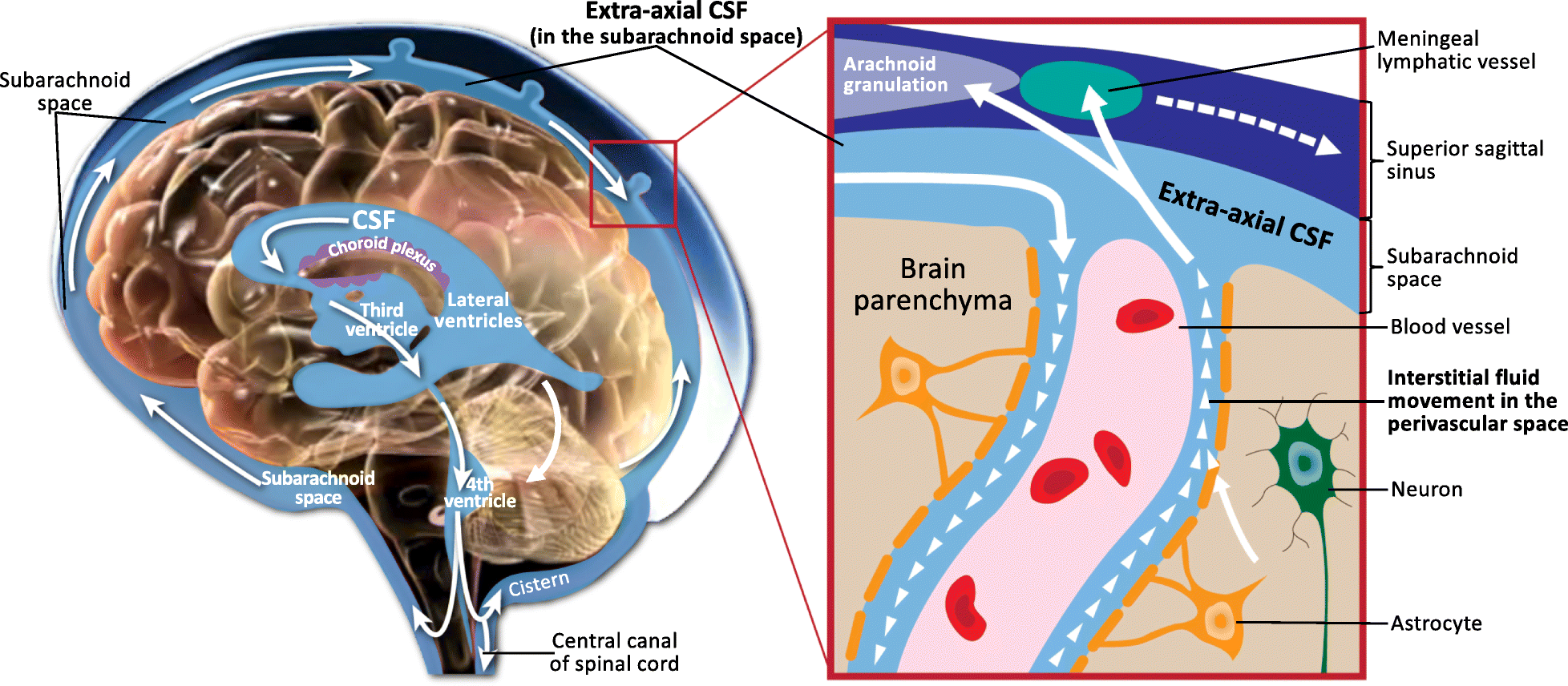
The median aperture (median aperture of fourth ventricle or foramen of Magendie) is an opening at the caudal portion of the roof of the fourth ventricle. It allows the flow of cerebrospinal fluid (CSF) from the fourth ventricle into the cisterna magna. The other openings of the fourth ventricle are the lateral apertures - one on either side. The median aperture varies in size but accounts for most of the outflow of CSF from the fourth ventricle. Structure Relations The median foramen on axial images is posterior to the pons and anterior to the caudal cerebellum. It is surrounded by the obex and gracile tubercles of the medulla, tela choroidea of the fourth ventricle and its choroid plexus, which is attached to the cerebellar vermis The cerebellar vermis (from Latin ''vermis,'' "worm") is located in the medial, cortico-nuclear zone of the cerebellum, which is in the posterior cranial fossa, posterior fossa of the cranium. The primary fissure in the vermis curves ventr ... [...More Info...] [...Related Items...] OR: [Wikipedia] [Google] [Baidu] |
Fourth Ventricle
The fourth ventricle is one of the four connected fluid-filled cavities within the human brain. These cavities, known collectively as the ventricular system, consist of the left and right lateral ventricles, the third ventricle, and the fourth ventricle. The fourth ventricle extends from the cerebral aqueduct (''aqueduct of Sylvius'') to the obex, and is filled with cerebrospinal fluid (CSF). The fourth ventricle has a characteristic diamond shape in cross-sections of the human brain. It is located within the pons or in the upper part of the medulla oblongata. CSF entering the fourth ventricle through the cerebral aqueduct can exit to the subarachnoid space of the spinal cord through two lateral apertures and a single, midline median aperture. Boundaries The fourth ventricle has a roof at its ''upper'' (posterior) surface and a floor at its ''lower'' (anterior) surface, and side walls formed by the cerebellar peduncles (nerve bundles joining the structure on the post ... [...More Info...] [...Related Items...] OR: [Wikipedia] [Google] [Baidu] |
Roof Of The Fourth Ventricle
The roof of fourth ventricle is the dorsal surface of the fourth ventricle. It corresponds to the ventral surface of the cerebellum The cerebellum (: cerebella or cerebellums; Latin for 'little brain') is a major feature of the hindbrain of all vertebrates. Although usually smaller than the cerebrum, in some animals such as the mormyrid fishes it may be as large as it or eve .... The upper portion of the roof is formed by the cerebellum. The roof of ventricle is diamond shaped and can be divided into superior and inferior parts. The superior part or cranial part is formed by superior cerebellar peduncles and superior medullary velum. The inferior or caudal part is formed by ventricular ependyma and double fold of pia mater. Additional images File:Gray649.png, Hind-brain of a human embryo of three months—viewed from behind and partly from left side. References Brainstem {{Portal bar, Anatomy ... [...More Info...] [...Related Items...] OR: [Wikipedia] [Google] [Baidu] |
Cerebrospinal Fluid
Cerebrospinal fluid (CSF) is a clear, colorless Extracellular fluid#Transcellular fluid, transcellular body fluid found within the meninges, meningeal tissue that surrounds the vertebrate brain and spinal cord, and in the ventricular system, ventricles of the brain. CSF is mostly produced by specialized Ependyma, ependymal cells in the choroid plexuses of the ventricles of the brain, and absorbed in the arachnoid granulations. It is also produced by ependymal cells in the lining of the ventricles. In humans, there is about 125 mL of CSF at any one time, and about 500 mL is generated every day. CSF acts as a shock absorber, cushion or buffer, providing basic mechanical and immune system, immunological protection to the brain inside the Human skull, skull. CSF also serves a vital function in the cerebral autoregulation of cerebral blood flow. CSF occupies the subarachnoid space (between the arachnoid mater and the pia mater) and the ventricular system around and inside t ... [...More Info...] [...Related Items...] OR: [Wikipedia] [Google] [Baidu] |
Cisterna Magna
The cisterna magna (posterior cerebellomedullary cistern, or cerebellomedullary cistern) is the largest of the subarachnoid cisterns. It occupies the space created by the angle between the caudal/inferior surface of the cerebellum, and the dorsal/posterior surface of the medulla oblongata (it is created by the arachnoidea that bridges this angle). The fourth ventricle communicates with the cistern via the unpaired midline median aperture. It is continuous inferiorly with the subarachnoid space of the spinal canal. The cisterna magna contains the two vertebral arteries, the origins of the two posterior inferior cerebellar arteries, the glossopharyngeal nerve (CN IX), vagus nerve (CN X), accessory nerve (CN XI), hypoglossal nerve (XII), and choroid plexus. The vertebral artery and posterior inferior cerebellar artery of either side pass traverse either lateral portion of the cistern. Etymology The '' Terminologia Anatomica'' classifies the terms ''cisterna magna'' and ' ... [...More Info...] [...Related Items...] OR: [Wikipedia] [Google] [Baidu] |
Lateral Aperture
The lateral aperture, lateral aperture of fourth ventricle or foramen of Luschka (after anatomist Hubert von Luschka) at whonamedit.com is an opening at the lateral extremity of either of the opening anteriorly into (sources differ) the pontine cistern/ lateral cerebellomedullary cistern at [...More Info...] [...Related Items...] OR: [Wikipedia] [Google] [Baidu] |
Pons
The pons (from Latin , "bridge") is part of the brainstem that in humans and other mammals, lies inferior to the midbrain, superior to the medulla oblongata and anterior to the cerebellum. The pons is also called the pons Varolii ("bridge of Varolius"), after the Italian anatomist and surgeon Costanzo Varolio (1543–75). This region of the brainstem includes neural pathways and tracts that conduct signals from the brain down to the cerebellum and medulla, and tracts that carry the sensory signals up into the thalamus. Structure The pons in humans measures about in length. It is the part of the brainstem situated between the midbrain and the medulla oblongata. The horizontal ''medullopontine sulcus'' demarcates the boundary between the pons and medulla oblongata on the ventral aspect of the brainstem, and the roots of cranial nerves VI/VII/VIII emerge from the brainstem along this groove. The junction of pons, medulla oblongata, and cerebellum forms the cerebellopontine ... [...More Info...] [...Related Items...] OR: [Wikipedia] [Google] [Baidu] |
Caudal (anatomical Term)
Standard anatomical terms of location are used to describe unambiguously the anatomy of humans and other animals. The terms, typically derived from Latin or Greek roots, describe something in its standard anatomical position. This position provides a definition of what is at the front ("anterior"), behind ("posterior") and so on. As part of defining and describing terms, the body is described through the use of anatomical planes and axes. The meaning of terms that are used can change depending on whether a vertebrate is a biped or a quadruped, due to the difference in the neuraxis, or if an invertebrate is a non-bilaterian. A non-bilaterian has no anterior or posterior surface for example but can still have a descriptor used such as proximal or distal in relation to a body part that is nearest to, or furthest from its middle. International organisations have determined vocabularies that are often used as standards for subdisciplines of anatomy. For example, '' Terminologia ... [...More Info...] [...Related Items...] OR: [Wikipedia] [Google] [Baidu] |
Cerebellum
The cerebellum (: cerebella or cerebellums; Latin for 'little brain') is a major feature of the hindbrain of all vertebrates. Although usually smaller than the cerebrum, in some animals such as the mormyrid fishes it may be as large as it or even larger. In humans, the cerebellum plays an important role in motor control and cognition, cognitive functions such as attention and language as well as emotion, emotional control such as regulating fear and pleasure responses, but its movement-related functions are the most solidly established. The human cerebellum does not initiate movement, but contributes to motor coordination, coordination, precision, and accurate timing: it receives input from sensory systems of the spinal cord and from other parts of the brain, and integrates these inputs to fine-tune motor activity. Cerebellar damage produces disorders in fine motor skill, fine movement, sense of balance, equilibrium, list of human positions, posture, and motor learning in humans. ... [...More Info...] [...Related Items...] OR: [Wikipedia] [Google] [Baidu] |
Obex
The obex () is the point in the human brain at which the fourth ventricle narrows to become the central canal of the spinal cord. Cerebrospinal fluid can flow from the fourth ventricle into the obex. In anatomical studies, the obex has been found to occur approximately 10–12 mm above the level of the foramen magnum. In patients with low tonsillar position, the obex has been found at or below the plane of the foramen magnum. The obex occurs in the caudal medulla. The decussation of sensory fibers happens at this point. Clinical significance Lesions at the location can result in obstructive hydrocephalus. The most common lesion at this location is a subependymoma, a benign tumor. Hemangioblastoma has been observed in this location. Detection of prions Immunohistochemistry (IHC) to test brain, lymph, and neuroendocrine tissues for the presence of the abnormal prion protein to diagnose wasting diseases like chronic wasting disease in deer A deer (: deer) or ... [...More Info...] [...Related Items...] OR: [Wikipedia] [Google] [Baidu] |
Gracile Tubercle
The dorsal column nuclei are a pair of nuclei in the dorsal columns of the dorsal column–medial lemniscus pathway (DCML) in the brainstem. The name refers collectively to the cuneate nucleus and gracile nucleus, which are situated at the lower end of the medulla oblongata. Both nuclei contain second-order neurons of the DCML, which convey fine touch and proprioceptive information from the body to the brain via the thalamus. Structure Nerve pathways The dorsal column nuclei each have an associated nerve tract in the spinal cord, the gracile fasciculus and the cuneate fasciculus, together forming the dorsal columns. Both dorsal column nuclei contain synapses from afferent nerve fibers that have travelled in the spinal cord. They then send on second-order neurons of the dorsal column–medial lemniscal pathway. Neurons of the dorsal column nuclei eventually reach the midbrain and the thalamus. They send axons that form the internal arcuate fibers. These cross over at t ... [...More Info...] [...Related Items...] OR: [Wikipedia] [Google] [Baidu] |
Medulla Oblongata
The medulla oblongata or simply medulla is a long stem-like structure which makes up the lower part of the brainstem. It is anterior and partially inferior to the cerebellum. It is a cone-shaped neuronal mass responsible for autonomic (involuntary) functions, ranging from vomiting to sneezing. The medulla contains the cardiovascular center, the respiratory center, vomiting and vasomotor centers, responsible for the autonomic functions of breathing, heart rate and blood pressure as well as the sleep–wake cycle. "Medulla" is from Latin, ‘pith or marrow’. And "oblongata" is from Latin, ‘lengthened or longish or elongated'. During embryonic development, the medulla oblongata develops from the myelencephalon. The myelencephalon is a secondary brain vesicle which forms during the maturation of the rhombencephalon, also referred to as the hindbrain. The bulb is an archaic term for the medulla oblongata. In modern clinical usage, the word bulbar (as in bulbar palsy) is r ... [...More Info...] [...Related Items...] OR: [Wikipedia] [Google] [Baidu] |
Tela Choroidea
The tela choroidea (or tela chorioidea) is a region of meninges, meningeal pia mater that adheres to the underlying ependyma, and gives rise to the choroid plexus in each of the brain’s Ventricular system, four ventricles. ''Tela'' is Latin for ''woven'' and is used to describe a web-like membrane or layer. The tela choroidea is a very thin part of the loose connective tissue of pia mater overlying and closely adhering to the ependyma. It has a rich blood supply. The ependyma and capillary, vascular pia mater – the tela choroidea, form regions of minute projections known as a choroid plexus that projects into each ventricle. The choroid plexus produces most of the cerebrospinal fluid of the central nervous system that circulates through the ventricles of the brain, the central canal of the spinal cord, and the subarachnoid space. The tela choroidea in the ventricles forms from different parts of the roof plate in the embryonic development, development of the embryo. Structure ... [...More Info...] [...Related Items...] OR: [Wikipedia] [Google] [Baidu] |


