|
OBEX
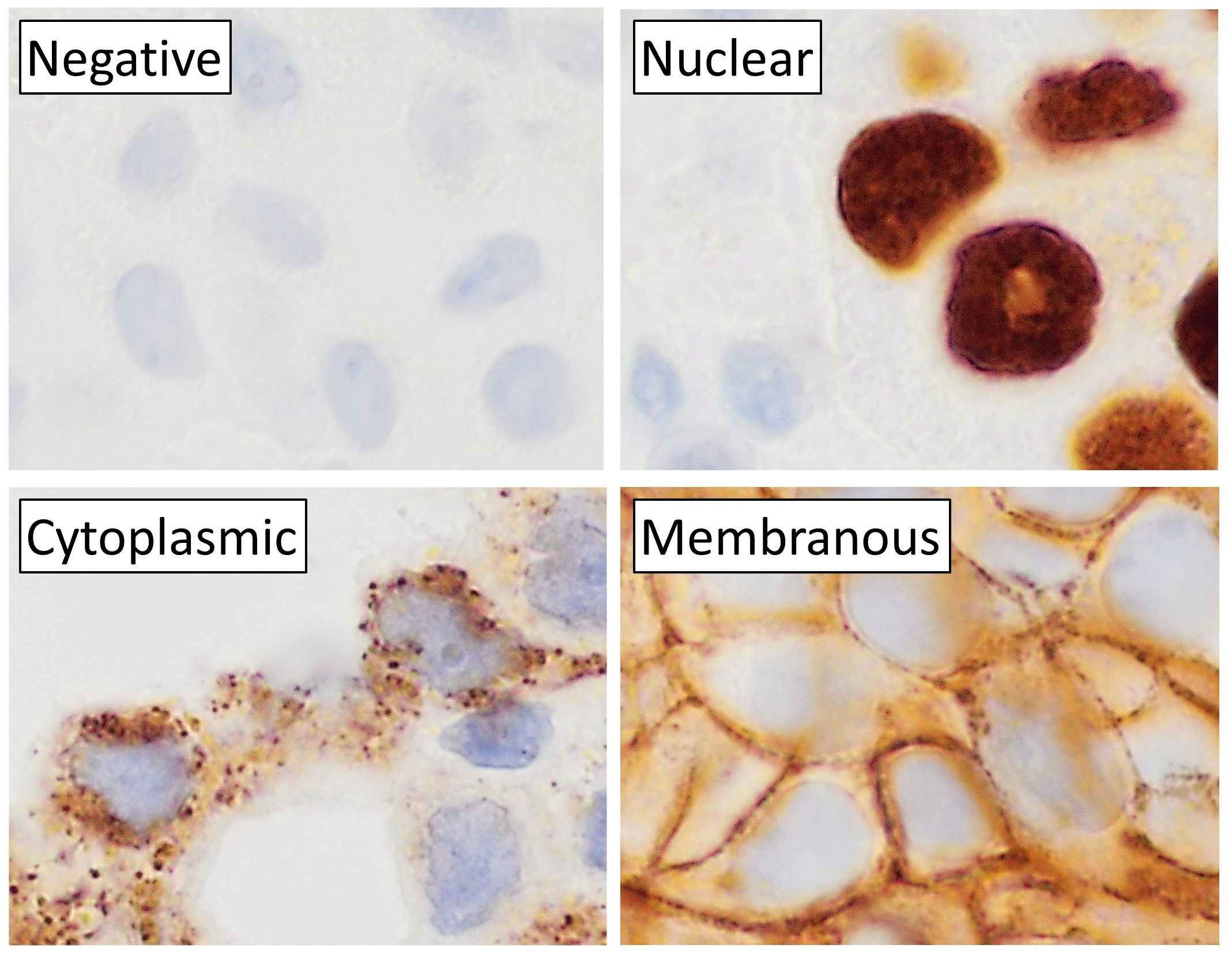
The obex () is the point in the human brain at which the fourth ventricle narrows to become the central canal of the spinal cord. Cerebrospinal fluid can flow from the fourth ventricle into the obex. In anatomical studies, the obex has been found to occur approximately 10–12 mm above the level of the foramen magnum. In patients with low tonsillar position, the obex has been found at or below the plane of the foramen magnum. The obex occurs in the caudal medulla. The decussation of sensory fibers happens at this point. Clinical significance Lesions at the location can result in obstructive hydrocephalus. The most common lesion at this location is a subependymoma, a benign tumor. Hemangioblastoma has been observed in this location. Detection of prions Immunohistochemistry (IHC) to test brain, lymph, and neuroendocrine tissues for the presence of the abnormal prion protein to diagnose wasting diseases like chronic wasting disease in deer A deer (: deer) or ... [...More Info...] [...Related Items...] OR: [Wikipedia] [Google] [Baidu] |
Human Brain
The human brain is the central organ (anatomy), organ of the nervous system, and with the spinal cord, comprises the central nervous system. It consists of the cerebrum, the brainstem and the cerebellum. The brain controls most of the activities of the human body, body, processing, integrating, and coordinating the information it receives from the sensory nervous system. The brain integrates sensory information and coordinates instructions sent to the rest of the body. The cerebrum, the largest part of the human brain, consists of two cerebral hemispheres. Each hemisphere has an inner core composed of white matter, and an outer surface – the cerebral cortex – composed of grey matter. The cortex has an outer layer, the neocortex, and an inner allocortex. The neocortex is made up of six Cerebral cortex#Layers of neocortex, neuronal layers, while the allocortex has three or four. Each hemisphere is divided into four lobes of the brain, lobes – the frontal lobe, frontal, pa ... [...More Info...] [...Related Items...] OR: [Wikipedia] [Google] [Baidu] |
Tumor
A neoplasm () is a type of abnormal and excessive growth of tissue. The process that occurs to form or produce a neoplasm is called neoplasia. The growth of a neoplasm is uncoordinated with that of the normal surrounding tissue, and persists in growing abnormally, even if the original trigger is removed. This abnormal growth usually forms a mass, which may be called a tumour or tumor.'' ICD-10 classifies neoplasms into four main groups: benign neoplasms, in situ neoplasms, malignant neoplasms, and neoplasms of uncertain or unknown behavior. Malignant neoplasms are also simply known as cancers and are the focus of oncology. Prior to the abnormal growth of tissue, such as neoplasia, cells often undergo an abnormal pattern of growth, such as metaplasia or dysplasia. However, metaplasia or dysplasia does not always progress to neoplasia and can occur in other conditions as well. The word neoplasm is from Ancient Greek 'new' and 'formation, creation'. Types A neopla ... [...More Info...] [...Related Items...] OR: [Wikipedia] [Google] [Baidu] |
Deer
A deer (: deer) or true deer is a hoofed ruminant ungulate of the family Cervidae (informally the deer family). Cervidae is divided into subfamilies Cervinae (which includes, among others, muntjac, elk (wapiti), red deer, and fallow deer) and Capreolinae (which includes, among others reindeer (caribou), white-tailed deer, roe deer, and moose). Male deer of almost all species (except the water deer), as well as female reindeer, grow and shed new antlers each year. These antlers are bony extensions of the skull and are often used for combat between males. The musk deer ( Moschidae) of Asia and chevrotains ( Tragulidae) of tropical African and Asian forests are separate families that are also in the ruminant clade Ruminantia; they are not especially closely related to Cervidae. Deer appear in art from Paleolithic cave paintings onwards, and they have played a role in mythology, religion, and literature throughout history, as well as in heraldry, such as red deer that app ... [...More Info...] [...Related Items...] OR: [Wikipedia] [Google] [Baidu] |
Chronic Wasting Disease
Chronic wasting disease (CWD), sometimes called zombie deer disease, is a transmissible spongiform encephalopathy (TSE) affecting deer. TSEs are a family of diseases thought to be caused by misfolded proteins called prions and include similar diseases such as BSE (mad cow disease) in cattle, Creutzfeldt–Jakob disease (CJD) in humans, and scrapie in sheep. Natural infection causing CWD affects members of the deer family. In the United States, CWD affects mule deer, white-tailed deer, red deer, sika deer, elk, bison, antelope, caribou, and moose. The transmission of CWD to other species such as squirrel monkeys and humanized mice has been observed in experimental settings. In 1967, CWD was first identified in mule deer at a government research facility in northern Colorado, United States. It was initially recognized as a clinical "wasting" syndrome and then in 1978, it was identified more specifically as a TSE disease. Since then, CWD has been found in free-rang ... [...More Info...] [...Related Items...] OR: [Wikipedia] [Google] [Baidu] |
Prion
A prion () is a Proteinopathy, misfolded protein that induces misfolding in normal variants of the same protein, leading to cellular death. Prions are responsible for prion diseases, known as transmissible spongiform encephalopathy (TSEs), which are fatal and transmissible neurodegenerative diseases affecting both humans and animals. These proteins can misfold sporadically, due to genetic mutations, or by exposure to an already misfolded protein, leading to an abnormal Protein tertiary structure, three-dimensional structure that can propagate misfolding in other proteins. The term ''prion'' comes from "proteinaceous infectious particle". Unlike other infectious agents such as viruses, bacteria, and fungi, prions do not contain nucleic acids (DNA or RNA). Prions are mainly twisted Protein isoform, isoforms of the major prion protein (PrP), a naturally occurring protein with an uncertain function. They are the hypothesized cause of various transmissible spongiform encephalopath ... [...More Info...] [...Related Items...] OR: [Wikipedia] [Google] [Baidu] |
Lymph
Lymph () is the fluid that flows through the lymphatic system, a system composed of lymph vessels (channels) and intervening lymph nodes whose function, like the venous system, is to return fluid from the tissues to be recirculated. At the origin of the fluid-return process, interstitial fluid—the fluid between the cells in all body tissues—enters the lymph capillaries. This lymphatic fluid is then transported via progressively larger lymphatic vessels through lymph nodes, where substances are removed by tissue lymphocytes and circulating lymphocytes are added to the fluid, before emptying ultimately into the right or the left subclavian vein, where it mixes with central venous blood. Because it is derived from interstitial fluid, with which blood and surrounding cells continually exchange substances, lymph undergoes continual change in composition. It is generally similar to blood plasma, which is the fluid component of blood. Lymph returns proteins and excess interstiti ... [...More Info...] [...Related Items...] OR: [Wikipedia] [Google] [Baidu] |
Brain
The brain is an organ (biology), organ that serves as the center of the nervous system in all vertebrate and most invertebrate animals. It consists of nervous tissue and is typically located in the head (cephalization), usually near organs for special senses such as visual perception, vision, hearing, and olfaction. Being the most specialized organ, it is responsible for receiving information from the sensory nervous system, processing that information (thought, cognition, and intelligence) and the coordination of motor control (muscle activity and endocrine system). While invertebrate brains arise from paired segmental ganglia (each of which is only responsible for the respective segmentation (biology), body segment) of the ventral nerve cord, vertebrate brains develop axially from the midline dorsal nerve cord as a brain vesicle, vesicular enlargement at the rostral (anatomical term), rostral end of the neural tube, with centralized control over all body segments. All vertebr ... [...More Info...] [...Related Items...] OR: [Wikipedia] [Google] [Baidu] |
Immunohistochemistry
Immunohistochemistry is a form of immunostaining. It involves the process of selectively identifying antigens in cells and tissue, by exploiting the principle of Antibody, antibodies binding specifically to antigens in biological tissues. Albert Coons, Albert Hewett Coons, Ernst Berliner, Ernest Berliner, Norman Jones and Hugh J Creech was the first to develop immunofluorescence in 1941. This led to the later development of immunohistochemistry. Immunohistochemical staining is widely used in the diagnosis of abnormal cells such as those found in cancerous tumors. In some cancer cells certain tumor antigens are expressed which make it possible to detect. Immunohistochemistry is also widely used in basic research, to understand the distribution and localization of biomarkers and differentially expressed proteins in different parts of a biological tissue. Sample preparation Immunohistochemistry can be performed on tissue that has been fixed and embedded in Paraffin wax, paraffin, ... [...More Info...] [...Related Items...] OR: [Wikipedia] [Google] [Baidu] |
Hemangioblastoma
Hemangioblastomas, or haemangioblastomas, are vascular tumors of the central nervous system that originate from the vascular system, usually during middle age. Sometimes, these tumors occur in other sites such as the spinal cord and retina. They may be associated with other diseases such as polycythemia (increased blood cell count), pancreatic cysts and Von Hippel–Lindau syndrome (VHL syndrome). Hemangioblastomas are most commonly composed of stromal cells in small blood vessels and usually occur in the cerebellum, brainstem or spinal cord. They are classed as grade I tumors under the World Health Organization's classification system. Presentation Complications Hemangioblastomas can cause an polycythemia, abnormally high number of red blood cells in the bloodstream due to ectopic production of the hormone erythropoietin as a paraneoplastic syndrome. Pathogenesis Hemangioblastomas are composed of endothelial cells, pericytes and stromal cells. In VHL syndrome the Von Hippel–Lin ... [...More Info...] [...Related Items...] OR: [Wikipedia] [Google] [Baidu] |
Subependymoma
A subependymoma is a type of brain tumor; specifically, it is a rare form of ependymal tumor. They are usually in middle aged people. Earlier, they were called subependymal astrocytomas. The prognosis for a subependymoma is better than for most ependymal tumors, and it is considered a grade I tumor in the World Health Organization (WHO) classification. They are classically found within the fourth ventricle, typically have a well demarcated interface to normal tissue and do not usually extend into the brain parenchyma, like ependymomas often do. Symptoms and signs Patients are often asymptomatic, and are incidentally diagnosed. Larger tumours are often with increased intracranial pressure. Pathology These tumours are small, no more than two centimeters across, coming from the ependyma. The best way to distinguish it from a subependymal giant cell astrocytoma is the size. Diagnosis The diagnosis is based on tissue, e.g. a biopsy. Histologically subependymomas consistent ... [...More Info...] [...Related Items...] OR: [Wikipedia] [Google] [Baidu] |
Fourth Ventricle
The fourth ventricle is one of the four connected fluid-filled cavities within the human brain. These cavities, known collectively as the ventricular system, consist of the left and right lateral ventricles, the third ventricle, and the fourth ventricle. The fourth ventricle extends from the cerebral aqueduct (''aqueduct of Sylvius'') to the obex, and is filled with cerebrospinal fluid (CSF). The fourth ventricle has a characteristic diamond shape in cross-sections of the human brain. It is located within the pons or in the upper part of the medulla oblongata. CSF entering the fourth ventricle through the cerebral aqueduct can exit to the subarachnoid space of the spinal cord through two lateral apertures and a single, midline median aperture. Boundaries The fourth ventricle has a roof at its ''upper'' (posterior) surface and a floor at its ''lower'' (anterior) surface, and side walls formed by the cerebellar peduncles (nerve bundles joining the structure on the post ... [...More Info...] [...Related Items...] OR: [Wikipedia] [Google] [Baidu] |
Obstructive Hydrocephalus
Hydrocephalus is a condition in which cerebrospinal fluid (CSF) builds up within the brain, which can cause pressure to increase in the skull. Symptoms may vary according to age. Headaches and double vision are common. Elderly adults with normal pressure hydrocephalus (NPH) may have poor balance, difficulty controlling urination, or mental impairment. In babies, there may be a rapid increase in head size. Other symptoms may include vomiting, sleepiness, seizures, and downward pointing of the eyes. Hydrocephalus can occur due to birth defects (primary) or can develop later in life (secondary). Hydrocephalus can be classified via mechanism into communicating, noncommunicating, ''ex vacuo'', and normal pressure hydrocephalus. Diagnosis is made by physical examination and medical imaging, such as a CT scan. Hydrocephalus is typically treated through surgery. One option is the placement of a shunt system. A procedure called an endoscopic third ventriculostomy has gained p ... [...More Info...] [...Related Items...] OR: [Wikipedia] [Google] [Baidu] |


