|
Medial Posterior Superior Nasal Nerves
The (medial and lateral) posterior superior nasal nerves are branches of the maxillary nerve (CN V2) that arise in the pterygopalatine fossa from pterygopalatine ganglion and pass through the sphenopalatine foramen into the nasal cavity to innervate the nasal septum (the medial nerves), and the posterosuperior portion of the lateral wall of the nasal cavity (the lateral nerves). Median posterior superior nasal nerves The median posterior superior nasal nerves traverse the roof of the nose to be distributed to the nasal septum. The largest of the median posterior superior nasal nerves is the nasopalatine nerve (which continues out of the nasal cavity to the hard palate The hard palate is a thin horizontal bony plate made up of two bones of the facial skeleton, located in the roof of the mouth. The bones are the palatine process of the maxilla and the horizontal plate of palatine bone. The hard palate spans ...). Lateral posterior superior nasal nerves The lateral poster ... [...More Info...] [...Related Items...] OR: [Wikipedia] [Google] [Baidu] |
Maxillary Nerve
In neuroanatomy, the maxillary nerve (V) is one of the three branches or divisions of the trigeminal nerve, the fifth (CN V) cranial nerve. It comprises the principal functions of Sense, sensation from the maxilla, nasal cavity, Sinus (anatomy), sinuses, the palate and subsequently that of the mid-face, and is intermediate, both in position and size, between the ophthalmic nerve and the mandibular nerve.Illustrated Anatomy of the Head and Neck, Fehrenbach and Herring, Elsevier, 2012, page 180 Structure It begins at the middle of the trigeminal ganglion as a flattened plexiform band then it passes through the lateral wall of the cavernous sinus. It leaves the skull through the foramen rotundum, where it becomes more cylindrical in form, and firmer in texture. After leaving foramen rotundum it gives two branches to the pterygopalatine ganglion. It then crosses the pterygopalatine fossa, inclines lateralward on the back of the maxilla, and enters the orbit through the inferior orb ... [...More Info...] [...Related Items...] OR: [Wikipedia] [Google] [Baidu] |
Pterygopalatine Fossa
In human anatomy, the pterygopalatine fossa (sphenopalatine fossa) is a fossa in the skull. A human skull contains two pterygopalatine fossae—one on the left side, and another on the right side. Each fossa is a cone-shaped paired depression deep to the infratemporal fossa and posterior to the maxilla on each side of the skull, located between the pterygoid process and the maxillary tuberosity close to the apex of the orbit. It is the indented area medial to the pterygomaxillary fissure leading into the sphenopalatine foramen. It communicates with the nasal and oral cavities, infratemporal fossa, orbit, pharynx, and middle cranial fossa through eight foramina. Structure Boundaries It has the following boundaries: * ''anterior'': superomedial part of the infratemporal surface of maxilla * ''posterior'': root of the pterygoid process and adjoining anterior surface of the greater wing of sphenoid bone * ''medial'': perpendicular plate of the palatine bone and its orbital an ... [...More Info...] [...Related Items...] OR: [Wikipedia] [Google] [Baidu] |
Sphenopalatine Foramen
The sphenopalatine foramen is a foramen of the skull that connects the nasal cavity and the pterygopalatine fossa. It gives passage to the sphenopalatine artery, nasopalatine nerve, and the superior nasal nerve (all passing from the pterygopalatine fossa into the nasal cavity). Structure The processes of the superior border of the palatine bone are separated by the ''sphenopalatine notch'', which is converted into the sphenopalatine foramen by the under surface of the body of the sphenoid. The sphenopalatine foramen is situated posterior to the middle nasal meatus orbital process of palatine bone, anterior to the sphenoidal process of palatine bone, inferior to the body and of the sphenoid bone, and superior to the superior margin of the perpendicular plate of palatine bone. Relations The ethmoid crest (a reliable surgical landmark A landmark is a recognizable natural or artificial feature used for navigation, a feature that stands out from its near environment and ... [...More Info...] [...Related Items...] OR: [Wikipedia] [Google] [Baidu] |
Nasal Cavity
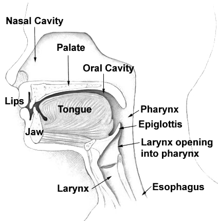
The nasal cavity is a large, air-filled space above and behind the nose in the middle of the face. The nasal septum divides the cavity into two cavities, also known as fossae. Each cavity is the continuation of one of the two nostrils. The nasal cavity is the uppermost part of the respiratory system and provides the nasal passage for inhaled air from the nostrils to the nasopharynx and rest of the respiratory tract. The paranasal sinuses surround and drain into the nasal cavity. Structure The term "nasal cavity" can refer to each of the two cavities of the nose, or to the two sides combined. The lateral wall of each nasal cavity mainly consists of the maxilla. However, there is a deficiency that is compensated for by the perpendicular plate of the palatine bone, the medial pterygoid plate, the labyrinth of ethmoid and the inferior concha. The paranasal sinuses are connected to the nasal cavity through small orifices called ostia. Most of these ostia communicat ... [...More Info...] [...Related Items...] OR: [Wikipedia] [Google] [Baidu] |
Nasal Septum
The nasal septum () separates the left and right airways of the Human nose, nasal cavity, dividing the two nostrils. It is Depression (kinesiology), depressed by the depressor septi nasi muscle. Structure The fleshy external end of the nasal septum is called the Human nose#Cartilages, columella or columella nasi, and is made up of cartilage and soft tissue. The nasal septum contains bone and hyaline cartilage. It is normally about 2 mm thick. The nasal septum is composed of four structures: * Maxillary bone (the crest) * Perpendicular plate of ethmoid bone * Septal nasal cartilage (ie, quandrangular cartilage) * Vomer bone The lowest part of the septum is a narrow strip of bone that projects from the maxilla and the palatine bones, and is the length of the septum. This strip of bone is called the maxillary crest; it articulates in front with the septal nasal cartilage, and at the back with the vomer. The maxillary crest is described in the anatomy of the nasal septum as h ... [...More Info...] [...Related Items...] OR: [Wikipedia] [Google] [Baidu] |
Nasopalatine Nerve
The nasopalatine nerve (also long sphenopalatine nerve) is a nerve of the head. It is a sensory branch of the maxillary nerve (CN V2) that passes through the pterygopalatine ganglion (without synapsing) and then through the sphenopalatine foramen to enter the nasal cavity, and finally out of the nasal cavity through the incisive canal and then the incisive fossa to enter the hard palate. It provides sensory innervation to the posteroinferior part of the nasal septum, and gingiva just posterior to the upper incisor teeth. The nasopalatine nerve is the largest of the medial posterior superior nasal nerves. Structure Course It exits the pterygopalatine fossa through the sphenopalatine foramen to enter the nasal cavity. It passes across the roof of the nasal cavity below the orifice of the sphenoidal sinus to reach the posterior part of the nasal septum. It passes anteroinferiorly upon the nasal septum along a groove upon the vomer, running between the periosteum and mucous membr ... [...More Info...] [...Related Items...] OR: [Wikipedia] [Google] [Baidu] |
Hard Palate
The hard palate is a thin horizontal bony plate made up of two bones of the facial skeleton, located in the roof of the mouth. The bones are the palatine process of the maxilla and the horizontal plate of palatine bone. The hard palate spans the alveolar arch formed by the alveolar process that holds the upper teeth (when these are developed). Structure The hard palate is formed by the palatine process of the maxilla and horizontal plate of palatine bone. It forms a partition between the nasal passages and the mouth. On the anterior portion of the hard palate are the ''plicae'', irregular ridges in the mucous membrane that help hold food while the teeth are biting into it while also facilitating the movement of food backward towards the larynx once pieces have been bitten off. This partition is continued deeper into the mouth by a fleshy extension called the soft palate. On the ventral surface of the hard palate, some projections or transverse ridges are present which ... [...More Info...] [...Related Items...] OR: [Wikipedia] [Google] [Baidu] |

