|
Interspinal Ligament
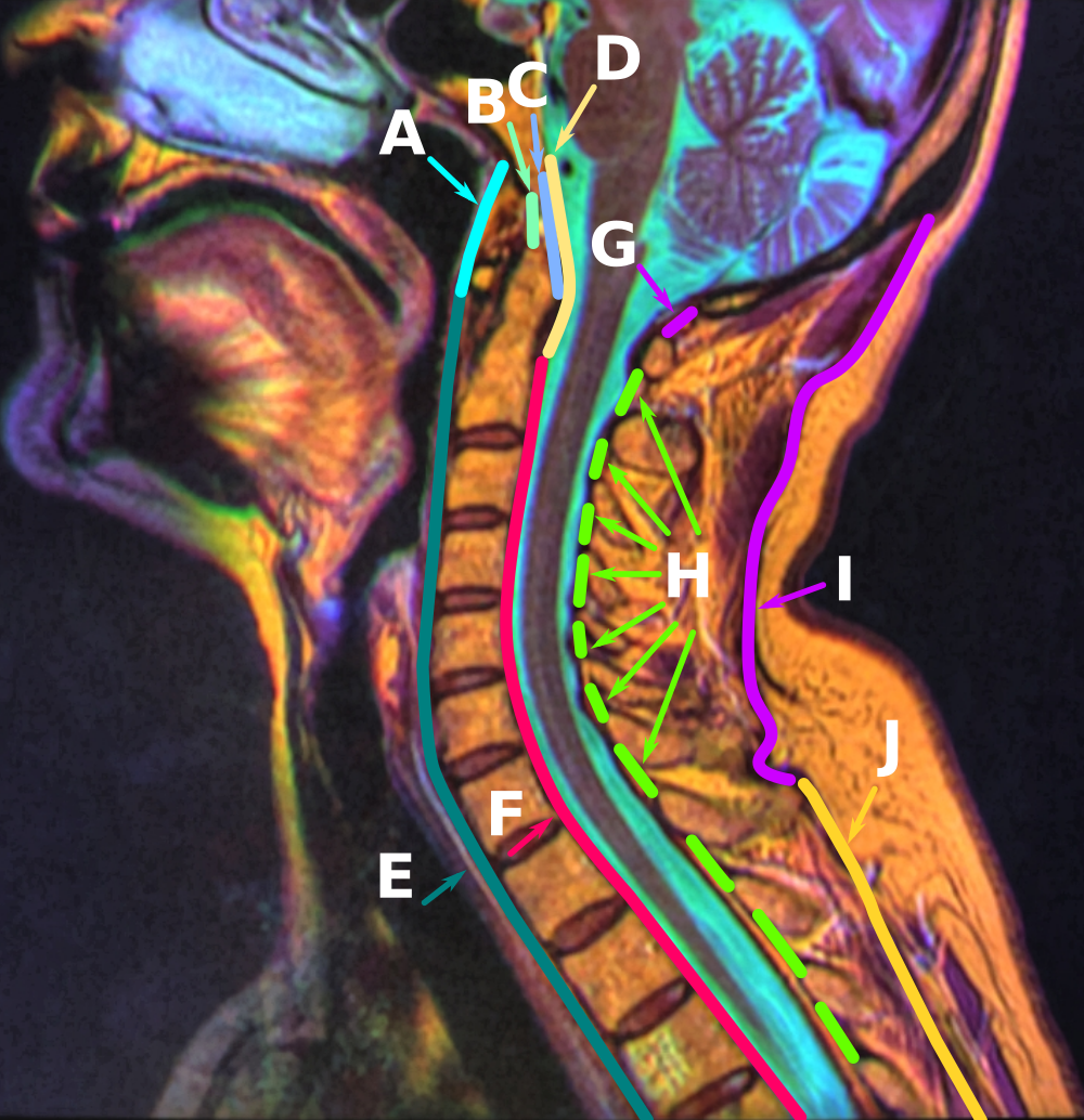
The interspinous ligaments (interspinal ligaments) are thin, membranous ligaments that connect adjoining spinous processes of the vertebra in the spine. They take the form of relatively weak sheets of fibrous tissue and are well developed only in the lumbar region. They extend from the root to the apex of each spinous process. They meet the ligamenta flava anteriorly, and blend with the supraspinous ligament posteriorly at the apexes of the spinal processes. The function of the interspinous ligaments is to limit ventral flexion of the spine and sliding movement of the vertebrae. The ligaments are narrow and elongated in the thoracic region. They are broader, thicker, and quadrilateral in form in the lumbar region. They are only slightly developed in the neck; in the neck, they are often considered part of the nuchal ligament The nuchal ligament is a ligament at the back of the neck that is continuous with the supraspinous ligament. Structure The nuchal ligament extends from ... [...More Info...] [...Related Items...] OR: [Wikipedia] [Google] [Baidu] |
Sagittal Section
The sagittal plane (; also known as the longitudinal plane) is an anatomical plane that divides the body into right and left sections. It is perpendicular to the transverse and coronal planes. The plane may be in the center of the body and divide it into two equal parts ( mid-sagittal), or away from the midline and divide it into unequal parts (para-sagittal). The term ''sagittal'' was coined by Gerard of Cremona. Variations in terminology Examples of sagittal planes include: * The terms '' median plane'' or ''mid-sagittal plane'' are sometimes used to describe the sagittal plane running through the midline. This plane cuts the body into halves (assuming bilateral symmetry), passing through midline structures such as the navel and spine. It is one of the planes which, combined with the umbilical plane, defines the four quadrants of the human abdomen. * The term ''parasagittal'' is used to describe any plane parallel or adjacent to a given sagittal plane. Specific named par ... [...More Info...] [...Related Items...] OR: [Wikipedia] [Google] [Baidu] |
Lumbar Vertebrae
The lumbar vertebrae are located between the thoracic vertebrae and pelvis. They form the lower part of the back in humans, and the tail end of the back in quadrupeds. In humans, there are five lumbar vertebrae. The term is used to describe the anatomy of humans and quadrupeds, such as horses, pigs, or cattle. These bones are found in particular cuts of meat, including tenderloin or sirloin steak. Human anatomy In human anatomy, the five vertebrae are between the rib cage and the pelvis. They are the largest segments of the vertebral column and are characterized by the absence of the foramen transversarium within the transverse process (since it is only found in the cervical region) and by the absence of facets on the sides of the body (as found only in the thoracic region). They are designated L1 to L5, starting at the top. The lumbar vertebrae help support the weight of the body, and permit movement. General characteristics The adjacent figure depicts the general cha ... [...More Info...] [...Related Items...] OR: [Wikipedia] [Google] [Baidu] |
Ligament
A ligament is a type of fibrous connective tissue in the body that connects bones to other bones. It also connects flight feathers to bones, in dinosaurs and birds. All 30,000 species of amniotes (land animals with internal bones) have ligaments. It is also known as ''articular ligament'', ''articular larua'', ''fibrous ligament'', or ''true ligament''. Comparative anatomy Ligaments are similar to tendons and fasciae as they are all made of connective tissue. The differences among them are in the connections that they make: ligaments connect one bone to another bone, tendons connect muscle to bone, and fasciae connect muscles to other muscles. These are all found in the skeletal system of the human body. Ligaments cannot usually be regenerated naturally; however, there are periodontal ligament stem cells located near the periodontal ligament which are involved in the adult regeneration of periodontist ligament. The study of ligaments is known as . Humans Other ligame ... [...More Info...] [...Related Items...] OR: [Wikipedia] [Google] [Baidu] |
Spinous Process
Each vertebra (: vertebrae) is an irregular bone with a complex structure composed of bone and some hyaline cartilage, that make up the vertebral column or spine, of vertebrates. The proportions of the vertebrae differ according to their spinal segment and the particular species. The basic configuration of a vertebra varies; the vertebral body (also ''centrum'') is of bone and bears the load of the vertebral column. The upper and lower surfaces of the vertebra body give attachment to the intervertebral discs. The posterior part of a vertebra forms a vertebral arch, in eleven parts, consisting of two pedicles (pedicle of vertebral arch), two laminae, and seven processes. The laminae give attachment to the ligamenta flava (ligaments of the spine). There are vertebral notches formed from the shape of the pedicles, which form the intervertebral foramina when the vertebrae articulate. These foramina are the entry and exit conduits for the spinal nerves. The body of the vertebra ... [...More Info...] [...Related Items...] OR: [Wikipedia] [Google] [Baidu] |
Spinous Processes
Each vertebra (: vertebrae) is an irregular bone with a complex structure composed of bone and some hyaline cartilage, that make up the vertebral column or spine, of vertebrates. The proportions of the vertebrae differ according to their spinal segment and the particular species. The basic configuration of a vertebra varies; the vertebral body (also ''centrum'') is of bone and bears the load of the vertebral column. The upper and lower surfaces of the vertebra body give attachment to the intervertebral discs. The posterior part of a vertebra forms a vertebral arch, in eleven parts, consisting of two pedicles (pedicle of vertebral arch), two laminae, and seven processes. The laminae give attachment to the ligamenta flava (ligaments of the spine). There are vertebral notches formed from the shape of the pedicles, which form the intervertebral foramina when the vertebrae articulate. These foramina are the entry and exit conduits for the spinal nerves. The body of the vertebra an ... [...More Info...] [...Related Items...] OR: [Wikipedia] [Google] [Baidu] |
Human Vertebral Column
The spinal column, also known as the vertebral column, spine or backbone, is the core part of the axial skeleton in vertebrates. The vertebral column is the defining and eponymous characteristic of the vertebrate. The spinal column is a segmented column of vertebrae that surrounds and protects the spinal cord. The vertebrae are separated by intervertebral discs in a series of cartilaginous joints. The dorsal portion of the spinal column houses the spinal canal, an elongated body cavity, cavity formed by the alignment of the vertebral neural arches that encloses and protects the spinal cord, with spinal nerves exiting via the intervertebral foramina to innervate each body segment. There are around 50,000 species of animals that have a vertebral column. The human spine is one of the most-studied examples, as the general structure of human vertebrae is fairly homology (biology), typical of that found in other mammals, reptiles, and birds. The shape of the vertebral body does, howev ... [...More Info...] [...Related Items...] OR: [Wikipedia] [Google] [Baidu] |
Ligamenta Flava
The ligamenta flava (: ligamentum flavum, Latin for ''yellow ligament'') are a series of ligaments that connect the ventral parts of the laminae of adjacent vertebrae. They help to preserve upright posture, preventing hyperflexion, and ensuring that the vertebral column straightens after flexion. Hypertrophy can cause spinal stenosis. They appear yellowish in colour due to their high elastic fibre content. Anatomy Each ligamentum flavum connects the laminae of two adjacent vertebrae. They attach to the anterior portion of the upper lamina above, and the posterior portion of the lower lamina below. They begin with the junction of the axis and third cervical vertebra, continuing down to the junction of the 5th lumbar vertebra and the sacrum. In the neck region the ligaments are thin, but broad and long; they are thicker in the thoracic region, and thickest in the lumbar region. They are thinnest between the atlas bone (C1) and the axis bone (C2), and may be absent in some ... [...More Info...] [...Related Items...] OR: [Wikipedia] [Google] [Baidu] |
Supraspinous Ligament
The supraspinous ligament (also known as the supraspinal ligament) is a ligament extending across the tips of the spinous processes of the vertebra of the vertebral column. Anatomy The supraspinous ligament connects the tips of the spinous processes from the seventh cervical vertebra to the sacrum. Superior to the 7th cervical vertebra, the supraspinous ligament is continuous with the nuchal ligament. It is thicker and broader in the lumbar region than in the thoracic region, and intimately blended with the neighboring fascia in both these regions. Inferior to L4, the supraspinous ligament becomes indistinct, lost amid the prominent lumbar fascia. Between the spinous processes, the supraspinous ligament is continuous with the interspinous ligaments. Structure The most superficial fibers of this ligament extend across 3-4 vertebrae, deeper fibres extend across 2-3 vertebrae, while the deepest connect the spinous processes of adjacent vertebrae. Function The supraspinous ligam ... [...More Info...] [...Related Items...] OR: [Wikipedia] [Google] [Baidu] |
Flexion
Motion, the process of movement, is described using specific anatomical terminology, anatomical terms. Motion includes movement of Organ (anatomy), organs, joints, Limb (anatomy), limbs, and specific sections of the body. The terminology used describes this motion according to its direction relative to the anatomical position of the body parts involved. Anatomy, Anatomists and others use a unified set of terms to describe most of the movements, although other, more specialized terms are necessary for describing unique movements such as those of the hands, feet, and eyes. In general, motion is classified according to the anatomical plane it occurs in. ''Flexion'' and ''extension'' are examples of ''angular'' motions, in which two axes of a joint are brought closer together or moved further apart. ''Rotational'' motion may occur at other joints, for example the shoulder, and are described as ''internal'' or ''external''. Other terms, such as ''elevation'' and ''depression'', descri ... [...More Info...] [...Related Items...] OR: [Wikipedia] [Google] [Baidu] |
Thoracic Vertebrae
In vertebrates, thoracic vertebrae compose the middle segment of the vertebral column, between the cervical vertebrae and the lumbar vertebrae. In humans, there are twelve thoracic vertebra (anatomy), vertebrae of intermediate size between the cervical and lumbar vertebrae; they increase in size going towards the lumbar vertebrae. They are distinguished by the presence of Zygapophysial joint, facets on the sides of the bodies for Articulation (anatomy), articulation with the head of rib, heads of the ribs, as well as facets on the transverse processes of all, except the eleventh and twelfth, for articulation with the tubercle (rib), tubercles of the ribs. By convention, the human thoracic vertebrae are numbered T1–T12, with the first one (T1) located closest to the skull and the others going down the spine toward the lumbar region. General characteristics These are the general characteristics of the second through eighth thoracic vertebrae. The first and ninth through twelfth v ... [...More Info...] [...Related Items...] OR: [Wikipedia] [Google] [Baidu] |


