|
Inferior Pharyngeal Constrictor Muscle
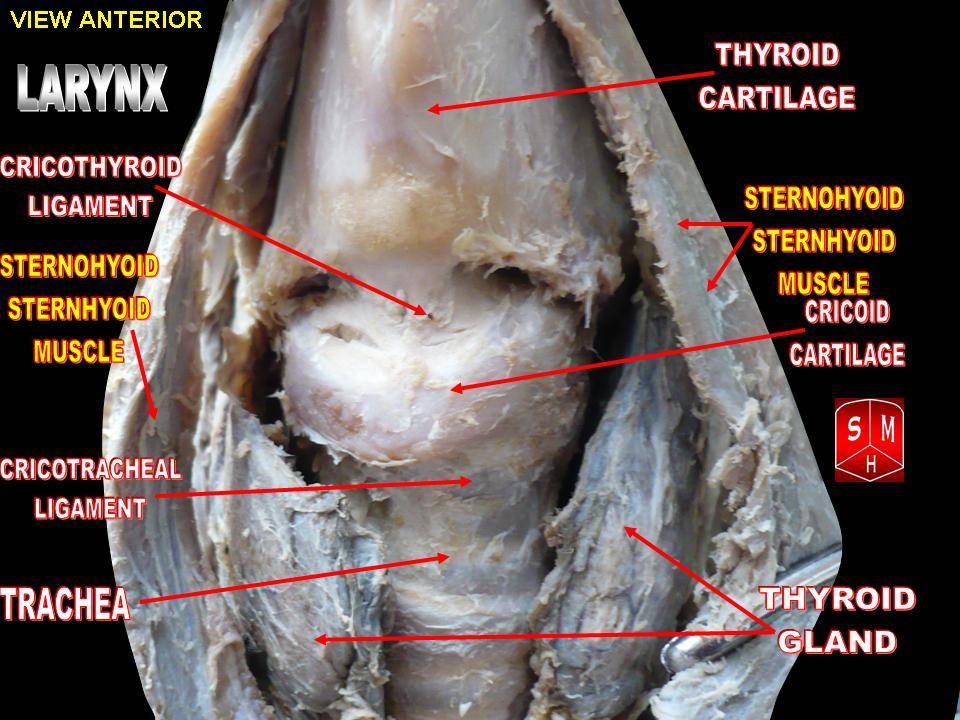
The inferior pharyngeal constrictor muscle is a skeletal muscle of the neck. It is the thickest of the three outer pharyngeal muscles. It arises from the sides of the cricoid cartilage and the thyroid cartilage. It is supplied by the vagus nerve (CN X). It is active during swallowing, and partially during breathing and speech. It may be affected by Zenker's diverticulum. Structure The inferior pharyngeal constrictor muscle is composed of two parts. The first part (and more superior) arises from the thyroid cartilage (thyropharyngeal part), and the second part arises from the cricoid cartilage (cricopharyngeal part). * On the ''thyroid cartilage'', it arises from the oblique line on the side of the lamina, from the surface behind this nearly as far as the posterior border and from the inferior horn of the thyroid cartilage. * From the ''cricoid cartilage'', it arises in the interval between the cricothyroid muscle in front, and the articular facet for the inferior horn of ... [...More Info...] [...Related Items...] OR: [Wikipedia] [Google] [Baidu] |
Cricoid Cartilage
The cricoid cartilage , or simply cricoid (from the Greek ''krikoeides'' meaning "ring-shaped") or cricoid ring, is the only complete ring of cartilage around the trachea. It forms the back part of the voice box and functions as an attachment site for muscles, cartilages, and ligaments involved in opening and closing the airway and in producing speech. Anatomy The cricoid cartilage is the only laryngeal cartilage to form a complete circle around the airway. It is smaller yet thicker and tougher than the thyroid cartilage above. It articulates superiorly with the thyroid cartilage, and the paired arytenoid cartilage. Inferiorly, the trachea attaches onto it. It occurs at the level of the C6 vertebra. Structure The posterior part of the cricoid cartilage (cricoid lamina) is somewhat broader than the anterior and lateral part (cricoid arch). Its shape is said to resemble a signet ring. Cricoid arch The cricoid arch is the curved and vertically narrow anterior portion of ... [...More Info...] [...Related Items...] OR: [Wikipedia] [Google] [Baidu] |
Inferior Horn Of Thyroid Cartilage
The thyroid cartilage is the largest of the nine cartilages that make up the ''laryngeal skeleton'', the cartilage structure in and around the trachea that contains the larynx. It does not completely encircle the larynx (only the cricoid cartilage encircles it). Structure The thyroid cartilage is a hyaline cartilage structure that sits in front of the larynx and above the thyroid gland. The cartilage is composed of two halves, which meet in the middle at a peak called the laryngeal prominence, also called the Adam's apple, which is more prominent in males. In the midline above the prominence is the superior thyroid notch. A counterpart notch at the bottom of the cartilage is called the inferior thyroid notch. The two halves of the cartilage that make out the outer surfaces extend obliquely to cover the sides of the trachea. The posterior edge of each half articulates with the cricoid cartilage inferiorly at a joint called the cricothyroid joint. The most posterior part of the c ... [...More Info...] [...Related Items...] OR: [Wikipedia] [Google] [Baidu] |
Sleep
Sleep is a state of reduced mental and physical activity in which consciousness is altered and certain Sensory nervous system, sensory activity is inhibited. During sleep, there is a marked decrease in muscle activity and interactions with the surrounding environment. While sleep differs from wakefulness in terms of the ability to react to Stimulus (physiology), stimuli, it still involves active Human brain, brain patterns, making it more reactive than a coma or disorders of consciousness. Sleep occurs in sleep cycle, repeating periods, during which the body alternates between two distinct modes: rapid eye movement sleep (REM) and Non-rapid eye movement sleep, non-REM sleep. Although REM stands for "rapid eye movement", this mode of sleep has many other aspects, including virtual Rapid eye movement sleep#Muscle, paralysis of the body. Dreams are a succession of images, ideas, emotions, and sensations that usually occur involuntarily in the mind during certain stages of sleep. ... [...More Info...] [...Related Items...] OR: [Wikipedia] [Google] [Baidu] |
Peristalsis
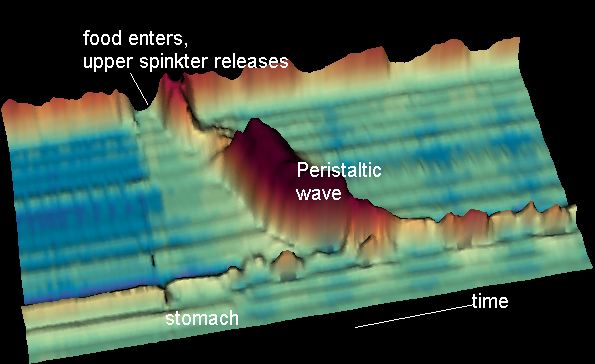
Peristalsis ( , ) is a type of intestinal motility, characterized by symmetry in biology#Radial symmetry, radially symmetrical contraction and relaxation of muscles that propagate in a wave down a tube, in an wikt:anterograde, anterograde direction. Peristalsis is progression of coordinated contraction of involuntary circular muscles, which is preceded by a simultaneous contraction of the longitudinal muscle and relaxation of the circular muscle in the lining of the gut. In much of a digestive tract, such as the human gastrointestinal tract, smooth muscle tissue contracts in sequence to produce a peristaltic wave, which propels a ball of food (called a bolus (digestion), bolus before being transformed into chyme in the stomach) along the tract. The peristaltic movement comprises relaxation of circular smooth muscles, then their contraction behind the chewed material to keep it from moving backward, then longitudinal contraction to push it forward. Earthworms use a similar mec ... [...More Info...] [...Related Items...] OR: [Wikipedia] [Google] [Baidu] |
Bolus (digestion)
In digestion, a bolus () is a ball-like mixture of food and saliva that forms in the mouth during the process of chewing (which is largely an adaptation for plant-eating mammals). It has the same color as the food being eaten, and the saliva gives it an alkaline pH. Under normal circumstances, the bolus is swallowed, and travels down the esophagus to the stomach for digestion. See also * Chyme * Chyle References Digestive system {{Digestive-stub ... [...More Info...] [...Related Items...] OR: [Wikipedia] [Google] [Baidu] |
Pharynx
The pharynx (: pharynges) is the part of the throat behind the human mouth, mouth and nasal cavity, and above the esophagus and trachea (the tubes going down to the stomach and the lungs respectively). It is found in vertebrates and invertebrates, though its structure varies across species. The pharynx carries food to the esophagus and air to the larynx. The flap of cartilage called the epiglottis stops food from entering the larynx. In humans, the pharynx is part of the Digestion, digestive system and the conducting zone of the respiratory system. (The conducting zone—which also includes the nostrils of the Human nose, nose, the larynx, trachea, bronchus, bronchi, and bronchioles—filters, warms, and moistens air and conducts it into the lungs). The human pharynx is conventionally divided into three sections: the nasopharynx, oropharynx, and laryngopharynx (hypopharynx). In humans, two sets of pharyngeal muscles form the pharynx and determine the shape of its lumen (anatomy), ... [...More Info...] [...Related Items...] OR: [Wikipedia] [Google] [Baidu] |
Pharyngobasilar Fascia
The pharyngobasilar fascia is a fascia of the pharynx. It is situated between the mucous and muscular layers of the pharynx. It is formed as a thickening of the pharyngeal mucosa superior to the superior pharyngeal constrictor muscle. It attaches to the basilar part of occipital bone, the petrous part of the temporal bone The petrous part of the temporal bone is pyramid-shaped and is wedged in at the base of the skull between the sphenoid and occipital bones. Directed medially, forward, and a little upward, it presents a base, an apex, three surfaces, and three ... (medial to the pharyngotympanic tube), the (posterior border of the) medial pterygoid plate, and the pterygomandibular raphe. It diminishes in thickness inferiorly. Posteriorly, it is reinforced by the pharyngeal raphe. It reinforces the pharyngeal wall where muscle is deficient. Additional images File:Slide1kuku.JPG, Larynx, pharynx and tongue. Deep dissection, posterior view. File:Slide2kuku.JPG, Larynx, p ... [...More Info...] [...Related Items...] OR: [Wikipedia] [Google] [Baidu] |
Langenbecks Arch Surg
''Langenbeck's Archives of Surgery'' (or in German, ''Langenbecks Archiv für Chirurgie''), established in 1860 as ''Archiv für Klinische Chirurgie'' by founding editor Bernhard von Langenbeck, is the oldest medical journal of surgery in the world. It is the official journal of several European surgical societies: German Society of Surgery, German Society of General and Visceral Surgery, German Association of Endocrine Surgeons, European Society of Endocrine Surgeons, and the German, Austrian and Swiss Surgical Associations for Minimal Invasive Surgery. The journal is currently published by Springer Science+Business Media and the editor-in-chief is Markus W. Büchler (University of Heidelberg). The journal was original published in German, but is exclusively in English since 1998, when it obtained its current English title. Abstracting and indexing The journal is indexed and abstracted in: According to the ''Journal Citation Reports'', the journal has a 2020 impact factor ... [...More Info...] [...Related Items...] OR: [Wikipedia] [Google] [Baidu] |
The Anatomical Record
''The Anatomical Record'' is a peer-reviewed scientific journal covering anatomy. It was established by the American Association of Anatomists in 1906 and is published by John Wiley & Sons. According to the ''Journal Citation Reports'', the journal has a 2020 impact factor of 2.064. See also * List of biology journals This is a list of articles about scientific journals in biology and its various subfields. General Agriculture * Advances in Agronomy * '' Agriculture, Ecosystems & Environment'' * '' Agroecology and Sustainable Food Systems'' * ''Animal'' ... References External links * Anatomy journals Wiley (publisher) academic journals Publications established in 1906 Monthly journals English-language journals 1906 establishments in the United States {{biology-journal-stub ... [...More Info...] [...Related Items...] OR: [Wikipedia] [Google] [Baidu] |
Upper Esophageal Sphincter
The esophagus (American English), oesophagus (British English), or œsophagus ( archaic spelling) ( see spelling difference) all ; : ((o)e)(œ)sophagi or ((o)e)(œ)sophaguses), colloquially known also as the food pipe, food tube, or gullet, is an organ in vertebrates through which food passes, aided by peristaltic contractions, from the pharynx to the stomach. The esophagus is a fibromuscular tube, about long in adults, that travels behind the trachea and heart, passes through the diaphragm, and empties into the uppermost region of the stomach. During swallowing, the epiglottis tilts backwards to prevent food from going down the larynx and lungs. The word ''esophagus'' is from Ancient Greek οἰσοφάγος (oisophágos), from οἴσω (oísō), future form of φέρω (phérō, "I carry") + ἔφαγον (éphagon, "I ate"). The wall of the esophagus from the lumen outwards consists of mucosa, submucosa (connective tissue), layers of muscle fibers between layers of ... [...More Info...] [...Related Items...] OR: [Wikipedia] [Google] [Baidu] |
Middle Pharyngeal Constrictor Muscle
The middle pharyngeal constrictor is a fan-shaped muscle located in the neck. It is one of three pharyngeal constrictor muscles. It is smaller than the inferior pharyngeal constrictor muscle. The middle pharyngeal constrictor originates from the greater cornu and lesser cornu of the hyoid bone, and the stylohyoid ligament. It inserts onto the pharyngeal raphe. It is innervated by a branch of the vagus nerve through the pharyngeal plexus. It acts to propel a bolus downwards along the pharynx towards the esophagus, facilitating swallowing. Structure The middle pharyngeal constrictor is a sheet-like, fan-shaped muscle. The muscle's fibers diverge from their origin: the more inferior fibres descend deep to the inferior pharyngeal constrictor muscle; the middle portion of fibres pass transversely; the more superior fibers ascend and overlap the superior pharyngeal constrictor muscle. Origin Two parts of the middle pharyngeal constrictor muscle are distinguished according to i ... [...More Info...] [...Related Items...] OR: [Wikipedia] [Google] [Baidu] |
Esophagus
The esophagus (American English), oesophagus (British English), or œsophagus (Œ, archaic spelling) (American and British English spelling differences#ae and oe, see spelling difference) all ; : ((o)e)(œ)sophagi or ((o)e)(œ)sophaguses), colloquially known also as the food pipe, food tube, or gullet, is an Organ (anatomy), organ in vertebrates through which food passes, aided by Peristalsis, peristaltic contractions, from the Human pharynx, pharynx to the stomach. The esophagus is a :wiktionary:fibromuscular, fibromuscular tube, about long in adults, that travels behind the trachea and human heart, heart, passes through the Thoracic diaphragm, diaphragm, and empties into the uppermost region of the stomach. During swallowing, the epiglottis tilts backwards to prevent food from going down the larynx and lungs. The word ''esophagus'' is from Ancient Greek οἰσοφάγος (oisophágos), from οἴσω (oísō), future form of φέρω (phérō, "I carry") + ἔφαγον ( ... [...More Info...] [...Related Items...] OR: [Wikipedia] [Google] [Baidu] |


