|
Inferior Gluteal Artery
The inferior gluteal artery (sciatic artery) is a terminal branch of the anterior trunk of the internal iliac artery. It exits the pelvis through the greater sciatic foramen. It is distributed chiefly to the buttock and the back of the thigh. Anatomy Origin It is the smaller of the two terminal branches of the anterior trunk of the internal iliac artery. Course It passes posterior-ward within parietal pelvic fascia. It travels in between the S1 nerve and S2 (or S2-S3) nerve(s). It descends upon the nerves of the sacral plexus and the piriformis muscle, posterior to the internal pudendal artery. It passes through the inferior part of the greater sciatic foramen. It exits the pelvis inferior to the piriformis muscle, between piriformis muscle and coccygeus muscle. It then descends in the interval between the greater trochanter of the femur and tuberosity of the ischium. It is accompanied by the sciatic nerve and the posterior femoral cutaneous nerves, and covered by the glu ... [...More Info...] [...Related Items...] OR: [Wikipedia] [Google] [Baidu] |
Sciatic Nerve
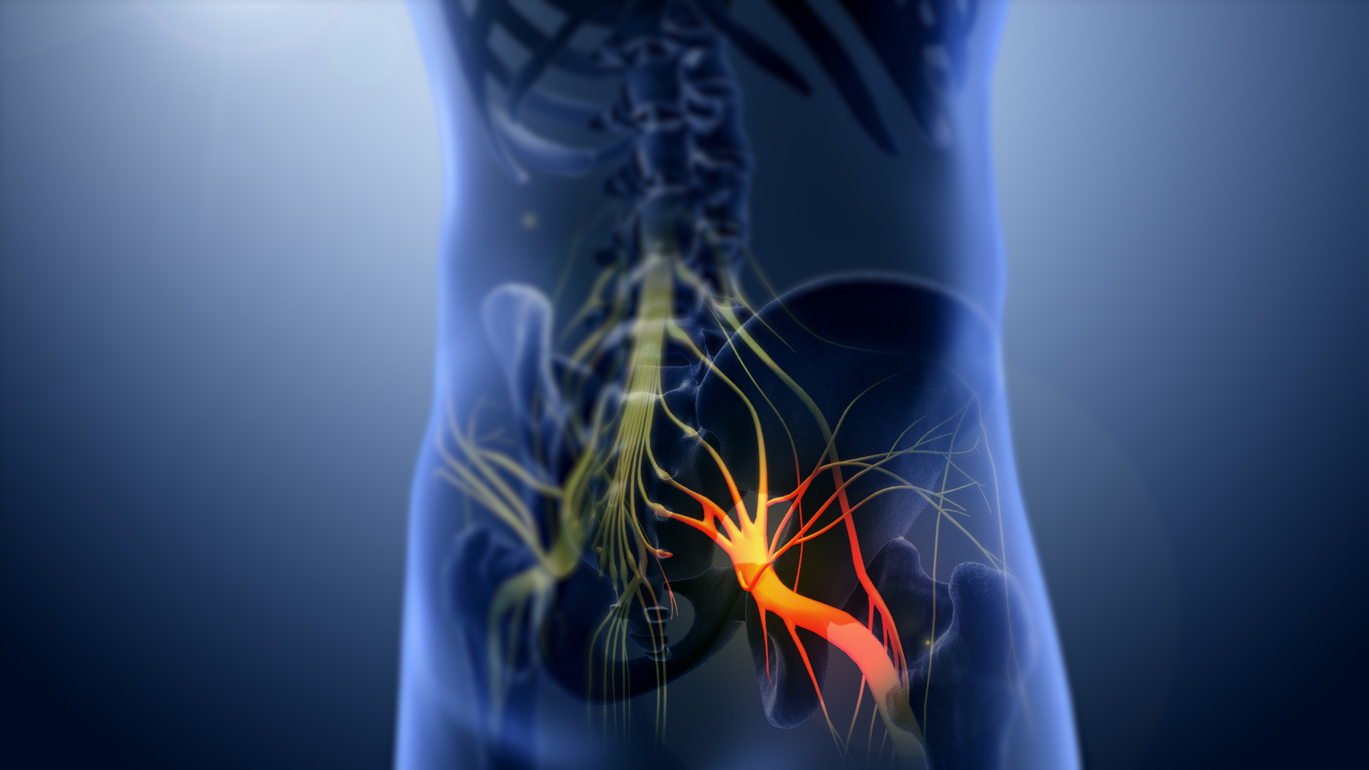
The sciatic nerve, also called the ischiadic nerve, is a large nerve in humans and other vertebrate animals. It is the largest branch of the sacral plexus and runs alongside the hip joint and down the right lower limb. It is the longest and widest single nerve in the human body, going from the top of the leg to the foot on the posterior aspect. The sciatic nerve has no cutaneous branches for the thigh. This nerve provides the connection to the nervous system for the skin of the lateral leg and the whole foot, the muscles of the back of the thigh, and those of the leg and foot. It is derived from Spinal nerve, spinal nerves Lumbar spinal nerve 4, L4 to Sacral spinal nerve 3, S3. It contains Axon, fibres from both the anterior and posterior divisions of the lumbosacral plexus. Structure In humans, the sciatic nerve is formed from the L4 to S3 segments of the sacral plexus, a collection of nerve fibres that emerge from the Sacrum, sacral part of the spinal cord. The lumbosacral trunk ... [...More Info...] [...Related Items...] OR: [Wikipedia] [Google] [Baidu] |
Greater Trochanter
The greater trochanter of the femur is a large, irregular, quadrilateral eminence and a part of the skeletal system. It is directed lateral and medially and slightly posterior. In the adult it is about 2–4 cm lower than the femoral head.Standring, Susan, editor. ''Gray’s Anatomy: The Anatomical Basis of Clinical Practice''. Forty-First edition, Elsevier Limited, 2016, p. 1327. Because the pelvic outlet in the female is larger than in the male, there is a greater distance between the greater trochanters in the female. It has two surfaces and four borders. It is a traction epiphysis. Surfaces The ''lateral surface'', quadrilateral in form, is broad, rough, convex, and marked by a diagonal impression, which extends from the postero-superior to the antero-inferior angle, and serves for the insertion of the tendon of the gluteus medius. Above the impression is a triangular surface, sometimes rough for part of the tendon of the same muscle, sometimes smooth for the interp ... [...More Info...] [...Related Items...] OR: [Wikipedia] [Google] [Baidu] |
Cruciate Anastomosis
The cruciate anastomosis is a circulatory anastomosis in the upper thigh formed by the inferior gluteal artery, the lateral and medial circumflex femoral arteries, the first perforating artery of the deep femoral artery, and the anastomotic branch of the posterior branch of the obturator artery.Henri Rouviere 11Ed The cruciate anastomosis is clinically relevant because if there is a blockage between the femoral artery and external iliac artery, blood can reach the popliteal artery by means of the anastomosis. The route of blood is through the internal iliac, to the inferior gluteal artery, to a perforating branch of the deep femoral artery, to the lateral circumflex femoral artery, then to its descending branch into the superior lateral genicular artery and thus into the popliteal artery. Structure The cruciate anastomosis is so-called because it resembles a cross. Its four components are: * Inferior gluteal artery * Transverse branches of the lateral circumflex femoral ar ... [...More Info...] [...Related Items...] OR: [Wikipedia] [Google] [Baidu] |
Superior Gluteal Artery
The superior gluteal artery is the terminal branch of the posterior division of the internal iliac artery. It exits the pelvis through the greater sciatic foramen before splitting into a superficial branch and a deep branch. Structure Origin The superior gluteal artery is the largest and final branch of the internal iliac artery. It branches from the posterior division of the internal iliac artery; it represents the continuation of the posterior division. Course, relations and branches It is a short artery. It passes posterior-ward between the lumbosacral trunk and the first sacral nerve (S1). Within the pelvis, it gives branches to the iliacus, piriformis, and obturator internus muscles. Just prior to exiting the pelvic cavity, it also gives off a nutrient artery which enters the ilium. It exits the pelvis through the greater sciatic foramen superior to the piriformis muscle, then promptly divides into a superficial branch and a deep branch. Superficial branch The superfi ... [...More Info...] [...Related Items...] OR: [Wikipedia] [Google] [Baidu] |
Biceps Femoris
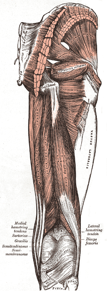
The biceps femoris () is a muscle of the thigh located to the posterior, or back. As its name implies, it consists of two heads; the long head is considered part of the hamstring muscle group, while the short head is sometimes excluded from this characterization, as it only causes knee flexion (but not hip extension) and is activated by a separate nerve (the peroneal, as opposed to the tibial branch of the sciatic nerve). Structure It has two heads of origin: *the ''long head'' arises from the lower and inner impression on the posterior part of the tuberosity of the ischium. This is a common tendon origin with the semitendinosus muscle, and from the lower part of the sacrotuberous ligament. *the ''short head'', arises from the lateral lip of the linea aspera, between the adductor magnus and vastus lateralis extending up almost as high as the insertion of the gluteus maximus, from the lateral prolongation of the linea aspera to within 5 cm. of the lateral condyle; and from ... [...More Info...] [...Related Items...] OR: [Wikipedia] [Google] [Baidu] |
Semitendinosus
The semitendinosus () is a long superficial muscle in the back of the thigh. It is so named because it has a very long tendon of insertion. It lies posteromedially in the thigh, superficial to the semimembranosus. Structure The semitendinosus, remarkable for the great length of its tendon of insertion, is situated at the posterior and medial aspect of the thigh. It arises from the lower and medial impression on the upper part of the tuberosity of the ischium, by a tendon common to it and the long head of the biceps femoris; it also arises from an aponeurosis which connects the adjacent surfaces of the two muscles to the extent of about 7.5 cm. from their origin. The muscle is fusiform and ends a little below the middle of the thigh in a long round tendon which lies along the medial side of the popliteal fossa; it then curves around the medial condyle of the tibia and passes over the medial collateral ligament of the knee-joint In humans and other primates, the knee j ... [...More Info...] [...Related Items...] OR: [Wikipedia] [Google] [Baidu] |
Semimembranosus
The semimembranosus muscle () is the most medial of the three hamstring muscles in the thigh. It is so named because it has a flat tendon of origin. It lies posteromedially in the thigh, deep to the semitendinosus muscle. It extends the hip joint and flexes the knee joint. Structure The semimembranosus muscle, so called from its membranous tendon of origin, is situated at the back and medial side of the thigh. It is wider, flatter, and deeper than the semitendinosus (with which it shares very close insertion and attachment points). The muscle overlaps the upper part of the popliteal vessels. Origin The semimembranosus muscle originates by a thick tendon from the superolateral aspect of the ischial tuberosity. It arises above and medial to the biceps femoris muscle and semitendinosus muscle. The tendon of origin expands into an aponeurosis, which covers the upper part of the anterior surface of the muscle; from this aponeurosis, muscular fibers arise, and converge to another ... [...More Info...] [...Related Items...] OR: [Wikipedia] [Google] [Baidu] |
Hamstring Muscles
A hamstring () is any one of the three posterior thigh muscles in human anatomy between the hip and the knee: from medial to lateral, the semimembranosus, semitendinosus and biceps femoris. Etymology The word "ham" is derived from the Old English “ham” or “hom” meaning the hollow or bend of the knee, from a Germanic base where it meant "crooked". It gained the meaning of the leg of an animal around the 15th century. ''String'' refers to tendons, and thus the hamstrings' string-like tendons felt on either side of the back of the knee. Criteria The common criteria of any hamstring muscles are: # Muscles should originate from ischial tuberosity. # Muscles should be inserted over the knee joint, in the tibia or in the fibula. # Muscles will be innervated by the tibial branch of the sciatic nerve. # Muscle will participate in flexion of the knee joint and extension of the hip joint. Those muscles which fulfill all of the four criteria are called true hamstrings. The adduct ... [...More Info...] [...Related Items...] OR: [Wikipedia] [Google] [Baidu] |
Obturator Internus Muscle
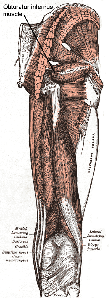
The internal obturator muscle or obturator internus muscle originates on the medial surface of the obturator membrane, the ischium near the membrane, and the rim of the pubis. It exits the pelvic cavity through the lesser sciatic foramen. The internal obturator is situated partly within the lesser pelvis, and partly at the back of the hip-joint. It functions to help laterally rotate femur with hip extension and abduct femur with hip flexion, as well as to steady the femoral head in the acetabulum. Structure Origin The internal obturator muscle arises from the inner surface of the antero-lateral wall of the pelvis. It surrounds the obturator foramen. It is attached to the inferior pubic ramus and ischium, and at the side to the inner surface of the hip bone below and behind the pelvic brim. It reaches from the upper part of the greater sciatic foramen above and behind to the obturator foramen below and in front. It also arises from the pelvic surface of the obturator mem ... [...More Info...] [...Related Items...] OR: [Wikipedia] [Google] [Baidu] |
Perforating Arteries
The perforating arteries are branches of the deep artery of the thigh, usually three in number, so named because they perforate the tendon of the adductor magnus to reach the back of the thigh. They pass backward near the linea aspera of the femur underneath the small tendinous arches of the adductor magnus muscle. The first perforating artery arises from the deep artery of the thigh above the adductor brevis, the second in front of this muscle, and the third immediately below it. First The first perforating artery (''a. perforans prima'') passes posteriorly between the p ectineus and adductor brevis (sometimes it perforates the latter); it then pierces the adductor magnus close to the linea aspera. It gives branches to the adductores brevis and magnus, biceps femoris, and gluteus maximus The gluteus maximus is the main extensor muscle of the hip in humans. It is the largest and outermost of the three gluteal muscles and makes up a large part of the shape and appearance of ea ... [...More Info...] [...Related Items...] OR: [Wikipedia] [Google] [Baidu] |
Gluteus Maximus
The gluteus maximus is the main extensor muscle of the hip in humans. It is the largest and outermost of the three gluteal muscles and makes up a large part of the shape and appearance of each side of the hips. It is the single largest muscle in the human body. Its thick fleshy mass, in a quadrilateral shape, forms the prominence of the buttocks. The other gluteal muscles are the medius and minimus, and sometimes informally these are collectively referred to as the glutes. Its large size is one of the most characteristic features of the muscular system in humans,Norman Eizenberg et al., ''General Anatomy: Principles and Applications'' (2008), p. 17. connected as it is with the power of maintaining the trunk in the erect posture. Other primates have much flatter hips and cannot sustain standing erectly. The muscle is made up of muscle fascicles lying parallel with one another, and are collected together into larger bundles separated by fibrous septa. Structure The gluteus maxi ... [...More Info...] [...Related Items...] OR: [Wikipedia] [Google] [Baidu] |
Posterior Femoral Cutaneous Nerves
The posterior cutaneous nerve of the thigh (also called the posterior femoral cutaneous nerve) is a sensory nerve of the thigh. It is a branch of the sacral plexus. It supplies the skin of the posterior surface of the thigh, leg, buttock, and also the perineum. Unlike most nerves termed "cutaneous" which are subcutaneous, only the terminal branches of this nerve pass into subcutaneous tissue before being distributed to the skin, with most of the nerve itself situated deep to the deep fascia. Structure Origin The posterior cutaneous nerve of the thigh is a branch of the sacral plexus. It arises from the posterior divisions of the S1- S2, and the anterior divisions of S2- S3 sacral spinal nerves. Course It leaves the pelvis through the greater sciatic foramen inferior to the piriformis muscle. It then descends deep to the gluteus maximus muscle, medial or posterior to the sciatic nerve, and alongside the inferior gluteal artery. It descends within the posterior thigh deep ... [...More Info...] [...Related Items...] OR: [Wikipedia] [Google] [Baidu] |


