|
Indocyanine Green Angiography
Indocyanine green angiography (ICGA) is a diagnostic procedure used to examine choroidal blood flow and associated pathology. Indocyanine green (ICG) is a water soluble cyanine dye which shows fluorescence in near-infrared (790–805 nm) range, with peak spectral absorption of 800-810 nm in blood. The near infrared light used in ICGA penetrates ocular pigments such as melanin and xanthophyll, as well as exudates and thin layers of sub-retinal vessels. Age-related macular degeneration is the third main cause of blindness worldwide, and it is the leading cause of blindness in industrialized countries. Indocyanine green angiography is widely used to study choroidal neovascularization in patients with exudative age-related macular degeneration. In nonexudative AMD, ICGA is used in classification of drusen and associated subretinal deposits. Indications Indications for indocyanine green angiography include: * Choroidal neovascularisation (CNV): Indocyanine green angiography i ... [...More Info...] [...Related Items...] OR: [Wikipedia] [Google] [Baidu] |
Choroid
The choroid, also known as the choroidea or choroid coat, is a part of the uvea, the vascular layer of the eye. It contains connective tissues, and lies between the retina and the sclera. The human choroid is thickest at the far extreme rear of the eye (at 0.2 mm), while in the outlying areas it narrows to 0.1 mm. The choroid provides oxygen and nourishment to the outer layers of the retina. Along with the ciliary body and iris, the choroid forms the uveal tract. The structure of the choroid is generally divided into four layers (classified in order of furthest away from the retina to closest): *Haller's layer – outermost layer of the choroid consisting of larger diameter blood vessels; * Sattler's layer – layer of medium diameter blood vessels; * Choriocapillaris – layer of capillaries; and * Bruch's membrane (synonyms: Lamina basalis, Complexus basalis, Lamina vitra) – innermost layer of the choroid. Blood supply There are two circulations of the eye: ... [...More Info...] [...Related Items...] OR: [Wikipedia] [Google] [Baidu] |
Angioid Streaks
Angioid streaks, also called Knapp streaks or Knapp striae, are small breaks in Bruch's membrane, an elastic tissue containing membrane of the retina that may become calcified and crack. Up to 50% of angioid streak cases are idiopathic. It may occur secondary to blunt trauma, or it may be associated with many systemic diseases. The condition is usually asymptomatic, but decrease in vision may occur due to choroidal neovascularization. Clinical features Angioid streaks are often associated with pseudoxanthoma elasticum, but have been found to occur in conjunction with other disorders, including Paget's disease, sickle cell disease and Ehlers–Danlos syndrome. These streaks can have a negative impact on vision due to choroidal neovascularization or choroidal rupture. Also, vision can be impaired if the streaks progress to the fovea and damage the retinal pigment epithelium. Signs Retinal fundus examination may reveal grey or dark red spoke like lesions around optic disk and rad ... [...More Info...] [...Related Items...] OR: [Wikipedia] [Google] [Baidu] |
Eye Procedures
An eye is a sensory organ that allows an organism to perceive visual information. It detects light and converts it into electro-chemical impulses in neurons (neurones). It is part of an organism's visual system. In higher organisms, the eye is a complex optical system that collects light from the surrounding environment, regulates its intensity through a diaphragm, focuses it through an adjustable assembly of lenses to form an image, converts this image into a set of electrical signals, and transmits these signals to the brain through neural pathways that connect the eye via the optic nerve to the visual cortex and other areas of the brain. Eyes with resolving power have come in ten fundamentally different forms, classified into compound eyes and non-compound eyes. Compound eyes are made up of multiple small visual units, and are common on insects and crustaceans. Non-compound eyes have a single lens and focus light onto the retina to form a single image. This type of ey ... [...More Info...] [...Related Items...] OR: [Wikipedia] [Google] [Baidu] |
Eye Examination
An eye examination, commonly known as an eye test, is a series of tests performed to assess Visual acuity, vision and ability to Focus (optics), focus on and discern objects. It also includes other tests and examinations of the human eye, eyes. Eye examinations are primarily performed by an optometrist, ophthalmologist, or an orthoptist. Health care professionals often recommend that all people should have periodic and thorough eye examinations as part of routine primary care, especially since many eye diseases are asymptomatic. Typically, a healthy individual who otherwise has no concerns with their eyes receives an eye exam once in their 20s and twice in their 30s. Eye examinations may detect potentially treatable blindness, blinding eye diseases, ocular manifestation of systemic disease, ocular manifestations of systemic disease, or signs of tumour, tumors or other anomalies of the Human brain, brain. A full eye examination consists of a comprehensive evaluation of medical h ... [...More Info...] [...Related Items...] OR: [Wikipedia] [Google] [Baidu] |
Fluorescein Angiography
Fluorescein angiography (FA), fluorescent angiography (FAG), or fundus fluorescein angiography (FFA) is a technique for examining the circulation of the retina and choroid (parts of the fundus) using a fluorescent dye and a specialized camera. Sodium fluorescein is added into the systemic circulation, the retina is illuminated with blue-green light at a wavelength of 490 nanometers, and an angiogram is obtained by photographing the fluorescent green light that is emitted by the dye. The fluorescein is administered intravenously in intravenous fluorescein angiography (IVFA) and orally in oral fluorescein angiography (OFA). The test is a dye tracing method. The fluorescein dye also reappears in the patient urine, causing the urine to appear darker, and sometimes orange. It can also cause discolouration of the saliva. Fluorescein angiography is one of several health care applications of this dye, all of which have a risk of severe adverse effects. See fluorescein safety in h ... [...More Info...] [...Related Items...] OR: [Wikipedia] [Google] [Baidu] |
Fundus Photography
Fundus photography involves photographing the rear of an eye, also known as the fundus (eye), fundus. Specialized fundus cameras consisting of an intricate microscope attached to a flash (photography), flash enabled camera are used in fundus photography. The main structures that can be visualized on a fundus photo are the central and peripheral retina, optic disc and Macula of retina, macula. Fundus photography can be performed with colored filters, or with specialized dyes including Fluorescein angiography, fluorescein and indocyanine green. The models and technology of fundus photography have advanced and evolved rapidly over the last century. History The concept of fundus photography was first introduced in the mid 19th century, after the introduction of photography in 1839. In 1851, Hermann von Helmholtz introduced the Ophthalmoscopy, Ophthalmoscope, and James Clerk Maxwell presented a colour photography method in 1861. In the early 1860s, Henry Noyes and Abner Mulhollan ... [...More Info...] [...Related Items...] OR: [Wikipedia] [Google] [Baidu] |
Bolus (medicine)
In medicine, a bolus (from Latin '' bolus'', ball) is the administration of a discrete amount of medication, drug, or other compound within a specific time, generally 1–30 minutes, to raise its concentration in blood to an effective level. The administration can be given by injection: intravenously, intramuscularly, intrathecally, subcutaneously, or by inhalation. The article on routes of administration provides more information, as the preceding list of ROAs is not exhaustive. Placement The placement of the bolus dose depends on the systemic levels of the contents desired throughout the body. An intramuscular injection of vaccines allows for a slow release of the antigen to stimulate the body's immune system and to allow time for developing antibodies. Subcutaneous injections are used by heroin addicts (called 'skin popping', referring to the bump formed by the bolus of heroin), to sustain a slow release that staves off withdrawal symptoms without producing euphoria. A ... [...More Info...] [...Related Items...] OR: [Wikipedia] [Google] [Baidu] |
Antecubital Vein
In human anatomy, the cephalic vein (also called the antecubital vein) is a superficial vein in the arm. It is the longest vein of the upper limb. It starts at the anatomical snuffbox from the radial end of the dorsal venous network of hand, and ascends along the radial (lateral) side of the arm before emptying into the axillary vein. At the elbow, it communicates with the basilic vein via the median cubital vein. Anatomy The cephalic vein is situated within the superficial fascia along the anterolateral surface of the biceps. Origin The cephalic vein forms at the roof of the anatomical snuffbox at the radial end of the dorsal venous network of hand. Course and relations From its origin, it ascends up the lateral aspect of the radius. Near the shoulder, the cephalic vein passes between the deltoid and pectoralis major muscles (deltopectoral groove) through the clavipectoral triangle, where it empties into the axillary vein. Anastomoses It communicates with the basili ... [...More Info...] [...Related Items...] OR: [Wikipedia] [Google] [Baidu] |
Pupil
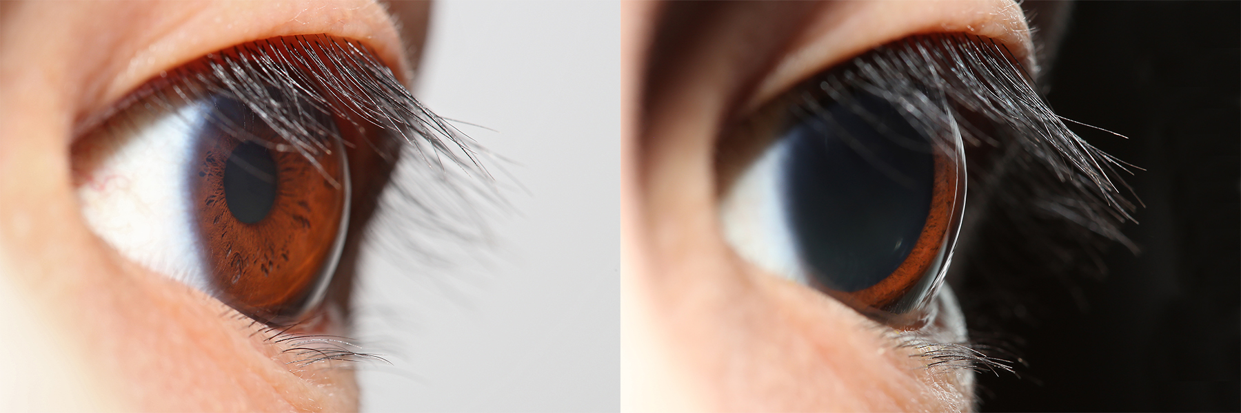
The pupil is a hole located in the center of the iris of the eye that allows light to strike the retina.Cassin, B. and Solomon, S. (1990) ''Dictionary of Eye Terminology''. Gainesville, Florida: Triad Publishing Company. It appears black because light rays entering the pupil are either absorbed by the tissues inside the eye directly, or absorbed after diffuse reflections within the eye that mostly miss exiting the narrow pupil. The size of the pupil is controlled by the iris, and varies depending on many factors, the most significant being the amount of light in the environment. The term "pupil" was coined by Gerard of Cremona. In humans, the pupil is circular, but its shape varies between species; some cats, reptiles, and foxes have vertical slit pupils, goats and sheep have horizontally oriented pupils, and some catfish have annular types. In optical terms, the anatomical pupil is the eye's aperture and the iris is the aperture stop. The image of the pupil as seen from o ... [...More Info...] [...Related Items...] OR: [Wikipedia] [Google] [Baidu] |
Infracyanine Green
Infracyanine green (IFCG) is a cyanine dye used in medical diagnostics especially in ophthalmology. Unlike Indocyanine green (ICG) it is an iodine free dye. Properties Pharmacological properties of infracyanine green are similar to ICG. Since IFCG is iodine free, instead of ICG, it is used in patients with iodine allergy. Though it is impossible to be allergic to iodine as this would be incompatible with human life. It has a peak spectral absorption between 600 nm to 700 nm. IFCG can be dissolved in 5% glucose solution instead of pure water, which makes it less cytotoxic in rabbits macular applications. Uses Infracyanine green which stains the Internal limiting membrane layer of retina, is used to see structures to be removed during vitreoretinal surgery. Toxicity At a concentration above 0.05% IFCG may induce acute and chronic toxicities. But the retinal phototoxicity Phototoxicity, also called photoirritation, is a chemically induced skin irritation, requiring ... [...More Info...] [...Related Items...] OR: [Wikipedia] [Google] [Baidu] |
Optic Disc
The optic disc or optic nerve head is the point of exit for ganglion cell axons leaving the eye. Because there are no rods or cones overlying the optic disc, it corresponds to a small blind spot in each eye. The ganglion cell axons form the optic nerve after they leave the eye. The optic disc represents the beginning of the optic nerve and is the point where the axons of retinal ganglion cells come together. The optic disc in a normal human eye carries 1–1.2 million afferent nerve fibers from the eye toward the brain. The optic disc is also the entry point for the major arteries that supply the retina with blood, and the exit point for the veins from the retina. Structure The optic disc is located 3 to 4 mm to the nasal side of the fovea. It is a vertical oval, with average dimensions of 1.76mm horizontally by 1.92mm vertically. There is a central depression, of variable size, called the optic cup. This depression can be a variety of shapes from a shallow indent ... [...More Info...] [...Related Items...] OR: [Wikipedia] [Google] [Baidu] |
Acute Idiopathic Blind Spot Enlargement Syndrome
Acute idiopathic blind spot enlargement syndrome (AIBSE) is a rare eye disease affecting the retina of the eye. It is basically a type of retinopathy which affects females more than males. Currently there is no treatment for this condition, but, it is usually self limiting. Cause Though exact etiology of AIBSE syndrome is unknown, studies shows that viral illness like influenza and vaccinations like MMR may trigger the condition. Demographics AIBSE syndrome affects females more than males. Higher incidence is seen in Caucasian people. Signs and Symptoms Enlargement of blind spot area in the visual field of the eye is the main sign and acute onset photopsia is the main symptom of AIBSE syndrome. Other symptoms include monocular scotoma and reduced light perception. Diagnosis Diagnostic techniques like ophthalmoscopy, visual field test, optical coherence tomography, fluorescein angiography, multifocal electroretinography and electrophysiology may be used in diagnosing AIBSE syn ... [...More Info...] [...Related Items...] OR: [Wikipedia] [Google] [Baidu] |


