|
Iliolumbar Ligament
The iliolumbar ligament is a strong ligament which attaches medially to the transverse process of the 5th lumbar vertebra, and laterally to back of the inner lip of the iliac crest (upper margin of ilium). Anatomy The ligament extends inferolaterally from its medial attachment, radiating laterally. It represents the thickened inferior border of anterior and middle layers of thoracolumbar fascia. Inferiorly, the ligament is partially continuous with the lumbosacral ligament (which may be considered an inferior subdivision of the iliolumbar ligament). Attachments Medial The ligament's medial attachment is at the apex and anteroinferior aspect of the transverse process of lumbar vertebra L5 (and occasionally an additional weak attachment at the transverse process of L4). Lateral Laterally, the ligament attaches onto the posterior part of the inner lip of the iliac crest. More precisely, its lateral attachment is by two main bands: * the superior band (which is one of th ... [...More Info...] [...Related Items...] OR: [Wikipedia] [Google] [Baidu] |
Pelvis
The pelvis (: pelves or pelvises) is the lower part of an Anatomy, anatomical Trunk (anatomy), trunk, between the human abdomen, abdomen and the thighs (sometimes also called pelvic region), together with its embedded skeleton (sometimes also called bony pelvis or pelvic skeleton). The pelvic region of the trunk includes the bony pelvis, the pelvic cavity (the space enclosed by the bony pelvis), the pelvic floor, below the pelvic cavity, and the perineum, below the pelvic floor. The pelvic skeleton is formed in the area of the back, by the sacrum and the coccyx and anteriorly and to the left and right sides, by a pair of hip bones. The two hip bones connect the spine with the lower limbs. They are attached to the sacrum posteriorly, connected to each other anteriorly, and joined with the two femurs at the hip joints. The gap enclosed by the bony pelvis, called the pelvic cavity, is the section of the body underneath the abdomen and mainly consists of the reproductive organs and ... [...More Info...] [...Related Items...] OR: [Wikipedia] [Google] [Baidu] |
Psoas Major
The psoas major ( or ; from ) is a long fusiform muscle located in the lateral lumbar region between the vertebral column and the brim of the lesser pelvis. It joins the iliacus muscle to form the iliopsoas. In other animals, this muscle is equivalent to the tenderloin. Structure The psoas major is divided into a superficial and a deep part. The deep part originates from the transverse processes of lumbar vertebrae L1–L5. The superficial part originates from the lateral surfaces of the last thoracic vertebra, lumbar vertebrae L1–L4, and the neighboring intervertebral discs. The lumbar plexus lies between the two layers. Together, the iliacus muscle and the psoas major form the iliopsoas, which is surrounded by the iliac fascia. The iliopsoas runs across the iliopubic eminence through the muscular lacuna to its insertion on the lesser trochanter of the femur. The iliopectineal bursa separates the tendon of the iliopsoas muscle from the external surface of the hip-j ... [...More Info...] [...Related Items...] OR: [Wikipedia] [Google] [Baidu] |
Interspinous Ligament
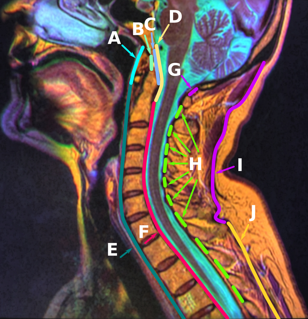
The interspinous ligaments (interspinal ligaments) are thin, membranous ligaments that connect adjoining spinous processes of the vertebra in the spine. They take the form of relatively weak sheets of fibrous tissue and are well developed only in the lumbar region. They extend from the root to the apex of each spinous process. They meet the ligamenta flava anteriorly, and blend with the supraspinous ligament posteriorly at the apexes of the spinal processes. The function of the interspinous ligaments is to limit ventral flexion of the spine and sliding movement of the vertebrae. The ligaments are narrow and elongated in the thoracic region. They are broader, thicker, and quadrilateral in form in the lumbar region. They are only slightly developed in the neck; in the neck, they are often considered part of the nuchal ligament The nuchal ligament is a ligament at the back of the neck that is continuous with the supraspinous ligament. Structure The nuchal ligament extends fr ... [...More Info...] [...Related Items...] OR: [Wikipedia] [Google] [Baidu] |
Ligamenta Flava
The ligamenta flava (: ligamentum flavum, Latin for ''yellow ligament'') are a series of ligaments that connect the ventral parts of the laminae of adjacent vertebrae. They help to preserve upright posture, preventing hyperflexion, and ensuring that the vertebral column straightens after flexion. Hypertrophy can cause spinal stenosis. They appear yellowish in colour due to their high elastic fibre content. Anatomy Each ligamentum flavum connects the laminae of two adjacent vertebrae. They attach to the anterior portion of the upper lamina above, and the posterior portion of the lower lamina below. They begin with the junction of the axis and third cervical vertebra, continuing down to the junction of the 5th lumbar vertebra and the sacrum. In the neck region the ligaments are thin, but broad and long; they are thicker in the thoracic region, and thickest in the lumbar region. They are thinnest between the atlas bone (C1) and the axis bone (C2), and may be absent in some ... [...More Info...] [...Related Items...] OR: [Wikipedia] [Google] [Baidu] |
Anterior Longitudinal Ligament
The anterior longitudinal ligament is a ligament that extends across the anterior/ventral aspect of the vertebral bodies and intervertebral discs the spine. It may be partially cut to treat certain abnormal curvatures in the vertebral column, such as kyphosis. Anatomy The anterior longitudinal ligament extends superoinferiorly between the basiocciput of the skull and the anterior tubercle of the atlas (cervical vertebra C1) superiorly, and the superior part of the sacrum inferiorly; inferiorly, it ends at the sacral promontory. It broadens inferiorly. Inferiorly, it becomes continuous with the anterior sacrococcygeal ligament. Superiorly, between the skull and atlas, the ligament is continuous laterally with the anterior atlantooccipital membrane. The ligament is thick and slightly more narrow over the vertebral bodies and thinner but slightly wider over the intervertebral discs. It tends to be narrower and thicker around thoracic vertebrae, and wider and thinner around ... [...More Info...] [...Related Items...] OR: [Wikipedia] [Google] [Baidu] |
Posterior Longitudinal Ligament
The posterior longitudinal ligament is a ligament connecting the posterior surfaces of the vertebral bodies of all of the vertebrae of humans. It weakly prevents hyperflexion of the vertebral column. It also prevents posterior spinal disc herniation, although problems with the ligament can cause it. Anatomy The posterior longitudinal ligament is situated within the vertebral canal. It extends across the posterior surfaces of the bodies of the vertebrae. It extends superoinferiorly between the body of the axis superiorly, and (sources differ) the sacrum and possibly the coccyx or upper sacral canal inferiorly. It is continuous with the tectorial membrane of atlanto-axial joint superiorly, and with the deep dorsal sacrococcygeal ligament inferiorly. The ligament gradually grows narrower inferiorly. The ligament is thicker in the thoracic than in the cervical and lumbar regions. In the thoracic and lumbar regions, it presents a series of dentations with intervening concave ma ... [...More Info...] [...Related Items...] OR: [Wikipedia] [Google] [Baidu] |
Lateral Lumbosacral Ligament
The lumbosacral ligament or lateral lumbosacral ligament is a ligament that helps to stabilise the lumbosacral joint. The ligament's medial attachment is at (the inferior border of) transverse process of lumbar vertebra L5; its lateral attachment is at the ala of sacrum. The lumbosacral ligament extends obliquely inferior-ward from its medial attachment. Superiorly, it is partially continuous with the inferior margin of the iliolumbar ligament (the lumbosarcal ligament can be considered an inferior subdivision of the iliolumbar ligament). Research According to a cadaveric study, the lumbosacral ligament forms a roof of a lumbosacral tunnel which is traversed by the (ipsilateral) lumbar spinal nerve L5; the tunnel may be the site of extraforaminal nerve entrapment due to mass effect ''Mass Effect'' is a military science fiction media franchise created by Casey Hudson. The franchise depicts a distant future where humanity and several alien civilizations have colonized the ... [...More Info...] [...Related Items...] OR: [Wikipedia] [Google] [Baidu] |
Lumbosacral Joint
The lumbosacral joint is a joint of the body, between the last lumbar vertebra and the first sacral segment of the vertebral column. In some ways, calling it a "joint" (singular) is a misnomer, since the lumbosacral junction includes a disc between the lower lumbar vertebral body and the uppermost sacral vertebral body, as well as two lumbosacral facet joints (right and left zygapophysial joint The facet joints (also zygapophysial joints, zygapophyseal, apophyseal, or Z-joints) are a set of synovial, plane joints between the articular processes of two adjacent vertebrae. There are two facet joints in each spinal motion segment and ...s). References Bones of the vertebral column Sacrum {{musculoskeletal-stub ... [...More Info...] [...Related Items...] OR: [Wikipedia] [Google] [Baidu] |
Iliopectineal Line
The iliopectineal line is the border of the iliopubic eminence. It can be defined as a compound structure of the arcuate line (from the ilium) and pectineal line (from the pubis). With the sacral promontory, it makes up the linea terminalis The linea terminalis or innominate line consists of the pubic crest, pectineal line (pecten pubis), the arcuate line, the sacral ala, and the sacral promontory. It is the pelvic brim, which is the edge of the pelvic inlet. The pelvic inlet i .... The Iliopectineal line divides the pelvis into the pelvis major (false pelvis) above and the pelvis minor (true pelvis) below. References {{Pelvis Bones of the pelvis ... [...More Info...] [...Related Items...] OR: [Wikipedia] [Google] [Baidu] |
Iliac Fossa
The iliac fossa is a large, smooth, concave surface on the internal surface of the Ilium (bone), ilium (part of the three fused bones making the hip bone). Structure The iliac fossa is bounded above by the iliac crest, and below by the Arcuate line (ilium), arcuate line. It is bordered in front and behind by the anterior and posterior borders of the Ilium (bone), ilium. The iliac fossa gives origin to the iliacus muscle. The obturator nerve passes around the iliac fossa. It is perforated at its inner part by a nutrient canal. Below it there is a smooth, rounded border, the arcuate line (ilium), arcuate line, which runs anterior, inferior, and medial. When the "left" or "right" adjective is used (e.g. "right iliac fossa"), the iliac fossa usually means one of the groin, inguinal regions of the Quadrants and regions of abdomen, nine regions of the abdomen. Additional images File:Anterior Hip Muscles 2.PNG, The iliacus and nearby muscles File:Slide5AA.JPG, Iliac fossa File:Slid ... [...More Info...] [...Related Items...] OR: [Wikipedia] [Google] [Baidu] |
Anterior Sacroiliac Ligament
The Anterior Sacroiliac Ligament (ASL) forms from a thickened part of the anterior joint capsule, and consists of numerous thin bands which form a smooth sheet of dense connective tissue spanning from the anterior surface of the lateral part of the sacrum to the margin of the auricular surface of the ilium and to thpreauricular sulcus It is also called the Ventral Sacroiliac Ligament. The Anterior Sacroiliac Ligament stretches between the ventral surfaces of the sacral alar and ilium. The ASL is the thinnest sacroiliac joint ligament and it is generally larger in males. See also *Posterior sacroiliac ligament The posterior sacroiliac ligament is situated in a deep depression between the sacrum and ilium behind; it is strong and forms the chief bond of union between the bone A bone is a rigid organ that constitutes part of the skeleton in mo ... References External links * () Ligaments of the torso Ligaments {{ligament-stub Pelvis ... [...More Info...] [...Related Items...] OR: [Wikipedia] [Google] [Baidu] |
Quadratus Lumborum
The quadratus lumborum muscle, informally called the ''QL'', is a paired muscle of the left and right posterior abdominal wall. It is the deepest abdominal muscle, and commonly referred to as a back muscle. Each muscle of the pair is an irregular quadrilateral in shape, hence the name. The quadratus lumborum muscles originate from the wings of the ilium; their insertions are on the transverse processes of the upper four lumbar vertebrae plus the lower posterior border of the twelfth rib. Contraction of one of the pair of muscles causes lateral flexion of the lumbar spine, ''elevation'' of the pelvis, or both. Contraction of both causes ''extension'' of the lumbar spine. A disorder of the quadratus lumborum muscles is pain due to muscle fatigue from constant contraction due to prolonged sitting, such as at a computer or in a car.Core Topics in Pain, p. 131, Anita Holdcraft and Sian Jaggar, 2005. Kyphosis and weak gluteal muscles can also contribute to the likelihood of quadratu ... [...More Info...] [...Related Items...] OR: [Wikipedia] [Google] [Baidu] |

