|
Hepatic Hydrothorax
Hepatic hydrothorax is a rare form of pleural effusion that occurs in people with Liver Cirrhosis, liver cirrhosis. It is defined as an effusion of over 500 mL in people with liver cirrhosis that is not caused by heart, lung, or pleural disease. It is found in 5–10% of people with liver cirrhosis and 2–3% of people with pleural effusions. In cases of decompensated liver cirrhosis, prevalence rises significantly up to 90%. Over 85% of cases occurring on the right, 13% on the left, and 2% on both. Although it is most common in people with severe ascites, it can also occur in people with mild or no ascites. Symptoms are not specific and mostly involve the respiratory system. The condition is diagnosed based on the existence of liver cirrhosis and fluid build-up in the abdomen (ascites) and analysis of the fluid. The fluid has a Transudate, low protein content. Mainly, the condition is treated by medical management, such as diet adjustment and usage of Diuretic, diuretics. Wh ... [...More Info...] [...Related Items...] OR: [Wikipedia] [Google] [Baidu] |
Pleural Effusion
A pleural effusion is accumulation of excessive fluid in the pleural space, the potential space that surrounds each lung. Under normal conditions, pleural fluid is secreted by the parietal pleural capillaries at a rate of 0.6 millilitre per kilogram weight per hour, and is cleared by lymphatic absorption leaving behind only 5–15 millilitres of fluid, which helps to maintain a functional vacuum between the parietal and visceral pleurae. Excess fluid within the pleural space can impair inhalation, inspiration by upsetting the functional vacuum and hydrostatically increasing the resistance against lung expansion, resulting in a fully or partially collapsed lung. Various kinds of fluid can accumulate in the pleural space, such as serous fluid (hydrothorax), blood (hemothorax), pus (pyothorax, more commonly known as pleural empyema), chyle (chylothorax), or very rarely urine (urinothorax) or feces (coprothorax). When unspecified, the term "pleural effusion" normally refers to hydro ... [...More Info...] [...Related Items...] OR: [Wikipedia] [Google] [Baidu] |
Scintigraphy
Scintigraphy (from Latin ''scintilla'', "spark"), also known as a gamma scan, is a diagnostic test in nuclear medicine, where radioisotopes attached to drugs that travel to a specific organ or tissue (radiopharmaceuticals) are taken internally and the emitted gamma ray, gamma radiation is captured by gamma cameras, which are external detectors that form two-dimensional images in a process similar to the capture of radiography, x-ray images. In contrast, Single photon emission computed tomography, SPECT and positron emission tomography (PET) form 3-dimensional images and are therefore classified as separate techniques from scintigraphy, although they also use gamma cameras to detect internal radiation. Scintigraphy is unlike a diagnostic X-ray where external radiation is passed through the body to form an image. Process Scintillography is an Molecular imaging, imaging method of nuclear events provoked by collisions or charged current interactions among nuclear particles or ioniz ... [...More Info...] [...Related Items...] OR: [Wikipedia] [Google] [Baidu] |
MELD Score
The Model for End-Stage Liver Disease, or MELD, is a scoring system for assessing the severity of chronic liver disease. It was initially developed to predict mortality within three months of surgery in patients who had undergone a transjugular intrahepatic portosystemic shunt (TIPS) procedure, and was subsequently found to be useful in determining prognosis and prioritizing for receipt of a liver transplant. This score is now used by the United Network for Organ Sharing (UNOS) and Eurotransplant for prioritizing allocation of liver transplants instead of the older Child-Pugh score. Determination MELD uses the patient's values for serum bilirubin, serum creatinine, and the international normalized ratio for prothrombin time (INR) to predict survival. It is calculated according to the following formula: \mathrm \overset 3.78\times \ln \left(\text\right) + 11.2 \times \ln(\text) + 9.57\times \ln\left( \text\right) + 6.43 MELD scores are reported as whole numbers, so the result ... [...More Info...] [...Related Items...] OR: [Wikipedia] [Google] [Baidu] |
Paracentesis
Paracentesis (from Ancient Greek, Greek κεντάω, "to pierce") is a form of body fluid sampling procedure, generally referring to peritoneocentesis (also called laparocentesis or abdominal paracentesis) in which the peritoneal cavity is punctured by a needle to sample peritoneal fluid. The procedure is used to remove fluid from the peritoneal cavity, particularly if this cannot be achieved with medication. The most common indication is ascites that has developed in people with cirrhosis. Indications It is used for a number of reasons: * to relieve abdominal pressure from ascites * to diagnose spontaneous bacterial peritonitis and other infections (e.g. abdominal TB) * to diagnose metastatic cancer * to diagnose blood in peritoneal space in trauma Paracentesis for ascites The procedure is often performed in a doctor's office or an outpatient clinic. In an expert's hands, it is usually very safe, although there is a small risk of infection, excessive bleeding or perforating ... [...More Info...] [...Related Items...] OR: [Wikipedia] [Google] [Baidu] |
Hemothorax
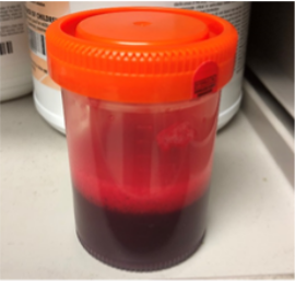
A hemothorax (derived from hemo- lood+ thorax hest plural ''hemothoraces'') is an accumulation of blood within the pleural cavity. The symptoms of a hemothorax may include chest pain and difficulty breathing, while the clinical signs may include reduced breath sounds on the affected side and a rapid heart rate. Hemothoraces are usually caused by an injury, but they may occur spontaneously due to cancer invading the pleural cavity, as a result of a blood clotting disorder, as an unusual manifestation of endometriosis, in response to pneumothorax, or rarely in association with other conditions. Hemothoraces are usually diagnosed using a chest X-ray, but they can be identified using other forms of imaging including ultrasound, a CT scan, or an MRI. They can be differentiated from other forms of fluid within the pleural cavity by analysing a sample of the fluid, and are defined as having a hematocrit of greater than 50% that of the person's blood. Hemothoraces may be treated b ... [...More Info...] [...Related Items...] OR: [Wikipedia] [Google] [Baidu] |
UKELD
The United Kingdom Model for End-Stage Liver Disease or UKELD is a medical scoring system used to predict the prognosis of patients with chronic liver disease. It is used in the United Kingdom to help determine the need for liver transplantation. It was developed from the MELD score, incorporating the serum sodium level. Determination The UKELD score is calculated from the patient's INR, serum creatinine, serum bilirubin and serum sodium, according to the formula: (5.395 \times \ln INR) + (1.485 \times \ln creatinine) + (3.13 \times \ln bilirubin) - (81.565 \times \ln Na) + 435 Interpretation Higher UKELD scores equate to higher one-year mortality risk. A UKELD score of 49 indicates a 9% one-year risk of mortality, and is the minimum score required to be added to the liver transplant waiting list in the U.K. A UKELD score of 60 indicates a 50% chance of one-year survival. History The UKELD score was developed in 2008 to aid in the selection of patients for liver transplan ... [...More Info...] [...Related Items...] OR: [Wikipedia] [Google] [Baidu] |
Fluoroscopic Image Of Transjugular Intrahepatic Portosystemic Shunt (TIPS) In Progress
Fluoroscopy (), informally referred to as "fluoro", is an imaging technique that uses X-rays to obtain real-time moving images of the interior of an object. In its primary application of medical imaging, a fluoroscope () allows a surgeon to see the internal structure and function of a patient, so that the pumping action of the heart or the motion of swallowing, for example, can be watched. This is useful for both diagnosis and therapy and occurs in general radiology, interventional radiology, and image-guided surgery. In its simplest form, a fluoroscope consists of an X-ray source and a fluorescent screen, between which a patient is placed. However, since the 1950s most fluoroscopes have included X-ray image intensifiers and cameras as well, to improve the image's visibility and make it available on a remote display screen. For many decades, fluoroscopy tended to produce live pictures that were not recorded, but since the 1960s, as technology improved, recording and playback beca ... [...More Info...] [...Related Items...] OR: [Wikipedia] [Google] [Baidu] |
Hepatic Hydrothorax After Treatment
The liver is a major metabolic organ exclusively found in vertebrates, which performs many essential biological functions such as detoxification of the organism, and the synthesis of various proteins and various other biochemicals necessary for digestion and growth. In humans, it is located in the right upper quadrant of the abdomen, below the diaphragm and mostly shielded by the lower right rib cage. Its other metabolic roles include carbohydrate metabolism, the production of a number of hormones, conversion and storage of nutrients such as glucose and glycogen, and the decomposition of red blood cells. Anatomical and medical terminology often use the prefix ''hepat-'' from ἡπατο-, from the Greek word for liver, such as hepatology, and hepatitis The liver is also an accessory digestive organ that produces bile, an alkaline fluid containing cholesterol and bile acids, which emulsifies and aids the breakdown of dietary fat. The gallbladder, a small hollow pouch that sit ... [...More Info...] [...Related Items...] OR: [Wikipedia] [Google] [Baidu] |


