|
Hemangioblastoma
Hemangioblastomas, or haemangioblastomas, are vascular tumors of the central nervous system that originate from the vascular system, usually during middle age. Sometimes, these tumors occur in other sites such as the spinal cord and retina. They may be associated with other diseases such as polycythemia (increased blood cell count), pancreatic cysts and Von Hippel–Lindau syndrome (VHL syndrome). Hemangioblastomas are most commonly composed of stromal cells in small blood vessels and usually occur in the cerebellum, brainstem or spinal cord. They are classed as grade I tumors under the World Health Organization's classification system. Presentation Complications Hemangioblastomas can cause an polycythemia, abnormally high number of red blood cells in the bloodstream due to ectopic production of the hormone erythropoietin as a paraneoplastic syndrome. Pathogenesis Hemangioblastomas are composed of endothelial cells, pericytes and stromal cells. In VHL syndrome the Von Hippel–Lin ... [...More Info...] [...Related Items...] OR: [Wikipedia] [Google] [Baidu] |
Vascular Tumor
A vascular tumor is a vascular anomaly where a tumor forms from cells that make blood or lymph vessels; a soft tissue growth that can be either benign or malignant. Examples of vascular tumors include hemangiomas, hemangioendotheliomas, Kaposi's sarcomas, angiosarcomas, and hemangioblastomas. An angioma refers to any type of benign vascular tumor. Some vascular tumors can be associated with serious blood-clotting disorders, making correct diagnosis critical. A vascular tumor may be described in terms of being ''highly vascularized'', or ''poorly vascularized'', referring to the degree of blood supply to the tumor. Classification Vascular tumors make up one of the classifications of vascular anomalies. The other grouping is vascular malformations. Vascular tumors can be further subclassified as being benign, borderline or aggressive, and malignant. Vascular tumors are described as ''proliferative'', and vascular malformations as ''nonproliferative''. Types A vascular t ... [...More Info...] [...Related Items...] OR: [Wikipedia] [Google] [Baidu] |
Micrograph
A micrograph is an image, captured photographically or digitally, taken through a microscope or similar device to show a magnify, magnified image of an object. This is opposed to a macrograph or photomacrograph, an image which is also taken on a microscope but is only slightly magnified, usually less than 10 times. Micrography is the practice or art of using microscopes to make photographs. A photographic micrograph is a photomicrograph, and one taken with an electron microscope is an electron micrograph. A micrograph contains extensive details of microstructure. A wealth of information can be obtained from a simple micrograph like behavior of the material under different conditions, the phases found in the system, failure analysis, grain size estimation, elemental analysis and so on. Micrographs are widely used in all fields of microscopy. Types Photomicrograph A light micrograph or photomicrograph is a micrograph prepared using an optical microscope, a process referred to ... [...More Info...] [...Related Items...] OR: [Wikipedia] [Google] [Baidu] |
Hormone
A hormone (from the Ancient Greek, Greek participle , "setting in motion") is a class of cell signaling, signaling molecules in multicellular organisms that are sent to distant organs or tissues by complex biological processes to regulate physiology and behavior. Hormones are required for the normal development of animals, plants and fungi. Due to the broad definition of a hormone (as a signaling molecule that exerts its effects far from its site of production), numerous kinds of molecules can be classified as hormones. Among the substances that can be considered hormones, are eicosanoids (e.g. prostaglandins and thromboxanes), steroids (e.g. Estrogen, oestrogen and brassinosteroid), amino acid derivatives (e.g. epinephrine and auxin), protein or peptides (e.g. insulin and CLE peptides), and gases (e.g. ethylene and nitric oxide). Hormones are used to communicate between organ (anatomy), organs and Tissue (biology), tissues. In vertebrates, hormones are responsible for regulating ... [...More Info...] [...Related Items...] OR: [Wikipedia] [Google] [Baidu] |
Transforming Growth Factor Alpha
Transforming growth factor alpha (TGF-α) is a protein that in humans is encoded by the TGFA gene. As a member of the epidermal growth factor (EGF) family, TGF-α is a mitogenic polypeptide. The protein becomes activated when binding to receptors capable of protein kinase activity for cellular signaling. TGF-α is a transforming growth factor that is a ligand for the epidermal growth factor receptor, which activates a signaling pathway for cell proliferation, differentiation and development. This protein may act as either a transmembrane-bound ligand or a soluble ligand. This gene has been associated with many types of cancers, and it may also be involved in some cases of cleft lip/palate. Synthesis TGF-α is synthesized internally as part of a 160 (human) or 159 (rat) amino acid transmembrane precursor. The precursor is composed of an extracellular domain containing a hydrophobic transmembrane domain, 50 amino acids of TGF-α, and a 35-residue-long cytoplasmic domain. In its s ... [...More Info...] [...Related Items...] OR: [Wikipedia] [Google] [Baidu] |
PDGFRB
Platelet-derived growth factor receptor beta is a protein that in humans is encoded by the ''PDGFRB'' gene. Mutations in PDGFRB are mainly associated with the clonal eosinophilia class of malignancies. Gene The ''PDGFRB'' gene is located on human chromosome 5 at position q32 (designated as 5q32) and contains 25 exons. The gene is flanked by the genes for granulocyte-macrophage colony-stimulating factor and Colony stimulating factor 1 receptor (also termed macrophage-colony stimulating factor receptor), all three of which may be lost together by a single deletional mutation thereby causing development of the 5q-syndrome. Other genetic abnormalities in ''PDGFRB'' lead to various forms of potentially malignant bone marrow disorders: small deletions in and chromosome translocations causing fusions between ''PDGFRB'' and any one of at least 30 genes can cause Myeloproliferative neoplasms that commonly involve eosinophilia, eosinophil-induced organ injury, and possible progress ... [...More Info...] [...Related Items...] OR: [Wikipedia] [Google] [Baidu] |
VEGF
Vascular endothelial growth factor (VEGF, ), originally known as vascular permeability factor (VPF), is a signal protein produced by many cells that stimulates the formation of blood vessels. To be specific, VEGF is a sub-family of growth factors, the platelet-derived growth factor family of cystine-knot growth factors. They are important signaling proteins involved in both vasculogenesis (the '' de novo'' formation of the embryonic circulatory system) and angiogenesis (the growth of blood vessels from pre-existing vasculature). It is part of the system that restores the oxygen supply to tissues when blood circulation is inadequate such as in hypoxic conditions. Serum concentration of VEGF is high in bronchial asthma and diabetes mellitus. VEGF's normal function is to create new blood vessels during embryonic development, new blood vessels after injury, muscle following exercise, and new vessels (collateral circulation) to bypass blocked vessels. It can contribute to disease. So ... [...More Info...] [...Related Items...] OR: [Wikipedia] [Google] [Baidu] |
Proteosome
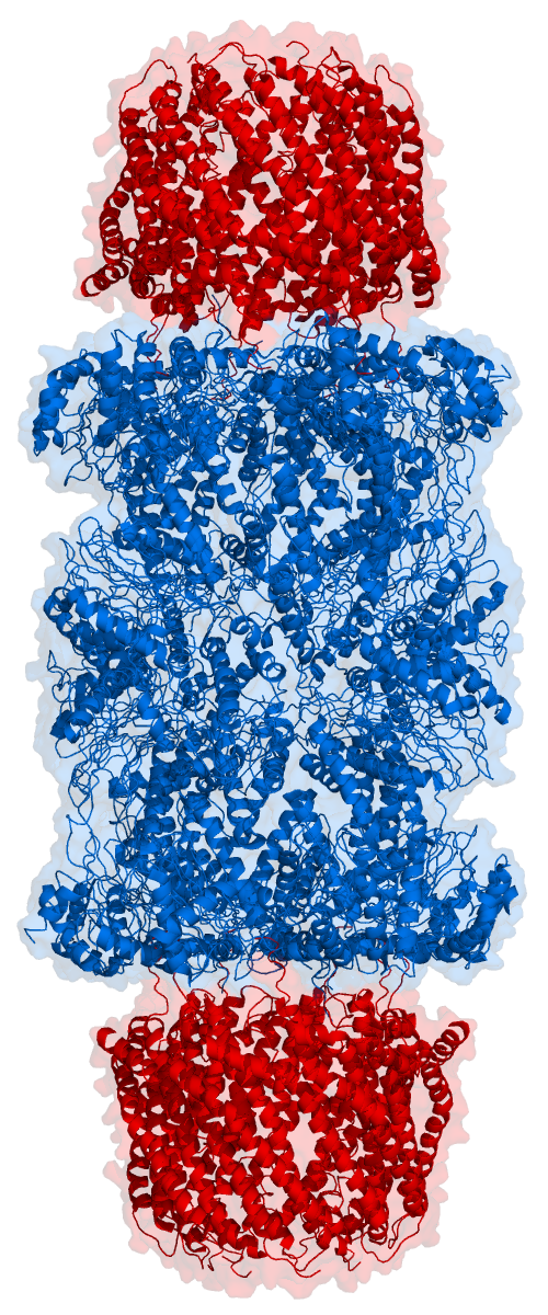
Proteasomes are essential protein complexes responsible for the degradation of proteins by proteolysis, a chemical reaction that breaks peptide bonds. Enzymes that help such reactions are called proteases. Proteasomes are found inside all eukaryotes and archaea, and in some bacteria. In eukaryotes, proteasomes are located both in the cell nucleus, nucleus and in the cytoplasm. The proteasomal degradation pathway is essential for many cellular processes, including the cell cycle, the regulation of gene expression, and responses to oxidative stress. The importance of proteolytic degradation inside cells and the role of ubiquitin in proteolytic pathways was acknowledged in the award of the 2004 Nobel Prize in Chemistry to Aaron Ciechanover, Avram Hershko and Irwin Rose. The core 20S proteasome (blue in the adjacent figure) is a cylindrical, compartmental protein complex of four stacked rings forming a central pore. Each ring is composed of seven individual proteins. The inner t ... [...More Info...] [...Related Items...] OR: [Wikipedia] [Google] [Baidu] |
Ubiquitin
Ubiquitin is a small (8.6 kDa) regulatory protein found in most tissues of eukaryotic organisms, i.e., it is found ''ubiquitously''. It was discovered in 1975 by Gideon Goldstein and further characterized throughout the late 1970s and 1980s. Four genes in the human genome code for ubiquitin: UBB, UBC, UBA52 and RPS27A. The addition of ubiquitin to a substrate protein is called ubiquitylation (or ubiquitination or ubiquitinylation). Ubiquitylation affects proteins in many ways: it can mark them for degradation via the 26S proteasome, alter their cellular location, affect their activity, and promote or prevent protein interactions. Ubiquitylation involves three main steps: activation, conjugation, and ligation, performed by ubiquitin-activating enzymes (E1s), ubiquitin-conjugating enzymes (E2s), and ubiquitin ligases (E3s), respectively. The result of this sequential cascade is to bind ubiquitin to lysine residues on the protein substrate via an isopeptide bond, ... [...More Info...] [...Related Items...] OR: [Wikipedia] [Google] [Baidu] |
HIF1A
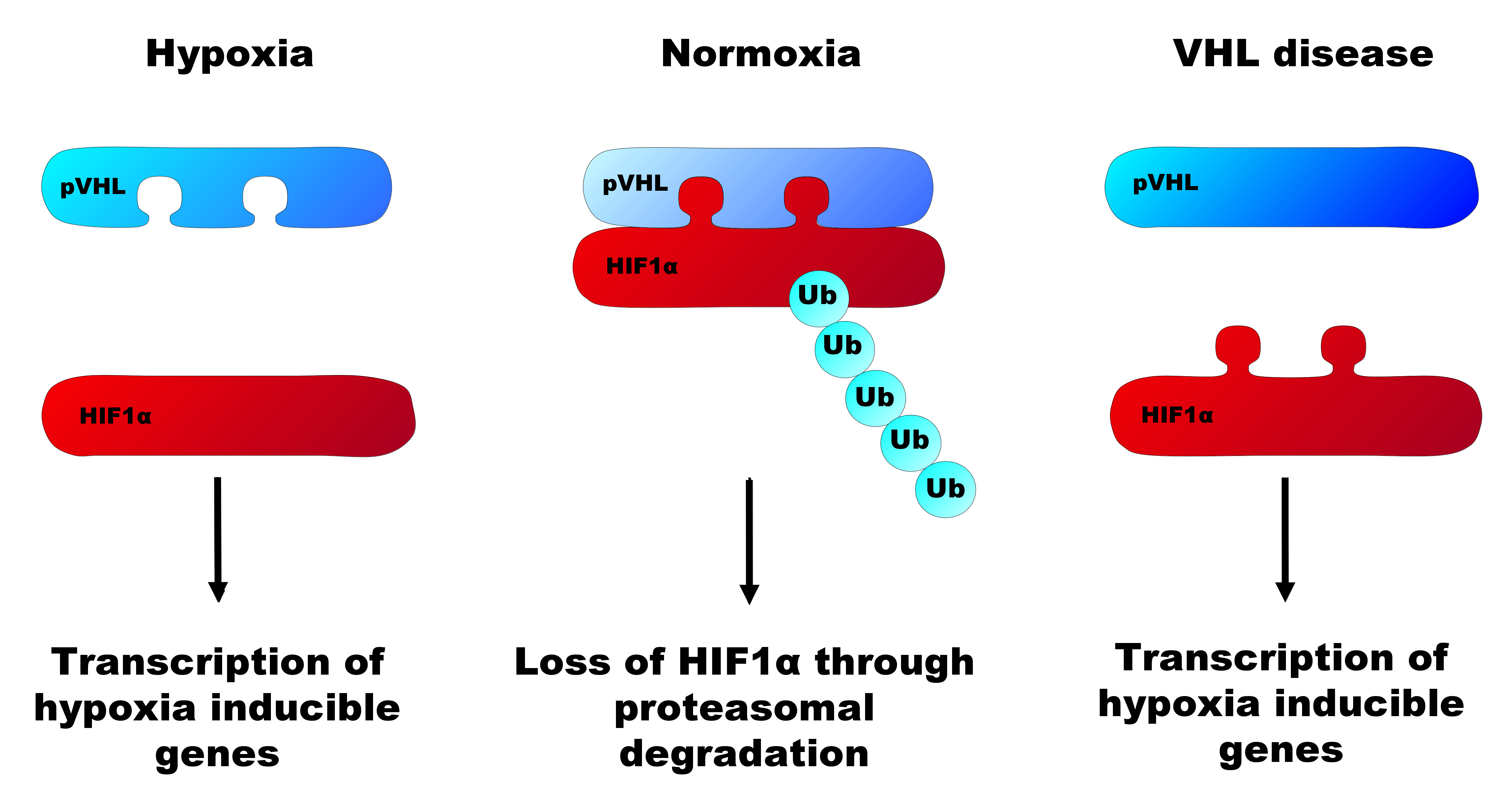
Hypoxia-inducible factor 1-alpha, also known as HIF-1-alpha, is a subunit of a heterodimeric transcription factor hypoxia-inducible factor 1 ( HIF-1) that is encoded by the ''HIF1A'' gene. The Nobel Prize in Physiology or Medicine 2019 was awarded for the discovery of HIF. HIF1A is a basic helix-loop-helix PAS domain containing protein, and is considered as the master transcriptional regulator of cellular and developmental response to hypoxia. The dysregulation and overexpression of ''HIF1A'' by either hypoxia or genetic alternations have been heavily implicated in cancer biology, as well as a number of other pathophysiologies, specifically in areas of vascularization and angiogenesis, energy metabolism, cell survival, and tumor invasion. The presence of HIF1A in a hypoxic environment is required to push forward normal placental development in early gestation. Two other alternative transcripts encoding different isoforms have been identified. Structure HIF1 is a het ... [...More Info...] [...Related Items...] OR: [Wikipedia] [Google] [Baidu] |
Von Hippel–Lindau Tumor Suppressor
The Von Hippel–Lindau tumor suppressor also known as pVHL is a protein that, in humans, is encoded by the ''VHL'' gene. Mutations of the VHL gene are associated with Von Hippel–Lindau disease, which is characterized by hemangioblastomas of the brain, spinal cord and retina. It is also associated with kidney and pancreatic lesions. Function The protein encoded by the VHL gene is the substrate recognition component of a protein complex that includes elongin B, elongin C, and cullin-2, and possesses E3 ubiquitin ligase activity. This complex is involved in the ubiquitination and subsequent degradation of hypoxia-inducible factors (HIFs), which are transcription factors that play a central role regulating gene expression in response to changing oxygen levels. RNA polymerase II subunit POLR2G/RPB7 is also reported to be a target of this protein. Alternatively spliced transcript variants encoding distinct isoforms have been observed. The resultant protein is produced in ... [...More Info...] [...Related Items...] OR: [Wikipedia] [Google] [Baidu] |


