|
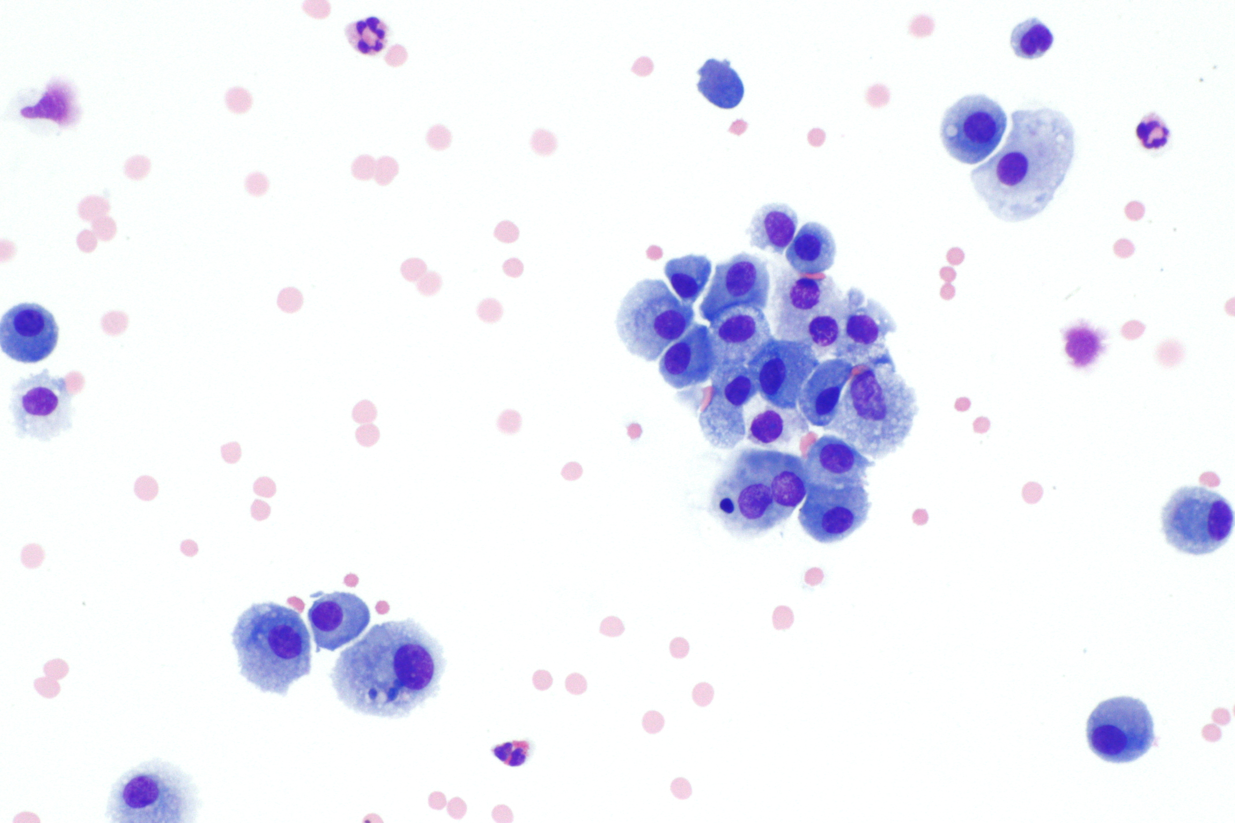
Fibroadenoma
Fibroadenomas are benign breast tumours characterized by an admixture of stromal and epithelial tissue. Breasts are made of lobules (milk producing glands) and ducts (tubes that carry the milk to the nipple). These are surrounded by glandular, fibrous and fatty tissues. Fibroadenomas develop from the lobules. The glandular tissue and ducts grow over the lobule to form a solid lump. Since both fibroadenomas and breast lumps as a sign of breast cancer can appear similar, it is recommended to perform ultrasound analyses and possibly tissue sampling with subsequent histopathologic analysis in order to make a proper diagnosis. Unlike typical lumps from breast cancer, fibroadenomas are easy to move, with clearly defined edges. Fibroadenomas are sometimes called breast mice or a breast mouse owing to their high mobility in the breast. Signs and symptoms Fibroadenomas are benign tumours of the breast, most often present in women in their 20s and 30s. Clinically, fibroadenomas are us ... [...More Info...] [...Related Items...] OR: [Wikipedia] [Google] [Baidu] |
Breast Lump
A breast mass, also known as a breast lump, is a localized Swelling (medical), swelling that feels different from the surrounding tissue (biology), tissue. Breast pain, nipple discharge, or skin changes may be present. Concerning findings include masses that are hard, do not move easily, are of an irregular shape, or are firmly attached to surrounding tissue. Causes include fibrocystic change, fibroadenomas, breast infection, galactoceles, and breast cancer. Breast cancer makes up about 10% of breast masses. Diagnosis is typically by breast examination, examination, medical imaging, and tissue biopsy. Tissue biopsy is often by fine needle aspiration biopsy. Repeated examination may be required. Treatment depends on the underlying cause. It may vary from simple pain medication to surgical removal. Some causes may resolve without treatment. Breast masses are relatively common. It is the most common breast complaint with the women's concern generally being that of cancer. Types ... [...More Info...] [...Related Items...] OR: [Wikipedia] [Google] [Baidu] |
Phyllodes Tumor
Phyllodes tumors (from Greek language, Greek: ), are a rare type of Biphasic tumor, biphasic Fibroepithelial neoplasm, fibroepithelial mass that form from the periductal stromal and epithelial cells of the breast. They account for less than 1% of all breast Neoplasia, neoplasms. They were previously termed cystosarcoma phyllodes, coined by Johannes Peter Müller, Johannes Müller in 1838, before being renamed to phyllodes tumor by the World Health Organization in 2003. , which means 'leaf' in Greek, describes the unique papillary projections characteristic of phyllodes tumors on histology. Diagnosis is made via a core-needle biopsy and treatment is typically surgical resection with wide margins (>1 cm), due to their propensity to recur. Signs and symptoms Phyllodes tumors typically present as a firm, mobile, and palpable mass that is painless in nature. On physical examination, the mass can demonstrate a smooth or nodular texture depending on its size. In addition, larger ma ... [...More Info...] [...Related Items...] OR: [Wikipedia] [Google] [Baidu] |
Fibroepithelial Tumor
A fibroepithelial neoplasm (or tumor) is a biphasic tumor. They consist of epithelial tissue, and stromal or mesenchymal tissue. They may be benign or malignant.Tavassoli, F.A., Devilee, P. (Eds). 2003. World Health Organization Classification of Tumours: Pathology & Genetics: Tumours of the breast and female genital organs. IARC Press: Lyon. Examples include: * Brenner tumor of the ovary * Fibroadenoma of the breast * Phyllodes tumor Phyllodes tumors (from Greek language, Greek: ), are a rare type of Biphasic tumor, biphasic Fibroepithelial neoplasm, fibroepithelial mass that form from the periductal stromal and epithelial cells of the breast. They account for less than 1% of ... of the breast Sometimes fibroepithelial polyps (FEPs) of the vulva may be misdiagnosed as cancers. However not much harm is caused because the treatment of both is excision. The consent for removal must however be completely informed. References External links * - "Premalignant Fibroepithelial ... [...More Info...] [...Related Items...] OR: [Wikipedia] [Google] [Baidu] |
Mixed Tumor
A mixed tumor is a tumor that derives from multiple tissue types. In turn citing:-For requiring neoplastic types: Miller-Keane Encyclopedia and Dictionary of Medicine, Nursing, and Allied Health, Seventh Edition- Without further specification:- Farlex Partner Medical Dictionary A biplastic tumor or biphasic tumor has two tissue types.For "biplastic tumor": For "biphasic tumor": True versus false *A ''true'' mixed tumor contains multiple types of neoplasia, neoplastic cells. Some sources require the included tissue types to be neoplastic for the definition of ''mixed tumor''. *A "false" mixed tumor contains one type of neoplastic cells, but which have more than one appearance. For example, benign pleomorphic salivary gland tumors may have some tumors cells that form pseudocartilage. Yet, all the tumor cells have similar myoepithelial profile on immunohistochemistry, and are thus classified as one cell type. Reactive or adaptive changes to a tumor does not count towards a classifica ... [...More Info...] [...Related Items...] OR: [Wikipedia] [Google] [Baidu] |
Needle Biopsy
Needle or Needles may refer to: Crafting * Crochet needle, a tool for making loops in thread or yarn * Knitting needle, a tool for knitting, not as sharp as a sewing needle * Sewing needle, a long slender tool with a pointed tip * Trussing needle, a long slender tool, sometimes with a flattened point, to tie poultry for cooking * Upholstery needle, a tool for upholstery, generally thick and curved Science and technology Botany * Needle (botany), of conifers Medicine * Acupuncture needle, in alternative medicine * Hypodermic needle, a hollow needle commonly used with a syringe to inject fluid into or extract fluid from the body * Surgical needle, several types of needles used for surgical suture * Tuohy needle, a needle used to administer epidural catheters Technology * Gramophone needle, used for playing records * Indicator needle, of a measuring instrument * Needle valve People * Dave Needle (1947–2016), American engineer * Sharon Needles (born 1981), American drag queen ... [...More Info...] [...Related Items...] OR: [Wikipedia] [Google] [Baidu] |
Stromal Cell
Stromal cells, or mesenchymal stromal cells, are differentiating cells found in abundance within bone marrow but can also be seen all around the body. Stromal cells can become connective tissue cells of any organ, for example in the uterine mucosa (endometrium), prostate, bone marrow, lymph node and the ovary. They are cells that support the function of the parenchymal cells of that organ. The most common stromal cells include fibroblasts and pericytes. The term ''stromal'' comes from Latin , "bed covering", and Ancient Greek , , "bed". Stromal cells are an important part of the body's immune response and modulate inflammation through multiple pathways. They also aid in differentiation of hematopoietic cells and forming necessary blood elements. The interaction between stromal cells and tumor cells is known to play a major role in cancer growth and progression. In addition, by regulating local cytokine networks (e.g. M-CSF, LIF), bone marrow stromal cells have been describ ... [...More Info...] [...Related Items...] OR: [Wikipedia] [Google] [Baidu] |
Metachromatic
{{inline citations needed, date=November 2024 Metachromasia (var. metachromasy) is a characteristic change in the color of staining carried out in biological tissues, exhibited by certain dyes when they bind to particular substances present in these tissues, called chromotropes. For example, toluidine blue becomes dark blue (with a colour range from blue-red dependent on glycosaminoglycan content) when bound to cartilage. Other widely used metachromatic stains include the family of Romanowsky stains that also contain thiazine dyes: the white cell nucleus stains purple, basophil granules intense magenta, whilst the cytoplasm (of mononuclear cells) stains blue, which is called the ''Romanowsky effect''. The absence of color change in staining is named orthochromasia. The underlying mechanism for metachromasia requires the presence of polyanions within the tissue. When these tissues are stained with a concentrated basic dye solution, such as toluidine blue, the bound dye molecules ... [...More Info...] [...Related Items...] OR: [Wikipedia] [Google] [Baidu] |
H&E Stain
Hematoxylin and eosin stain ( or haematoxylin and eosin stain or hematoxylin–eosin stain; often abbreviated as H&E stain or HE stain) is one of the principal tissue stains used in histology. It is the most widely used stain in medical diagnosis and is often the ''gold standard.'' For example, when a pathologist looks at a biopsy of a suspected cancer, the histological section is likely to be stained with H&E. H&E is the combination of two histological stains: hematoxylin and eosin. The hematoxylin stains cell nuclei a purplish blue, and eosin stains the extracellular matrix and cytoplasm pink, with other structures taking on different shades, hues, and combinations of these colors. Hence a pathologist can easily differentiate between the nuclear and cytoplasmic parts of a cell, and additionally, the overall patterns of coloration from the stain show the general layout and distribution of cells and provides a general overview of a tissue sample's structure. Thus, patte ... [...More Info...] [...Related Items...] OR: [Wikipedia] [Google] [Baidu] |
Diff-Quik
Diff-Quik is a commercial Romanowsky stain variant used to rapidly stain and differentiate a variety of pathology specimens. It is most frequently used for blood films and cytopathological smears, including fine needle aspirates. The Diff-Quik procedure is based on a modification of the Wright-Giemsa stain pioneered by Harleco in the 1970s, and has advantages over the routine Wright-Giemsa staining technique in that it reduces the 4-minute process into a much shorter operation and allows for selective increased eosinophilic or basophilic staining depending upon the time the smear is left in the staining solutions. There are generic brands of such stain, and the trade name is sometimes used loosely to refer to any such stain (much as "Coke" or "Band-Aid" are sometimes used imprecisely). Usage Diff-Quik may be utilized on material which is ''air-dried'' prior to alcohol fixation rather than immersed immediately (i.e. "wet-fixed"), although immediate alcohol fixation results in ... [...More Info...] [...Related Items...] OR: [Wikipedia] [Google] [Baidu] |
Foam Cell
Foam cells, also called lipid-laden macrophages, are a type of cell that contain cholesterol. These can form a plaque that can lead to atherosclerosis and trigger myocardial infarction and stroke. Foam cells are fat-laden cells with a M2 macrophage-like phenotype. They contain low density lipoproteins (LDL) and can be rapidly detected by examining a fatty plaque under a microscope after it is removed from the body. They are named because the lipoproteins give the cell a foamy appearance. Despite the connection with cardiovascular diseases they might not be inherently dangerous. Some foam cells are derived from smooth muscle cells and present a limited macrophage-like phenotype. Formation Foam cell formation is triggered by a number of factors including the uncontrolled uptake of modified low density lipoproteins (LDL), the upregulation of cholesterol esterification and the impairment of mechanisms associated with cholesterol release. Foam cells are a significant component ... [...More Info...] [...Related Items...] OR: [Wikipedia] [Google] [Baidu] |


