|
Drain (surgery)
A surgical drain is a tube used to remove pus, blood or other fluids from a wound, body cavity, or organ. They are commonly placed by surgeons or interventional radiologists after procedures or some types of injuries, but they can also be used as an intervention for decompression. There are several types of drains, and selection of which to use often depends on the placement site and how long the drain is needed. Use and Management Drains help to remove contents, usually fluids, from inside the body. This is beneficial since fluid accumulation may cause distension and pressure, which can lead to pain. For example, nasogastric (NG) tubes inserted through the nose and into the stomach can help remove stomach contents for patients who have a blockage further along in their gastrointestinal tract. After surgery, drains can be placed to remove blood, lymph, or other fluids that accumulate in the wound bed. This helps to promote wound healing and allows healthcare providers to moni ... [...More Info...] [...Related Items...] OR: [Wikipedia] [Google] [Baidu] |
Hand With Drain After Surgery (2)
A hand is a prehensile, multi-fingered appendage located at the end of the forearm or forelimb of primates such as humans, chimpanzees, monkeys, and lemurs. A few other vertebrates such as the Koala#Characteristics, koala (which has two thumb#Opposition and apposition, opposable thumbs on each "hand" and fingerprints extremely similar to human fingerprints) are often described as having "hands" instead of paws on their front limbs. The raccoon is usually described as having "hands" though opposable thumbs are lacking. Some evolutionary anatomists use the term ''hand'' to refer to the appendage of digits on the forelimb more generally—for example, in the context of whether the three Digit (anatomy), digits of the bird hand involved the same Homology (biology), homologous loss of two digits as in the dinosaur hand. The human hand usually has five digits: Finger numbering#Four-finger system, four fingers plus one thumb; however, these are often referred to collectively as Finger ... [...More Info...] [...Related Items...] OR: [Wikipedia] [Google] [Baidu] |
Granulation Tissue
Granulation tissue is new connective tissue and microscopic blood vessels that form on the surfaces of a wound during the healing process. Granulation tissue typically grows from the base of a wound and is able to fill wounds of almost any size. Examples of granulation tissue can be seen in pyogenic granulomas and pulp polyps. Its histological appearance is characterized by proliferation of fibroblasts and thin-walled, delicate capillaries (angiogenesis), and infiltrated inflammatory cells in a loose extracellular matrix. Appearance During the migratory phase of wound healing, granulation tissue is: * light red or dark pink, being perfused with new capillary loops or "buds"; * soft to the touch; * moist; * bumpy (granular) in appearance, due to punctate hemorrhages; * pulsatile on palpation; * painless when healthy; Structure Granulation tissue is composed of tissue matrix supporting a variety of cell types, most of which can be associated with one of the following function ... [...More Info...] [...Related Items...] OR: [Wikipedia] [Google] [Baidu] |
Incision And Drainage
Incision and drainage (I&D), also known as clinical lancing, are minor surgical procedures to release pus or pressure built up under the skin, such as from an abscess, boil, or infected paranasal sinus. It is performed by treating the area with an antiseptic, such as iodine-based solution, and then making a small incision to puncture the skin using a sterile instrument such as a sharp needle or a pointed scalpel. This allows the pus to escape by draining out through the incision. Good medical practice for large abdominal abscesses requires insertion of a drainage tube, preceded by insertion of a peripherally inserted central catheter line to enable readiness of treatment for possible septic shock. Adjunct antibiotics Uncomplicated cutaneous abscesses do not need antibiotics after successful drainage. In incisional abscesses For incisional abscesses, it is recommended that incision and drainage is followed by covering the area with a thin layer of gauze followed by sterile ... [...More Info...] [...Related Items...] OR: [Wikipedia] [Google] [Baidu] |
Wound Healing
Wound healing refers to a living organism's replacement of destroyed or damaged tissue by newly produced tissue. In undamaged skin, the epidermis (surface, epithelial layer) and dermis (deeper, connective layer) form a protective barrier against the external environment. When the barrier is broken, a regulated sequence of biochemical events is set into motion to repair the damage. This process is divided into predictable phases: blood clotting (hemostasis), inflammation, tissue growth ( cell proliferation), and tissue remodeling (maturation and cell differentiation). Blood clotting may be considered to be part of the inflammation stage instead of a separate stage. The wound-healing process is not only complex but fragile, and it is susceptible to interruption or failure leading to the formation of non-healing chronic wounds. Factors that contribute to non-healing chronic wounds are diabetes, venous or arterial disease, infection, and metabolic deficiencies of old age.Enoch, ... [...More Info...] [...Related Items...] OR: [Wikipedia] [Google] [Baidu] |
Mediastinum
The mediastinum (from ;: mediastina) is the central compartment of the thoracic cavity. Surrounded by loose connective tissue, it is a region that contains vital organs and structures within the thorax, mainly the heart and its vessels, the esophagus, the trachea, the vagus nerve, vagus, phrenic nerve, phrenic and cardiac nerves, the thoracic duct, the thymus and the lymph nodes of the central chest. Anatomy The mediastinum lies within the thorax and is enclosed on the right and left by pulmonary pleurae, pleurae. It is surrounded by the chest wall in front, the lungs to the sides and the Spine (anatomy), spine at the back. It extends from the sternum in front to the vertebral column behind. It contains all the organs of the thorax except the lungs. It is continuous with the loose connective tissue of the neck. The mediastinum can be divided into an upper (or superior) and lower (or inferior) part: * The superior mediastinum starts at the superior thoracic aperture and ends ... [...More Info...] [...Related Items...] OR: [Wikipedia] [Google] [Baidu] |
Pleural Cavity
The pleural cavity, or pleural space (or sometimes intrapleural space), is the potential space between the pleurae of the pleural sac that surrounds each lung. A small amount of serous pleural fluid is maintained in the pleural cavity to enable lubrication between the membranes, and also to create a pressure gradient. The serous membrane that covers the surface of the lung is the visceral pleura and is separated from the outer membrane, the parietal pleura, by just the film of pleural fluid in the pleural cavity. The visceral pleura follows the fissures of the lung and the root of the lung structures. The parietal pleura is attached to the mediastinum, the upper surface of the diaphragm, and to the inside of the ribcage. Structure In humans, the left and right lungs are completely separated by the mediastinum, and there is no communication between their pleural cavities. Therefore, in cases of a unilateral pneumothorax, the contralateral lung will remain functioning n ... [...More Info...] [...Related Items...] OR: [Wikipedia] [Google] [Baidu] |
Chest Tube
A chest tube (also chest drain, thoracic catheter, tube thoracostomy or intercostal drain) is a drain (surgery), surgical drain that is inserted through the chest wall and into the pleural space or the Mediastinum. The insertion of the tube is sometimes a lifesaving procedure. The tube can be used to remove clinically undesired substances such as air (pneumothorax), excess fluid (pleural effusion or hydrothorax), blood (hemothorax), chyle (chylothorax) or pus (empyema) from the intrathoracic space. An intrapleural chest tube is also known as a Bülau drain or an intercostal catheter (ICC), and can either be a thin, flexible silicone tube (known as a "pigtail" drain), or a larger, semi-rigid, fenestrated plastic tube, which often involves a flutter valve or trap (plumbing), underwater seal. The concept of chest drainage was first advocated by Hippocrates when he described the treatment of empyema by means of incision, cautery and insertion of metal tubes. However, the technique was ... [...More Info...] [...Related Items...] OR: [Wikipedia] [Google] [Baidu] |
Shirley Drain
The Shirley wound drain or sump drain is a suction Suction is the day-to-day term for the movement of gases or liquids along a pressure gradient with the implication that the movement occurs because the lower pressure pulls the gas or liquid. However, the forces acting in this case do not orig ... drain with an intake tube that provides air to the bottom of the main tube. This allows a continuous flow of suction so that the tube doesn't get blocked. The Shirley drain is a double-lumen drainage tube intended to aspirate efficiently the contents of a fresh surgical wound. It removes the blood oozing from the walls of the wound cavity before it clots. History The Shirley drain was invented in 1957 by surgeon and inventor Dr. Harold W. Andersen. References Medical equipment Medical pumps {{medical-equipment-stub ... [...More Info...] [...Related Items...] OR: [Wikipedia] [Google] [Baidu] |
Negative Pressure Wound Therapy
Negative-pressure wound therapy (NPWT), also known as a vacuum assisted closure (VAC), is a therapeutic technique using a suction pump, tubing, and a dressing to remove excess wound exudate and to promote healing in acute or chronic wounds and second- and third-degree burns. The use of this technique in wound management started in the 1990s and this technique is often recommended for treatment of a range of wounds including dehisced surgical wounds, closed surgical wounds, open abdominal wounds, open fractures, pressure injuries or pressure ulcers, diabetic foot ulcers, venous insufficiency ulcers, some types of skin grafts, burns, and sternal wounds. It may also be considered after a clean surgery in a person who is obese. NPWT is performed by applying a sub-atmospheric vacuum through a special sealed dressing. The continued vacuum draws out fluid from the wound and increases blood flow to the area. The vacuum may be applied continuously or intermittently, depending on the ... [...More Info...] [...Related Items...] OR: [Wikipedia] [Google] [Baidu] |
Drainage
Drainage is the natural or artificial removal of a surface's water and sub-surface water from an area with excess water. The internal drainage of most agricultural soils can prevent severe waterlogging (anaerobic conditions that harm root growth), but many soils need artificial drainage to improve production or to manage water supplies. History Early history The Indus Valley Civilization had sewerage and drainage systems. All houses in the major cities of Harappa and Mohenjo-daro had access to water and drainage facilities. Waste water was directed to covered gravity sewers, which lined the major streets. 18th and 19th century The invention of hollow-pipe drainage is credited to Sir Hugh Dalrymple, who died in 1753. Current practices Simple infrastructure such as open drains, pipes, and berms are still common. In modern times, more complex structures involving substantial earthworks and new technologies have been common as well. Geotextiles New storm water drainag ... [...More Info...] [...Related Items...] OR: [Wikipedia] [Google] [Baidu] |
Penrose Drain
A Penrose drain is a soft, flexible rubber tube used as a surgical drain, to prevent the buildup of fluid in a surgical site. It belongs to the "passive" type of drain, the other broad type being "active". The Penrose drain is named after American gynecologist Charles Bingham Penrose (1862–1925). See photo of a Penrose drain & its packaging in the following external link Common uses A Penrose drain removes fluid from a wound area. Frequently it is put in place by a surgeon after a procedure is complete to prevent the area from accumulating fluid, such as blood, which could serve as a medium for bacteria to grow in. In podiatry, a Penrose drain is often used as a tourniquet during a hallux nail avulsion procedure or ingrown toenail extraction. It can also be used to drain cerebrospinal fluid to treat a hydrocephalus patient. See also *Instruments used in general surgery There are many different Surgery, surgical specialties, some of which require specific kinds of Surgical ... [...More Info...] [...Related Items...] OR: [Wikipedia] [Google] [Baidu] |
Jackson-Pratt Drain
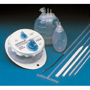
A Jackson-Pratt drain (also called a JP drain) is a Suction (medicine), closed-suction medical device that is commonly used as a drain (surgery), post-operative drain for collecting bodily fluids from surgical sites. The device consists of an internal drain connected to a grenade-shaped bulb or circular cylinder via plastic tubing. The purpose of a drain is to prevent fluid (blood or other) build-up in a closed ("dead") space,MedlinePlus Medical Dictionary, "Dead Space", http://www.merriam-webster.com/medlineplus/dead%20space which may cause either disruption of the wound and the healing process or become an infected abscess, with either scenario possibly requiring a formal drainage/repair procedure (and possibly another trip to the operating room). The drain is also used to evacuate an Abscess, internal abscess before surgery when an infection already exists.Patient.co.uk, "Surgical Drains - Indications, Management and Removal", http://www.patient.co.uk/doctor/Surgical-Drains-In ... [...More Info...] [...Related Items...] OR: [Wikipedia] [Google] [Baidu] |


