|
Dorsal Nerve Of The Penis
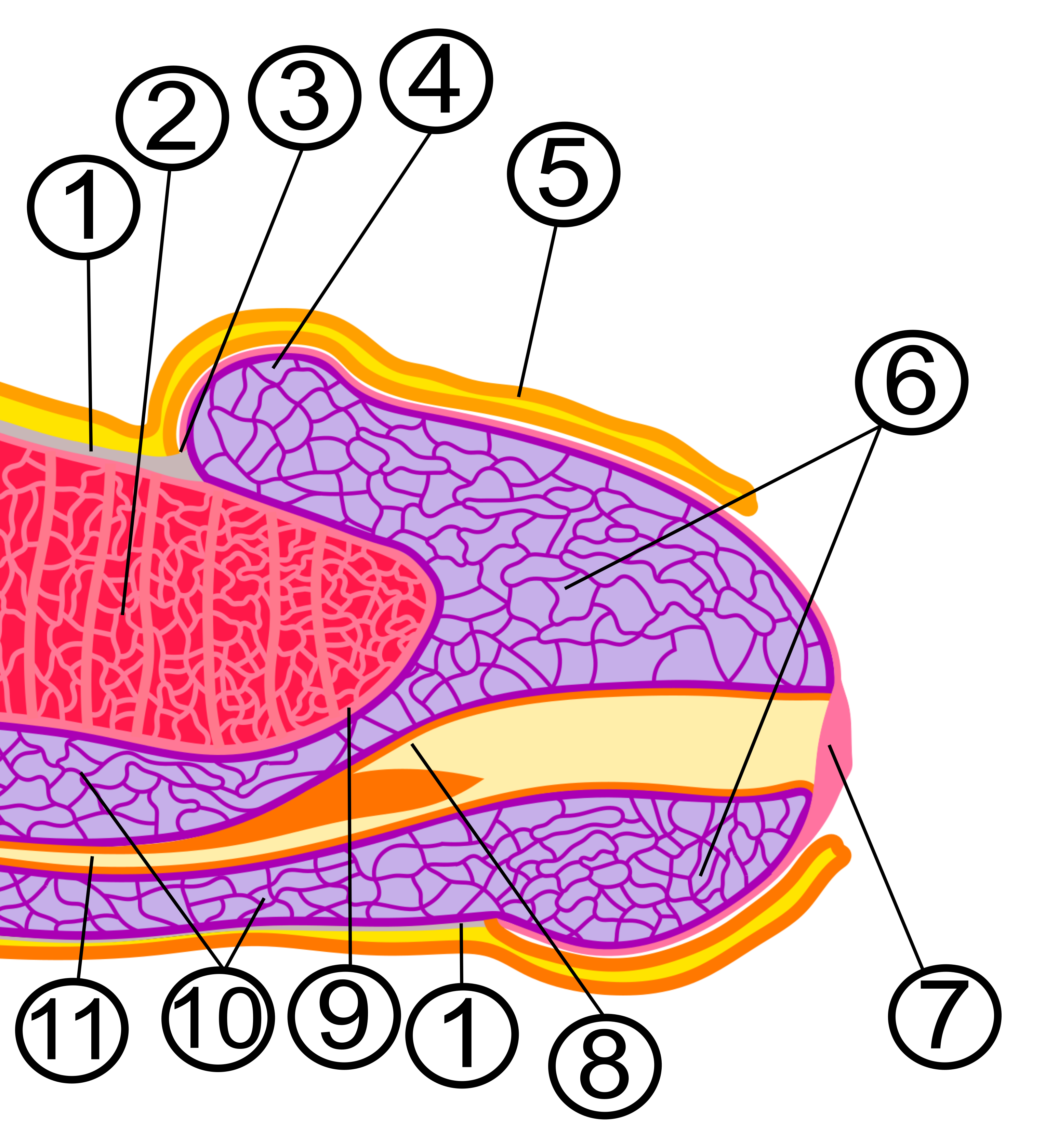
The dorsal nerve of the penis is the deepest of three divisions of the pudendal nerve; it accompanies the internal pudendal artery along the Ischium#Structure, ramus of the ischium; it then runs forward along the margin of the inferior ramus of the pubis, between the superior and inferior layers of the fascia of the urogenital diaphragm. Piercing the inferior layer it gives a branch to the corpus cavernosum penis, and passes forward, in company with the dorsal artery of the penis, between the layers of the Suspensory ligament of the penis, suspensory ligament, on to the dorsum of the penis, and ends on the glans penis. It innervates the skin of the penis. In humans, it has 8290 ± 2553 axons, in 25–45 loosely packed nerve bundles, half of which are myelinated. The researches were conducted on volunteers that donated tissues after death, therefore some of the nerve endings could not have been counted due to the natural degeneration of tissues found in old people. Gallery File ... [...More Info...] [...Related Items...] OR: [Wikipedia] [Google] [Baidu] |
Pudendal Nerve
The pudendal nerve is the main nerve of the perineum. It is a Mixed nerve, mixed (motor and sensory) nerve and also conveys Sympathetic nervous system, sympathetic Autonomic nervous system, autonomic fibers. It carries sensation from the external genitalia of both sexes and the skin around the Human anus, anus and perineum, as well as the Motor neuron, motor supply to various pelvic muscles, including the external sphincter muscle of male urethra, male or external sphincter muscle of female urethra, female external urethral sphincter and the external anal sphincter. If damaged, most commonly by childbirth, loss of sensation or fecal incontinence may result. The nerve may be temporarily anesthetized, called pudendal anesthesia or pudendal block. The pudendal canal that carries the pudendal nerve is also known by the eponymous term "Alcock's canal", after Benjamin Alcock, an Irish anatomist who documented the canal in 1836. Structure Origin The pudendal nerve is paired, me ... [...More Info...] [...Related Items...] OR: [Wikipedia] [Google] [Baidu] |
Corpus Cavernosum Penis
A corpus cavernosum penis (singular) (from Latin, characterised by "cavities/ hollows" of the penis, : corpora cavernosa) is one of a pair of sponge-like regions of erectile tissue, which contain most of the blood in the penis of several animals during an erection. It is homologous to the corpus cavernosum clitoridis in the female. Structure The corpora cavernosa are two expandable erectile tissues along the length of the penis, which fill with blood during penile erection. The two corpora cavernosa lie along the penile shaft, from the pubic bones to the head of the penis, where they join. These formations are made of a sponge-like tissue containing trabeculae, irregular blood-filled spaces lined by endothelium and separated by septum of the penis. The male anatomy has no vestibular bulbs, but instead a corpus spongiosum, a smaller region of erectile tissue along the bottom of the penis, which contains the urethra and forms the glans penis. Physiology In some circ ... [...More Info...] [...Related Items...] OR: [Wikipedia] [Google] [Baidu] |
Dorsal Nerve Of The Clitoris
The dorsal nerve of the clitoris is a nerve in females that branches off the pudendal nerve to innervate the clitoris. The nerve is important for female sexual pleasure, and it may play a role in clitoral erections. It travels from below the inferior pubic ramus to the suspensory ligament of the clitoris. At its thickest, the DNC is in diameter, visible to the naked eye during dissection. The DNC splits into two nerve branches on either side of the midline, closely following the crura of the clitoris. Some surgeries—for example, sling surgeries to treat female urinary incontinence—can damage the DNC, causing a loss of sensation in the clitoris. Understanding the nerve is important for urologists and gynecologists who may operate on organs near the DNC. The dorsal nerve of the clitoris is analogous to the dorsal nerve of the penis in males. It is a terminal branch of the pudendal nerve. See also * Posterior labial nerves * Perineal nerve The perineal nerve is a nerve ... [...More Info...] [...Related Items...] OR: [Wikipedia] [Google] [Baidu] |
Cavernous Nerves Of Penis
The cavernous nerves are post-ganglionic parasympathetic nerves that facilitate penile erection and clitoral erection. They arise from cell bodies in the inferior hypogastric plexus where they receive the pre-ganglionic pelvic splanchnic nerves (S2-S4). In the penis, there are both lesser cavernous nerves and a greater cavernous nerve. Clinical considerations These nerves are susceptible to injury following prostatectomy Prostatectomy (from the Ancient Greek language, Greek , "prostate" and , "excision") is the surgical removal of all or part of the prostate gland. This operation is done for benignity, benign conditions that cause urinary retention, as well as ... or genital surgery. Nerve-sparing prostatectomy was invented for surgeons to avoid injuring the nerves and causing erectile dysfunction complications. During surgery, a doctor may apply a small electrical stimulation to the nerve and measure the erectile function with a penile plethysmograph. This test ... [...More Info...] [...Related Items...] OR: [Wikipedia] [Google] [Baidu] |
Sacral Plexus
In human anatomy, the sacral plexus is a nerve plexus which provides motor and sensory nerves for the posterior thigh, most of the lower leg and foot, and part of the pelvis. It is part of the lumbosacral plexus and emerges from the lumbar vertebrae and sacral vertebrae (L4-S4).''Thieme Atlas of Anatomy'' (2006), pp 470-471 A sacral plexopathy is a disorder affecting the nerves of the sacral plexus, usually caused by trauma, nerve compression, vascular disease, or infection. Symptoms may include pain, loss of motor control, and sensory deficits. Structure The sacral plexus is formed by: * the lumbosacral trunk * the anterior division of the first sacral nerve * portions of the anterior divisions of the second and third sacral nerves The nerves forming the sacral plexus converge toward the lower part of the greater sciatic foramen, and unite to form a flattened band, from the anterior and posterior surfaces of which several branches arise. The band itself is continued as the ... [...More Info...] [...Related Items...] OR: [Wikipedia] [Google] [Baidu] |
Glans Penis
In male human anatomy, the glans penis or penile glans, commonly referred to as the glans, (; from Latin ''glans'' meaning "acorn") is the bulbous structure at the Anatomical terms of location#Proximal and distal, distal end of the human penis that is the human male's most sensitive erogenous zone and primary anatomical source of Human sexuality, sexual pleasure. The glans penis is present in the male reproductive system, reproductive organs of humans and most other mammals where it may appear smooth, spiny, elongated or divided. It is externally lined with Mucosa, mucosal tissue, which creates a smooth texture and glossy appearance. In humans, the glans is located over the distal ends of the Corpus cavernosum penis, corpora cavernosa and is a continuation of the Corpus spongiosum (penis), corpus spongiosum of the penis. At the summit appears the urinary meatus and at the base forms the Corona of glans penis, corona glandis. An elastic band of tissue, known as the Penile frenulum ... [...More Info...] [...Related Items...] OR: [Wikipedia] [Google] [Baidu] |
Suspensory Ligament Of The Penis
The suspensory ligament of the penis is a triangular midline structure anchoring the penis to the pubic symphysis, holding the penis close to the pubic bone and supporting it during erection. The ligament does not directly connect to the corpus cavernosum penis, but may still play a role in erectile dysfunction. The ligament can be surgically lengthened in a procedure known as ligamentolysis, which is a form of penis enlargement. Anatomy Structure The ligament is composed of a midline lamina, and two lateral laminae. Some of the fibres of the ligament come to constitute the fundiform ligament of the penis, extending into the scrotal septum. Attachments The ligament attaches by its apex onto the symphysis pubis and linea alba, and by its base onto the dorsal and lateral aspects of the corpora cavernosa penis. The midline lamina splits inferiorly/distally to attach onto each corpus cavernosus penis lateral to the groove of the deep dorsal vein of penis, whereas each ... [...More Info...] [...Related Items...] OR: [Wikipedia] [Google] [Baidu] |
Dorsal Artery Of The Penis
The dorsal artery of the penis is a bilaterally paired terminal branch of the internal pudendal artery which passes upon the dorsum of the penis to the base of the glans penis, where it unites with its contralateral partner and supply the glans and foreskin. The dorsal artery of the penis provides blood supply to the skin and fascia of the penis (including the foreskin), and the erective tissues of the penis (especially the glans penis). The dorsal artery of the penis may be damaged in traumatic amputation of the penis and repairing the dorsal artery surgically prevents skin loss, but it is not essential for sexual and urinary function. Its hemodynamics and blood pressure can be assessed to test for sexual impairment. Structure The homologous artery in the female is the dorsal artery of clitoris. Origin The dorsal artery of the penis is a terminal branch of the internal pudendal artery, arising at the inferior border of the symphysis pubis. Course and relations It ... [...More Info...] [...Related Items...] OR: [Wikipedia] [Google] [Baidu] |
Urogenital Diaphragm
Older texts have asserted the existence of a urogenital diaphragm, also called the triangular ligament, which was described as a layer of the pelvis that separates the deep perineal sac from the upper pelvis, lying between the inferior fascia of the urogenital diaphragm (perineal membrane) and superior fascia of the urogenital diaphragm. While this term is used to refer to a layer of the pelvis that separates the deep perineal sac from the upper pelvis, such a discrete border of the sac probably does not exist. While it has no official entry in Terminologia Anatomica, the term is still used occasionally to describe the muscular components of the deep perineal pouch. The urethra and the vagina, though part of the pouch, are usually said to be passing through the urogenital diaphragm, rather than part of the diaphragm itself. Some researchers still assert that such a diaphragm exists, and the term is still used in the literature. The urethral diaphragm is an anatomic landmark ... [...More Info...] [...Related Items...] OR: [Wikipedia] [Google] [Baidu] |
Lesser Sciatic Foramen
The lesser sciatic foramen is an opening (foramen) between the pelvis and the back of the thigh. The foramen is formed by the sacrotuberous ligament which runs between the sacrum and the ischial tuberosity and the sacrospinous ligament which runs between the sacrum and the ischial spine. Structure The lesser sciatic foramen has the following boundaries: * Anterior: the tuberosity of the ischium * Superior: the spine of the ischium and sacrospinous ligament * Posterior: the sacrotuberous ligament Alternatively, the foramen can be defined by the boundaries of the lesser sciatic notch and the two ligaments. Function The following pass through the foramen: * the tendon of the obturator internus * internal pudendal vessels * pudendal nerve * nerve to the obturator internus See also *Greater sciatic foramen The greater sciatic foramen is an opening (:wikt:foramen, foramen) in the posterior human pelvis. It is formed by the sacrotuberous ligament, sacrotuberous and sacrospino ... [...More Info...] [...Related Items...] OR: [Wikipedia] [Google] [Baidu] |
Fascia
A fascia (; : fasciae or fascias; adjective fascial; ) is a generic term for macroscopic membranous bodily structures. Fasciae are classified as superficial, visceral or deep, and further designated according to their anatomical location. The knowledge of fascial structures is essential in surgery, as they create borders for infectious processes (for example Psoas abscess) and haematoma. An increase in pressure may result in a compartment syndrome, where a prompt fasciotomy may be necessary. For this reason, profound descriptions of fascial structures are available in anatomical literature from the 19th century. Function Fasciae were traditionally thought of as passive structures that transmit mechanical tension generated by muscular activities or external forces throughout the body. An important function of muscle fasciae is to reduce friction of muscular force. In doing so, fasciae provide a supportive and movable wrapping for nerves and blood vessels as they pass thro ... [...More Info...] [...Related Items...] OR: [Wikipedia] [Google] [Baidu] |
Inferior Ramus Of The Pubis
In vertebrates, the pubis or pubic bone () forms the lower and anterior part of each side of the hip bone. The pubis is the most forward-facing (ventral and anterior) of the three bones that make up the hip bone. The left and right pubic bones are each made up of three sections; a superior ramus, an inferior ramus, and a body. Structure The pubic bone is made up of a ''body'', ''superior ramus'', and ''inferior ramus'' (). The left and right coxal bones join at the pubic symphysis. It is covered by a layer of Adipose tissue, fat – the mons pubis. The pubis is the lower limit of the suprapubic region. In the female, the pubis is anterior to the urethral sponge. Body The body of pubis has: * a superior border or the pubic crest * a pubic tubercle at the lateral end of the pubic crest * three surfaces (anterior, posterior and medial). The body forms the wide, strong, middle and flat part of the pubic bone. The bodies of the left and right pubic bones join at the pubic symphys ... [...More Info...] [...Related Items...] OR: [Wikipedia] [Google] [Baidu] |

