|
Congenital Tufting Enteropathy
''Congenital tufting enteropathy'' is an inherited disorder of the small intestine that presents with intractable diarrhea in young children. Signs and symptoms The infants present in the first few days of life with watery diarrhoea. This leads rapidly to dehydration and electrolyte imbalance and metabolic decompensation. Enteral feeding with a protein hydrolysate or amino acid based formulas worsen the diarrhoea and the children rapidly fail to thrive and develop protein energy malnutrition. In the majority of cases the severity of the malabsorption and diarrhoea make them dependent on daily long term total parenteral nutrition. Hepatic fibrosis and cirrhosis are known complications. Associated conditions *Choanal atresia *Nonspecific punctuated keratitis (60%) *Oesophageal atresia *Unperforated anus Genetics Two genes have been associated with this condition: Epithelial cell adhesion molecule (EpCAM) on chromosome 2 (2p21) and SPINT2 on chromosome 19. SPINT2 is a Kunitz-typ ... [...More Info...] [...Related Items...] OR: [Wikipedia] [Google] [Baidu] |
Cirrhosis
Cirrhosis, also known as liver cirrhosis or hepatic cirrhosis, and end-stage liver disease, is the impaired liver function caused by the formation of scar tissue known as fibrosis due to damage caused by liver disease. Damage causes tissue repair and subsequent formation of scar tissue, which over time can replace normal parenchyma, functioning tissue, leading to the impaired liver function of cirrhosis. The disease typically develops slowly over months or years. Early symptoms may include Fatigue (medicine), tiredness, Asthenia, weakness, Anorexia (symptom), loss of appetite, weight loss, unexplained weight loss, nausea and vomiting, and discomfort in the right upper quadrant of the abdomen. As the disease worsens, symptoms may include Pruritus, itchiness, peripheral edema, swelling in the lower legs, ascites, fluid build-up in the abdomen, jaundice, coagulopathy, bruising easily, and the development of spider angioma, spider-like blood vessels in the skin. The fluid build-up in ... [...More Info...] [...Related Items...] OR: [Wikipedia] [Google] [Baidu] |
Epithelial Cell Adhesion Molecule
Epithelial cell adhesion molecule (EpCAM), also known as CD326 among other names, is a transmembrane glycoprotein mediating Ca2+-independent homotypic cell–cell adhesion in epithelia. EpCAM is also involved in cell signaling, migration, proliferation, and differentiation. Additionally, EpCAM has oncogenic potential via its capacity to upregulate c-myc, e-fabp, and cyclins A & E. Since EpCAM is expressed exclusively in epithelia and epithelial-derived neoplasms, EpCAM can be used as diagnostic marker for various cancers. It appears to play a role in tumorigenesis and metastasis of carcinomas, so it can also act as a potential prognostic marker and as a potential target for immunotherapeutic strategies. Expression pattern First discovered in 1979, EpCAM was initially described as a dominant surface antigen on human colon carcinoma. Because of its prevalence on many carcinomas, it has been "discovered" many different times. EpCAM therefore has many aliases the most notable of w ... [...More Info...] [...Related Items...] OR: [Wikipedia] [Google] [Baidu] |
Chromosome 2
Chromosome 2 is one of the twenty-three pairs of chromosomes in humans. People normally have two copies of this chromosome. Chromosome 2 is the second-largest human chromosome, spanning more than 242 million base pairs and representing almost eight percent of the total DNA in human cells. Chromosome 2 contains the HOXD homeobox gene cluster. Chromosomes Humans have only twenty-three pairs of chromosomes, while all other extant members of Hominidae have twenty-four pairs. It is believed that Neanderthals and Denisovans had twenty-three pairs. Human chromosome 2 is a result of an end-to-end fusion of two ancestral chromosomes.It has been hypothesized that Human Chromosome 2 is a fusion of two ancestral chromosomes by Alec MacAndrew; accessed 18 May 2006. [...More Info...] [...Related Items...] OR: [Wikipedia] [Google] [Baidu] |
SPINT2
Kunitz-type protease inhibitor 2 is an enzyme inhibitor that in humans is encoded by the ''SPINT2'' gene. SPINT2 is a transmembrane protein with two extracellular Kunitz domains to inhibit serine proteases. This gene is a presumed tumor suppressor by inhibiting HGF activator which prevents the formation of active hepatocyte growth factor. Mutations in SPINT2 could result in congenital sodium diarrhea A birth defect, also known as a congenital disorder, is an abnormal condition that is present at birth regardless of its cause. Birth defects may result in disabilities that may be physical, intellectual, or developmental. The disabilities can r ... (CSD). References Further reading * * * * * * * * * * * * * * * * * External links * {{gene-19-stub ... [...More Info...] [...Related Items...] OR: [Wikipedia] [Google] [Baidu] |
Chromosome 19
Chromosome 19 is one of the 23 pairs of chromosomes in humans. People normally have two copies of this chromosome. Chromosome 19 spans more than 58.6 million base pairs, the building material of DNA. It is considered the most gene-rich chromosome containing roughly 1,500 genes, despite accounting for only 2 percent of the human genome. Genes Number of genes The following are some of the gene count estimates of human chromosome 19. Because researchers use different approaches to genome annotation, their predictions of the number of genes on each chromosome varies (for technical details, see gene prediction). Among various projects, the collaborative consensus coding sequence project ( CCDS) takes an extremely conservative strategy. So CCDS's gene number prediction represents a lower bound on the total number of human protein-coding genes. Gene list The following is a partial list of genes on human chromosome 19. For complete list, see the link in the infobox on the righ ... [...More Info...] [...Related Items...] OR: [Wikipedia] [Google] [Baidu] |
Protease
A protease (also called a peptidase, proteinase, or proteolytic enzyme) is an enzyme that catalyzes (increases reaction rate or "speeds up") proteolysis, breaking down proteins into smaller polypeptides or single amino acids, and spurring the formation of new protein products. They do this by cleaving the peptide bonds within proteins by hydrolysis, a reaction where water breaks bonds. Proteases are involved in many biological functions, including digestion of ingested proteins, protein catabolism (breakdown of old proteins), and cell signaling. In the absence of functional accelerants, proteolysis would be very slow, taking hundreds of years. Proteases can be found in all forms of life and viruses. They have independently evolved multiple times, and different classes of protease can perform the same reaction by completely different catalytic mechanisms. Hierarchy of proteases Based on catalytic residue Proteases can be classified into seven broad groups: * Serine prot ... [...More Info...] [...Related Items...] OR: [Wikipedia] [Google] [Baidu] |
Mononuclear Cell Infiltration
In immunology, agranulocytes (also known as nongranulocytes or mononuclear leukocytes) are one of the two types of leukocytes (white blood cells), the other type being granulocytes. Agranular cells are noted by the absence of granules in their cytoplasm, which distinguishes them from granulocytes. The two types of agranulocytes in the blood circulation are lymphocytes and monocytes. These make up about 35% of the hematologic blood values. The distinction between granulocytes and agranulocytes is not useful for several reasons. First, monocytes contain granules, which tend to be fine and weakly stained (see monocyte entry). Second, monocytes and the granulocytes are closely related cell types developmentally, physiologically and functionally. Third, this distinction is not used by haematologists; it is an erroneous separation that has no meaning. Lymphocytes are much more common in the lymphatic system and include natural killer T-cells. Blood has three types of lymphocytes: B c ... [...More Info...] [...Related Items...] OR: [Wikipedia] [Google] [Baidu] |
Lamina Propria
The lamina propria is a thin layer of connective tissue that forms part of the moist linings known as mucous membranes or mucosae, which line various tubes in the body, such as the respiratory tract, the gastrointestinal tract, and the urogenital tract. The lamina propria is a thin layer of loose (areolar) connective tissue, which lies beneath the epithelium, and together with the epithelium and basement membrane constitutes the mucosa. As its Latin name indicates, it is a characteristic component of the mucosa, or the mucosa's "own special layer." Thus, the term mucosa or mucous membrane refers to the combination of the epithelium and the lamina propria. The connective tissue of the lamina propria is loose and rich in cells. The cells of the lamina propria are variable and can include fibroblasts, lymphocytes, plasma cells, macrophages, eosinophilic leukocytes, and mast cells. It provides support and nutrition to the epithelium, as well as the means to bind to the underlyi ... [...More Info...] [...Related Items...] OR: [Wikipedia] [Google] [Baidu] |
Laminin
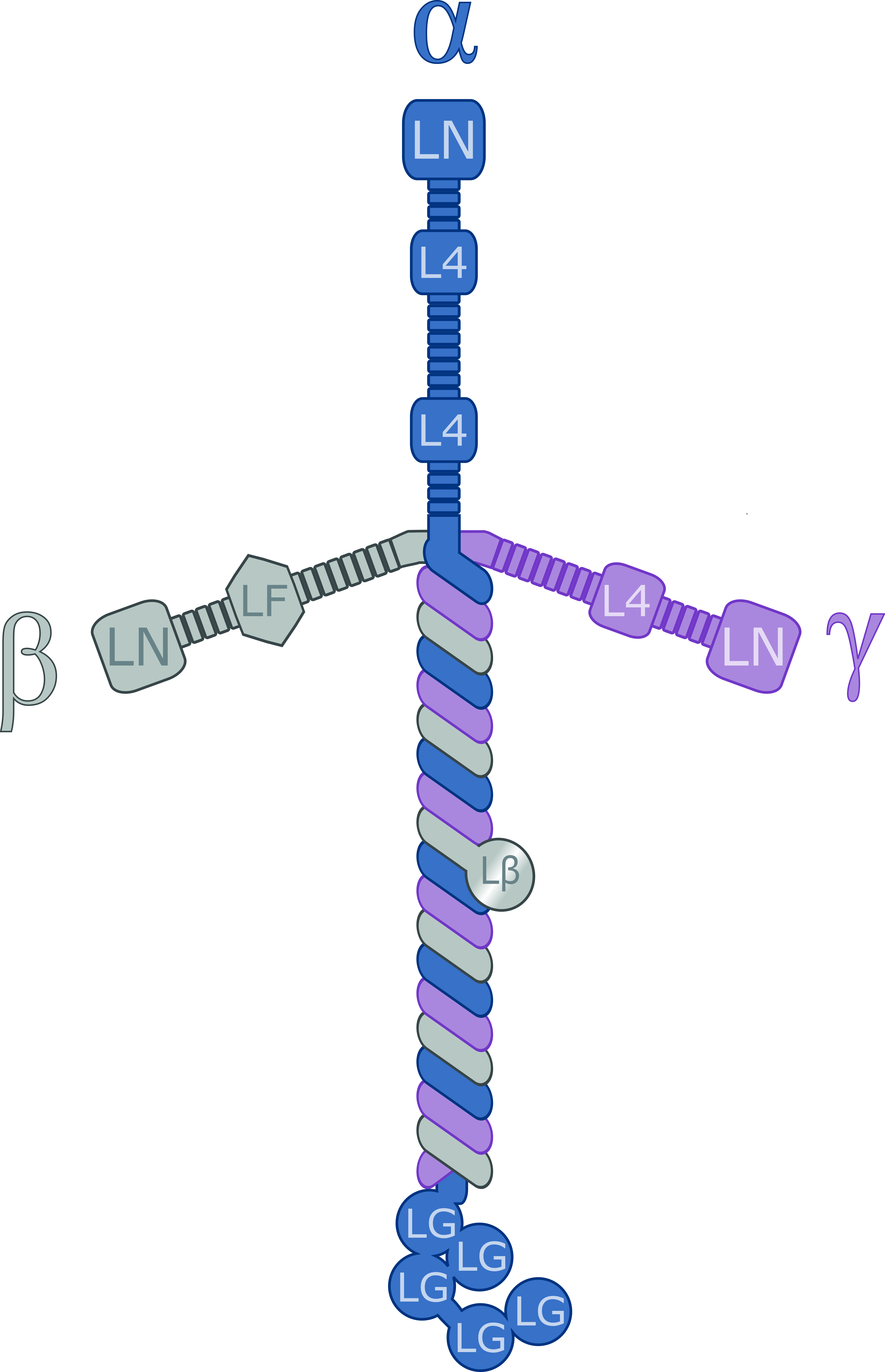
Laminins are a family of glycoproteins of the extracellular matrix of all animals. They are major components of the basal lamina (one of the layers of the basement membrane), the protein network foundation for most cells and organs. The laminins are an important and biologically active part of the basal lamina, influencing cell differentiation, migration, and adhesion. Laminins are heterotrimeric proteins with a high molecular mass (~400 to ~900 kDa). They contain three different chains (α, β and γ) encoded by five, four, and three paralogous genes in humans, respectively. The laminin molecules are named according to their chain composition. Thus, laminin-511 contains α5, β1, and γ1 chains. Fourteen other chain combinations have been identified ''in vivo''. The trimeric proteins intersect to form a cross-like structure that can bind to other cell membrane and extracellular matrix molecules. The three shorter arms are particularly good at binding to other laminin molecule ... [...More Info...] [...Related Items...] OR: [Wikipedia] [Google] [Baidu] |
Heparan Sulfate Proteoglycan
Heparan sulfate (HS) is a linear polysaccharide found in all animal tissues. It occurs as a proteoglycan (HSPG, i.e. Heparan Sulfate ProteoGlycan) in which two or three HS chains are attached in close proximity to cell surface or extracellular matrix proteins. It is in this form that HS binds to a variety of protein ligands, including Wnt, and regulates a wide range of biological activities, including developmental processes, angiogenesis, blood coagulation, abolishing detachment activity by GrB (Granzyme B), and tumour metastasis. HS has also been shown to serve as cellular receptor for a number of viruses, including the respiratory syncytial virus. One study suggests that cellular heparan sulfate has a role in SARS-CoV-2 Infection, particularly when the virus attaches with ACE2. Proteoglycans The major cell membrane HSPGs are the transmembrane syndecans and the glycosylphosphatidylinositol (GPI) anchored glypicans. Other minor forms of membrane HSPG include betaglycan and th ... [...More Info...] [...Related Items...] OR: [Wikipedia] [Google] [Baidu] |
Desmoglein
The desmogleins are a family of desmosomal cadherins consisting of proteins DSG1, DSG2, DSG3, and DSG4. They play a role in the formation of desmosomes that join cells to one another. Pathology Desmogleins are targeted in the autoimmune disease pemphigus. Desmoglein proteins are a type of cadherin, which is a transmembrane protein that binds with other cadherins to form junctions known as desmosomes between cells. These desmoglein proteins thus hold cells together, but, when the body starts producing antibodies against desmoglein, these junctions break down, and this results in subsequent blister A blister is a small pocket of body fluid ( lymph, serum, plasma, blood, or pus) within the upper layers of the skin, usually caused by forceful rubbing (friction), burning, freezing, chemical exposure or infection. Most blisters are filled ... or vesicle formation.Bolognia JL, Jorizzo JL, Schaffer JV, editors. Dermatology. 3rd ed. Philadelphia: Elsevier Saunders; 2012 Ref ... [...More Info...] [...Related Items...] OR: [Wikipedia] [Google] [Baidu] |
Congenital Chloride Diarrhoea
Congenital chloride diarrhea (CCD, also congenital chloridorrhea or Darrow Gamble syndrome) is a genetic disorder due to an autosomal recessive mutation on chromosome 7. The mutation is in downregulated-in-adenoma (DRA), a gene that encodes a membrane protein of intestinal cells. The protein belongs to the solute carrier 26 family of membrane transport proteins. More than 20 mutations in the gene are known to date. A rare disease, CCD occurs in all parts of the world but is more common in some populations with genetic founder effects, most notably in Finland. Symptoms and signs Chronic diarrhoea starting from early neonatal period. Failure to thrive is usually accompanying diarrhea. Pathophysiology CCD causes persistent secretory diarrhea. In a fetus, it leads to polyhydramnios and premature birth. Immediately after birth, it leads to dehydration, hypoelectrolytemia, hyperbilirubinemia, abdominal distention, and failure to thrive. Diagnosis CCD may be detectable on prena ... [...More Info...] [...Related Items...] OR: [Wikipedia] [Google] [Baidu] |


