|
Common Flexor Tendon
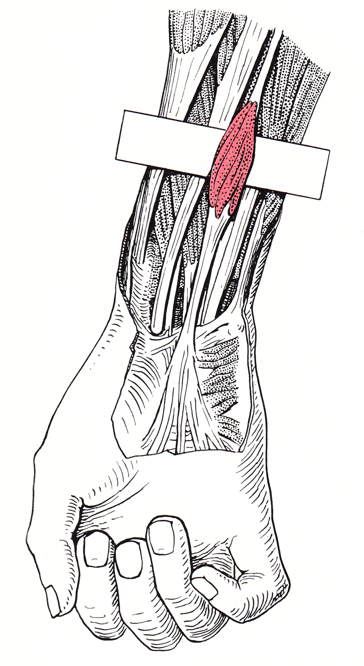
The common flexor tendon is a tendon that attaches to the medial epicondyle of the humerus (lower part of the bone of the upper arm that is near the elbow joint). It serves as the upper attachment point for the superficial muscles of the front of the forearm: *Flexor carpi ulnaris * Palmaris longus * Flexor carpi radialis In anatomy, flexor carpi radialis is a muscle of the human forearm that acts to flex and (radially) abduct the hand. The Latin ''carpus'' means wrist; hence flexor carpi is a flexor of the wrist. Origin and insertion The flexor carpi radialis is ... * Pronator teres * Flexor digitorum superficialis See also * Common extensor tendon References {{DEFAULTSORT:Common Flexor Tendon Tendons ... [...More Info...] [...Related Items...] OR: [Wikipedia] [Google] [Baidu] [Amazon] |
|
 |
Tendon
A tendon or sinew is a tough band of fibrous connective tissue, dense fibrous connective tissue that connects skeletal muscle, muscle to bone. It sends the mechanical forces of muscle contraction to the skeletal system, while withstanding tension (physics), tension. Tendons, like ligaments, are made of collagen. The difference is that ligaments connect bone to bone, while tendons connect muscle to bone. There are about 4,000 tendons in the adult human body. Structure A tendon is made of dense regular connective tissue, whose main cellular components are special fibroblasts called tendon cells (tenocytes). Tendon cells synthesize the tendon's extracellular matrix, which abounds with densely-packed collagen fibers. The collagen fibers run parallel to each other and are grouped into fascicles. Each fascicle is bound by an endotendineum, which is a delicate loose connective tissue containing thin collagen fibrils and elastic fibers. A set of fascicles is bound by an epitenon, whi ... [...More Info...] [...Related Items...] OR: [Wikipedia] [Google] [Baidu] [Amazon] |
|
Medial Epicondyle Of The Humerus
The medial epicondyle of the humerus is an epicondyle of the humerus bone of the upper arm in humans. It is larger and more prominent than the Lateral epicondyle of the humerus, lateral epicondyle and is directed slightly more posteriorly in the Anatomical position#Medical (human) anatomy, anatomical position. In birds, where the arm is somewhat rotated compared to other tetrapods, it is called the ventral epicondyle of the humerus. In comparative anatomy, the more neutral term entepicondyle is used. The medial epicondyle gives attachment to the ulnar collateral ligament of elbow joint, to the pronator teres, and to a common tendon of origin (the common flexor tendon) of some of the flexor muscles of the forearm: the flexor carpi radialis, the flexor carpi ulnaris, the flexor digitorum superficialis, and the palmaris longus. The medial epicondyle is located on the distal end of the humerus. Additionally, the medial epicondyle is inferior to the medial supracondylar ridge. It is ... [...More Info...] [...Related Items...] OR: [Wikipedia] [Google] [Baidu] [Amazon] |
|
 |
Humerus
The humerus (; : humeri) is a long bone in the arm that runs from the shoulder to the elbow. It connects the scapula and the two bones of the lower arm, the radius (bone), radius and ulna, and consists of three sections. The humeral upper extremity of humerus, upper extremity consists of a rounded head, a narrow neck, and two short processes (tubercles, sometimes called tuberosities). The body of humerus, body is cylindrical in its upper portion, and more prism (geometry), prismatic below. The lower extremity of humerus, lower extremity consists of 2 epicondyles, 2 processes (trochlea of the humerus, trochlea and capitulum of the humerus, capitulum), and 3 fossae (radial fossa, coronoid fossa, and olecranon fossa). As well as its true anatomical neck, the constriction below the greater and lesser tubercles of the humerus is referred to as its Surgical neck of the humerus, surgical neck due to its tendency to fracture, thus often becoming the focus of surgeons. Etymology The word ... [...More Info...] [...Related Items...] OR: [Wikipedia] [Google] [Baidu] [Amazon] |
 |
Forearm
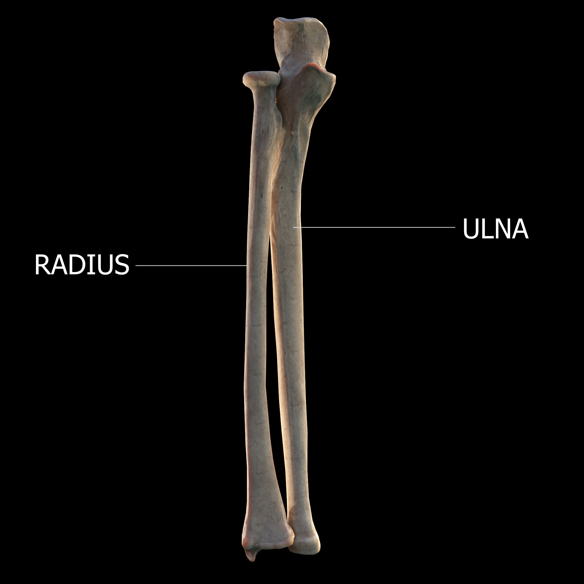
The forearm is the region of the upper limb between the elbow and the wrist. The term forearm is used in anatomy to distinguish it from the arm, a word which is used to describe the entire appendage of the upper limb, but which in anatomy, technically, means only the region of the upper arm, whereas the lower "arm" is called the forearm. It is homologous to the region of the leg that lies between the knee and the ankle joints, the crus. The forearm contains two long bones, the radius and the ulna, forming the two radioulnar joints. The interosseous membrane connects these bones. Ultimately, the forearm is covered by skin, the anterior surface usually being less hairy than the posterior surface. The forearm contains many muscles, including the flexors and extensors of the wrist, flexors and extensors of the digits, a flexor of the elbow ( brachioradialis), and pronators and supinators that turn the hand to face down or upwards, respectively. In cross-section, the forearm can ... [...More Info...] [...Related Items...] OR: [Wikipedia] [Google] [Baidu] [Amazon] |
|
Flexor Carpi Ulnaris
The flexor carpi ulnaris (FCU) is a skeletal muscle, muscle of the forearm that flexion, flexes and Adduction, adducts at the wrist joint. Structure Origin The flexor carpi ulnaris has two heads; a humeral head and ulnar head. The humeral head originates from the medial epicondyle of the humerus via the common flexor tendon. The ulnar head originates from the medial margin of the olecranon of the ulna and the upper two-thirds of the dorsal border of the ulna by an aponeurosis. Between the two heads passes the ulnar nerve and ulnar artery. Insertion The flexor carpi ulnaris inserts onto the pisiform bone, pisiform, hook of the hamate (via the pisohamate ligament) and the anterior surface of the base of the fifth metacarpal bone, fifth metacarpal (via the pisometacarpal ligament). Action The flexor carpi ulnaris flexes and adducts at the Wrist, wrist joint. Innervation The flexor carpi ulnaris is innervated by the ulnar nerve. The corresponding spinal nerves are Cervical spinal ... [...More Info...] [...Related Items...] OR: [Wikipedia] [Google] [Baidu] [Amazon] |
|
 |
Palmaris Longus
The palmaris longus is a muscle visible as a small tendon located between the flexor carpi radialis and the flexor carpi ulnaris, although it is not always present. Reviews report rates of absence in the general population ranging from 10–20%; however, the rate varies in different ethnic groups. Absence of the palmaris longus does not have an effect on grip strength. The lack of palmaris longus muscle does result in decreased pinch strength in fourth and fifth fingers. The absence of palmaris longus muscle is more prevalent in females than males. The palmaris longus muscle can be observed by touching the pads of the fourth finger and thumb and flexing the wrist. The tendon, if present, will be visible in the midline of the anterior wrist. Structure Palmaris longus is a slender, elongated, spindle shaped muscle, lying on the medial side of the flexor carpi radialis. It is widest in the middle, and narrowest at the proximal and distal attachments.''Gray's Anatomy'' (1918), see in ... [...More Info...] [...Related Items...] OR: [Wikipedia] [Google] [Baidu] [Amazon] |
|
Flexor Carpi Radialis
In anatomy, flexor carpi radialis is a muscle of the human forearm that acts to flex and (radially) abduct the hand. The Latin ''carpus'' means wrist; hence flexor carpi is a flexor of the wrist. Origin and insertion The flexor carpi radialis is one of four muscles in the superficial layer of the anterior compartment of the forearm. This muscle originates from the medial epicondyle of the humerus as part of the common flexor tendon. It runs just laterally of flexor digitorum superficialis and inserts on the anterior aspect of the base of the second metacarpal, and has small slips to both the third metacarpal and trapezium tuberosity. The tendon of the flexor carpi radialis is visible on the anterior surface of the forearm, just proximal to the wrist, when the wrist is flexed. It is the tendon seen most lateral, closest to the thumb. Nerve and artery Like most flexors of the anterior compartment of the forearm, FCR is innervated by the median nerve, specifically by axons fro ... [...More Info...] [...Related Items...] OR: [Wikipedia] [Google] [Baidu] [Amazon] |
|
|
Pronator Teres
The pronator teres is a muscle (located mainly in the forearm) that, along with the pronator quadratus, serves to pronate the forearm (turning it so that the palm faces posteriorly when from the anatomical position). It has two origins, at the medial humeral supracondylar ridge and the medial side of the coronoid process of the ulna and inserts near the middle of the radius. Structure The pronator teres has two heads—humeral and ulnar. * The humeral head, the larger and more superficial, arises from the medial supracondylar ridge immediately superior to the medial epicondyle of the humerus, and from the common flexor tendon (which arises from the medial epicondyle). * The ulnar head (or ulnar tuberosity) is a thin fasciculus, which arises from the medial side of the coronoid process of the ulna, and joins the preceding at an acute angle. The median nerve enters the forearm between the two heads of the muscle, and is separated from the ulnar artery by the ulnar head. T ... [...More Info...] [...Related Items...] OR: [Wikipedia] [Google] [Baidu] [Amazon] |
|
|
Flexor Digitorum Superficialis
Flexor digitorum superficialis (''flexor digitorum sublimis'') or flexor digitorum communis sublimis is an extrinsic flexor muscle of the fingers at the proximal interphalangeal joints. It is in the anterior compartment of the forearm. It is sometimes considered to be the deepest part of the superficial layer of this compartment, and sometimes considered to be a distinct, "intermediate layer" of this compartment. It is relatively common for the Flexor digitorum superficialis to be missing from the little finger, bilaterally and unilaterally, which can cause problems when diagnosing a little finger injury. Structure The muscle has two classically described heads – the humeroulnar and radial – and it is between these heads that the median nerve and ulnar artery pass. The ulnar collateral ligament of elbow joint gives its origin to part of this muscle. Four long tendons come off this muscle near the wrist and travel through the carpal tunnel formed by the flexor retinacu ... [...More Info...] [...Related Items...] OR: [Wikipedia] [Google] [Baidu] [Amazon] |
|
|
Common Extensor Tendon
The common extensor tendon is a tendon that attaches to the lateral epicondyle of the humerus. Structure The common extensor tendon serves as the upper attachment (in part) for the superficial muscles that are located on the posterior aspect of the forearm: * Extensor carpi radialis brevis * Extensor digitorum * Extensor digiti minimi * Extensor carpi ulnaris The tendon of extensor carpi radialis brevis is usually the most major tendon to which the other tendons merge. Function The common extensor tendon is the major attachment point for extensor muscles of the forearm. This enables finger extension and aids in forearm supination. Clinical significance Lateral elbow pain can be caused by various pathologies of the common extensor tendon. Overuse injuries can lead to inflammation Inflammation (from ) is part of the biological response of body tissues to harmful stimuli, such as pathogens, damaged cells, or irritants. The five cardinal signs are heat, pain, redn ... [...More Info...] [...Related Items...] OR: [Wikipedia] [Google] [Baidu] [Amazon] |