|
Brachial Vein
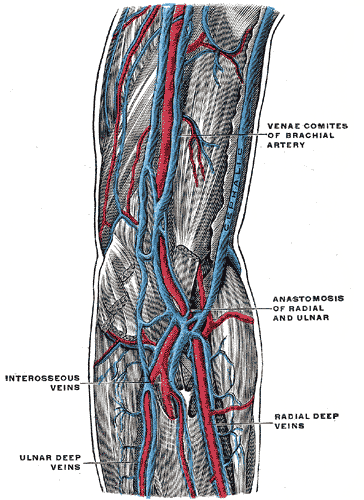
In human anatomy, the brachial veins are venae comitantes of the brachial artery in the arm proper. Because they are deep to muscle, they are considered deep veins. Their course is that of the brachial artery (in reverse): they begin where radial veins and ulnar veins join (corresponding to the bifurcation of the brachial artery). They end at the inferior border of the teres major muscle. At this point, the brachial veins join the basilic vein to form the axillary vein. The brachial veins also have small tributaries that drain the muscles of the upper arm, such as biceps brachii muscle and triceps brachii muscle. Additional images File:Slide8UUU.JPG, Brachial vein File:Gray576.png, The veins of the right axilla The axilla (: axillae or axillas; also known as the armpit, underarm or oxter) is the area on the human body directly under the shoulder joint. It includes the axillary space, an anatomical space within the shoulder girdle between the arm a ..., viewe ... [...More Info...] [...Related Items...] OR: [Wikipedia] [Google] [Baidu] |
Upper Limb
The upper Limb (anatomy), limbs or upper extremities are the forelimbs of an upright posture, upright-postured tetrapod vertebrate, extending from the scapulae and clavicles down to and including the digit (anatomy), digits, including all the musculatures and ligaments involved with the shoulder, elbow, wrist and knuckle joints. In humans, each upper limb is divided into the shoulder, arm, elbow, forearm, wrist and hand, and is primarily used for climbing, manual handling of loads, lifting and dexterity, manipulating objects. In anatomy, just as arm refers to the upper arm, leg refers to the lower leg. Definition In formal usage, the term "arm" only refers to the structures from the shoulder to the elbow, explicitly excluding the forearm, and thus "upper limb" and "arm" are not synonymous. However, in casual usage, the terms are often used interchangeably. The term "upper arm" is redundant in anatomy, but in informal usage is used to distinguish between the two terms. Structure I ... [...More Info...] [...Related Items...] OR: [Wikipedia] [Google] [Baidu] |
Radial Veins
In anatomy, the radial veins are paired veins that accompany the radial artery through the back of the hand and the lateral aspect of the forearm. They join the ulnar veins to form the brachial veins. They follow the same course as the radial artery In human anatomy, the radial artery is the main artery of the lateral aspect of the forearm. Structure The radial artery arises from the bifurcation of the brachial artery in the antecubital fossa. It runs distally on the anterior part of the .... Severing the radial artery can result in unconsciousness in as little as 30 seconds, and death in as little as two minutes. The Brachial artery runs along the inside of your arms. This artery is deep, but severing it will result in unconsciousness in as little as 15 seconds, and death in as little as 90 seconds. External links * Veins of the upper limb {{circulatory-stub ... [...More Info...] [...Related Items...] OR: [Wikipedia] [Google] [Baidu] |
Triceps Brachii Muscle
The triceps, or triceps brachii (Latin for "three-headed muscle of the arm"), is a large muscle on the back of the upper limb of many vertebrates. It consists of three parts: the medial, lateral, and long head. All three heads cross the elbow joint. However, the long head also crosses the shoulder joint. The triceps muscle contracts when the elbow is straightened and expands when the elbow is bent. The long head gets a further contraction when the arm is behind the torso due to how it crosses the shoulder joint. It is the muscle principally responsible for extension of the elbow joint (straightening of the arm). Structure * The long head arises from the infraglenoid tubercle of the scapula. It extends distally anterior to the teres minor and posterior to the teres major. * The medial head arises proximally in the humerus, just inferior to the groove of the radial nerve; from the dorsal (back) surface of the humerus; from the medial intermuscular septum; and its dista ... [...More Info...] [...Related Items...] OR: [Wikipedia] [Google] [Baidu] |
Biceps Brachii Muscle
The biceps or biceps brachii (, "two-headed muscle of the arm") is a large muscle that lies on the front of the upper arm between the shoulder and the elbow. Both Muscle head, heads of the muscle arise on the scapula and join to form a single muscle belly which is attached to the upper forearm. While the long head of the biceps crosses both the shoulder and elbow joints, its main function is at the elbow where it flexes and supination, supinates the forearm. Both these movements are used when opening a bottle with a corkscrew: first biceps screws in the cork (supination), then it pulls the cork out (flexion). Structure The biceps is one of three muscles in the Fascial compartments of arm#Anterior compartment, anterior compartment of the upper arm, along with the brachialis muscle and the coracobrachialis muscle, with which the biceps shares a nerve supply. The biceps muscle has two heads, the short head and the long head, distinguished according to their origin at the cor ... [...More Info...] [...Related Items...] OR: [Wikipedia] [Google] [Baidu] |
Muscle
Muscle is a soft tissue, one of the four basic types of animal tissue. There are three types of muscle tissue in vertebrates: skeletal muscle, cardiac muscle, and smooth muscle. Muscle tissue gives skeletal muscles the ability to muscle contraction, contract. Muscle tissue contains special Muscle contraction, contractile proteins called actin and myosin which interact to cause movement. Among many other muscle proteins, present are two regulatory proteins, troponin and tropomyosin. Muscle is formed during embryonic development, in a process known as myogenesis. Skeletal muscle tissue is striated consisting of elongated, multinucleate muscle cells called muscle fibers, and is responsible for movements of the body. Other tissues in skeletal muscle include tendons and perimysium. Smooth and cardiac muscle contract involuntarily, without conscious intervention. These muscle types may be activated both through the interaction of the central nervous system as well as by innervation ... [...More Info...] [...Related Items...] OR: [Wikipedia] [Google] [Baidu] |
Axillary Vein
In human anatomy, the axillary vein is a large blood vessel that conveys blood from the lateral aspect of the thorax, axilla (armpit) and upper limb toward the heart. There is one axillary vein on each side of the body. Structure Its origin is at the lower margin of the teres major muscle and a continuation of the brachial vein. This large vein is formed by the brachial vein and the basilic vein. At its terminal part, it is also joined by the cephalic vein. Other tributaries include the subscapular vein, circumflex humeral vein, lateral thoracic vein and thoraco-acromial vein. It terminates at the lateral margin of the first rib, at which it becomes the subclavian vein. It is accompanied along its course by a similarly named artery, the axillary artery In human anatomy, the axillary artery is a large blood vessel that conveys oxygenated blood to the lateral aspect of the thorax, the axilla (armpit) and the upper limb. Its origin is at the lateral margin of the fi ... [...More Info...] [...Related Items...] OR: [Wikipedia] [Google] [Baidu] |
Basilic Vein
The basilic vein is a large superficial vein of the upper limb that helps drain parts of the hand and forearm. It originates on the medial ( ulnar) side of the dorsal venous network of the hand and travels up the base of the forearm, where its course is generally visible through the skin as it travels in the subcutaneous fat and fascia lying superficial to the muscles. The basilic vein terminates by uniting with the brachial veins to form the axillary vein. Anatomy Course As it ascends the medial side of the biceps in the arm proper (between the elbow and shoulder), the basilic vein normally perforates the brachial fascia ( deep fascia) in the middle of the medial bicipital groove, and run upwards medial to the brachial artery to the lower border of teres major, continuing as the axillary vein. Tributaries and anastomoses Near the region anterior to the cubital fossa (in the bend of the elbow joint), the basilic vein usually communicates with the cephalic vein (th ... [...More Info...] [...Related Items...] OR: [Wikipedia] [Google] [Baidu] |
Teres Major
The teres major muscle is a muscle of the upper limb. It attaches to the scapula and the humerus and is one of the seven scapulohumeral muscles. It is a thick but somewhat flattened muscle. The teres major muscle (from Latin ''teres'', meaning "rounded") is positioned above the latissimus dorsi muscle and assists in the extension and medial rotation of the humerus. This muscle is commonly confused as a rotator cuff muscle, but it is not, because it does not attach to the capsule of the shoulder joint, unlike the teres minor muscle, for example. Structure The teres major muscle originates on the dorsal surface of the inferior angle and the lower part of the lateral border of the scapula. The fibers of teres major insert into the medial lip of the intertubercular sulcus of the humerus. Relations The tendon, at its insertion, lies behind that of the latissimus dorsi, from which it is separated by a bursa, the two tendons being, however, united along their lower borders for a ... [...More Info...] [...Related Items...] OR: [Wikipedia] [Google] [Baidu] |
Deep Vein
A deep vein is a vein that is deep in the body. This contrasts with superficial veins that are close to the body's surface. Deep veins are almost always beside an artery with the same name (e.g. the femoral vein is beside the femoral artery). Collectively, they carry the vast majority of the blood. Occlusion of a deep vein can be life-threatening and is most often caused by thrombosis. Occlusion of a deep vein by thrombosis is called ''deep vein thrombosis''. Because of their location deep within the body, operation on these veins can be difficult. List *Internal jugular vein Upper limb * Brachial vein * Axillary vein *Subclavian vein Lower limb *Common femoral vein *Femoral vein * Profunda femoris vein * Popliteal vein * Peroneal vein * Anterior tibial vein *Posterior tibial vein The posterior tibial veins are veins of the leg in humans. They drain the posterior compartment of the leg and the plantar surface of the foot to the popliteal vein. Structure The poste ... [...More Info...] [...Related Items...] OR: [Wikipedia] [Google] [Baidu] |
Radial Veins
In anatomy, the radial veins are paired veins that accompany the radial artery through the back of the hand and the lateral aspect of the forearm. They join the ulnar veins to form the brachial veins. They follow the same course as the radial artery In human anatomy, the radial artery is the main artery of the lateral aspect of the forearm. Structure The radial artery arises from the bifurcation of the brachial artery in the antecubital fossa. It runs distally on the anterior part of the .... Severing the radial artery can result in unconsciousness in as little as 30 seconds, and death in as little as two minutes. The Brachial artery runs along the inside of your arms. This artery is deep, but severing it will result in unconsciousness in as little as 15 seconds, and death in as little as 90 seconds. External links * Veins of the upper limb {{circulatory-stub ... [...More Info...] [...Related Items...] OR: [Wikipedia] [Google] [Baidu] |
Brachial Artery
The brachial artery is the major blood vessel of the (upper) arm. It is the continuation of the axillary artery beyond the lower margin of teres major muscle. It continues down the ventral surface of the arm until it reaches the cubital fossa at the Elbow-joint, elbow. It then divides into the radial artery, radial and ulnar artery, ulnar artery, arteries which run down the forearm. In some individuals, the bifurcation occurs much earlier and the ulnar and radial arteries extend through the upper arm. The pulse of the brachial artery is palpation, palpable on the anterior aspect of the elbow, medial to the tendon of the Biceps brachii muscle, biceps, and, with the use of a stethoscope and sphygmomanometer (blood pressure cuff), often used to measure the blood pressure. The brachial artery is closely related to the median nerve; in proximal regions, the median nerve is immediately lateral to the brachial artery. Distally, the median nerve crosses the medial side of the brachial ... [...More Info...] [...Related Items...] OR: [Wikipedia] [Google] [Baidu] |
Venae Comitantes
Vena comitans (Latin for accompanying vein, also known as a satellite vein) refers to a vein that is usually paired, with both veins lying on the sides of an artery. Because they are generally found in pairs, they are often referred to by their plural form: venae comitantes. Venae comitantes are usually found with certain smaller arteries, especially those in the extremities. Larger arteries, on the other hand, generally do not have venae comitantes. They usually have a single, similarly sized vein which is not as intimately associated with the artery. Function As the vein is found in close proximity to an artery the pulsations of the artery aid venous return. Claude Bernard suggested the interchange of heat between the arteries and adjacent veins might moderate cooling of the arterial blood, for which there is experimental evidence. Examples Examples of arteries and their venae comitantes: * Radial artery and radial veins * Ulnar artery and ulnar veins * Brachial artery a ... [...More Info...] [...Related Items...] OR: [Wikipedia] [Google] [Baidu] |

