|
Arachnoid Trabeculae
The arachnoid trabeculae (AT) are delicate strands of connective tissue that loosely connect the two innermost layers of the meninges – the arachnoid mater and the pia mater. " Encyclopædia Britannica. 2010. Encyclopædia Britannica Online. 09 Sep. 2010. They are found within the subarachnoid space where cerebrospinal fluid is also found. Arachnoid trabeculae are also known as subarachnoid trabeculae (SAT) or leptomeningeal trabeculae. Structure Human cranial arachnoid trabeculae are made mostly of[...More Info...] [...Related Items...] OR: [Wikipedia] [Google] [Baidu] |
Meninges
In anatomy, the meninges (; meninx ; ) are the three membranes that envelop the brain and spinal cord. In mammals, the meninges are the dura mater, the arachnoid mater, and the pia mater. Cerebrospinal fluid is located in the subarachnoid space between the arachnoid mater and the pia mater. The primary function of the meninges is to protect the central nervous system. Structure Dura mater The dura mater (), is a thick, durable membrane, closest to the Human skull, skull and vertebrae. The dura mater, the outermost part, is a loosely arranged, fibroelastic layer of cells, characterized by multiple interdigitating cell processes, no extracellular collagen, and significant extracellular spaces. The middle region is a mostly fibrous portion. It consists of two layers: the endosteal layer, which lies closest to the skull, and the inner meningeal layer, which lies closer to the brain. It contains larger blood vessels that split into the capillaries in the pia mater. It is composed ... [...More Info...] [...Related Items...] OR: [Wikipedia] [Google] [Baidu] |
Telencephalon
The cerebrum (: cerebra), telencephalon or endbrain is the largest part of the brain, containing the cerebral cortex (of the two cerebral hemispheres) as well as several subcortical structures, including the hippocampus, basal ganglia, and olfactory bulb. In the human brain, the cerebrum is the uppermost region of the central nervous system. The cerebrum develops prenatally from the forebrain (prosencephalon). In mammals, the dorsal telencephalon, or pallium, develops into the cerebral cortex, and the ventral telencephalon, or subpallium, becomes the basal ganglia. The cerebrum is also divided into approximately symmetric left and right cerebral hemispheres. With the assistance of the cerebellum, the cerebrum controls all voluntary actions in the human body. Structure The cerebrum is the largest part of the brain. Depending upon the position of the animal, it lies either in front or on top of the brainstem. In humans, the cerebrum is the largest and best-developed of ... [...More Info...] [...Related Items...] OR: [Wikipedia] [Google] [Baidu] |
Arachnoid Granulation
Arachnoid granulations (also arachnoid villi, and Pacchionian granulations or bodies) are small outpouchings of the arachnoid mater and subarachnoid space into the dural venous sinuses of the brain. The granulations are thought to mediate the draining of cerebrospinal fluid (CSF) from the subarachnoid space into the venous system. The largest and most numerous granulations lie along the superior sagittal sinus; they are however present along other dural sinuses as well. Anatomy The granulations are often situated near where cerebral veins drain into the dural sinuses. They are most prominent along the superior sagittal sinus, particularly those lodged within the lateral lacunae. In order of decreasing frequency, the granulations occur within the: superior sagittal sinus, transverse sinuses, superior petrosal sinuses, and straight sinus. The arachnoid granulations may be lodged within granular foveae — small pits upon the inner surface of the cranial bones. Structure ... [...More Info...] [...Related Items...] OR: [Wikipedia] [Google] [Baidu] |
Trabecula
A trabecula (: trabeculae, from Latin for 'small beam') is a small, often microscopic, biological tissue, tissue element in the form of a small Beam (structure), beam, strut or rod that supports or anchors a framework of parts within a body or organ. A trabecula generally has a mechanical function, and is usually composed of dense collagenous tissue (such as the trabeculae of spleen, trabecula of the spleen). It can be composed of other material such as muscle and bone. In the heart, muscles form trabeculae carneae and septomarginal trabecula, septomarginal trabeculae, and the left atrial appendage has a tubular trabeculated structure. Bone#Trabeculae, Cancellous bone is formed from groupings of trabeculated bone tissue. In cross section, trabeculae of a Bone#Trabeculae, cancellous bone can look like septum, septa, but in three dimensions they are topologically distinct, with trabeculae being roughly rod or pillar-shaped and septa being sheet-like. When crossing fluid-filled ... [...More Info...] [...Related Items...] OR: [Wikipedia] [Google] [Baidu] |
Subarachnoid Hemorrhage
Subarachnoid hemorrhage (SAH) is bleeding into the subarachnoid space—the area between the arachnoid (brain), arachnoid membrane and the pia mater surrounding the human brain, brain. Symptoms may include a thunderclap headache, severe headache of rapid onset, vomiting, decreased level of consciousness, fever, weakness, numbness, and sometimes seizures. Neck stiffness or neck pain are also relatively common. In about a quarter of people a small bleed with resolving symptoms occurs within a month of a larger bleed. SAH may occur as a result of a head injury or spontaneously, usually from a ruptured cerebral aneurysm. Risk factors for spontaneous cases include high blood pressure, smoking, family history, alcoholism, and cocaine use. Generally, the diagnosis can be determined by a computed tomography, CT scan of the head if done within six hours of symptom onset. Occasionally, a lumbar puncture is also required. After confirmation further tests are usually performed to determi ... [...More Info...] [...Related Items...] OR: [Wikipedia] [Google] [Baidu] |
Subarachnoid Cisterns
The subarachnoid cisterns are spaces formed by openings in the subarachnoid space, an anatomic space in the meninges of the brain. The space is situated between the two meninges, the arachnoid mater and the pia mater. These cisterns are filled with cerebrospinal fluid (CSF). Structure Although the pia mater adheres to the surface of the brain, closely following the contours of its gyri and sulci, the arachnoid mater only covers its superficial surface, bridging across the gyri. This leaves wider spaces between the pia and arachnoid and the cavities are known as the subarachnoid cisterns. Although they are often described as distinct compartments, the subarachnoid cisterns are not truly anatomically distinct. Rather, these subarachnoid cisterns are separated from each other by a trabeculated porous wall with various-sized openings. Cisterns There are many cisterns in the brain with several large ones noted with their own name. At the base of the spinal cord is another subarachno ... [...More Info...] [...Related Items...] OR: [Wikipedia] [Google] [Baidu] |
Oligodendrocyte
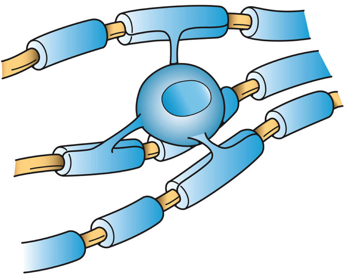
Oligodendrocytes (), also known as oligodendroglia, are a type of neuroglia whose main function is to provide the myelin sheath to neuronal axons in the central nervous system (CNS). Myelination gives metabolic support to, and insulates the axons of most vertebrates. A single oligodendrocyte can extend its Cellular extensions, processes to cover up to 40 axons, that can include multiple adjacent axons. The myelin sheath is segmented along the axon's length at gaps known as the nodes of Ranvier. In the peripheral nervous system the myelination of axons is carried out by Schwann cells. Oligodendrocytes are found exclusively in the CNS, which comprises the brain and spinal cord. They are the most widespread cell lineage, including oligodendrocyte progenitor cells, pre-myelinating cells, and mature myelinating oligodendrocytes in the CNS white matter. Non-myelinating oligodendrocytes are found in the grey matter surrounding and lying next to neuronal cell bodies. They are known as neu ... [...More Info...] [...Related Items...] OR: [Wikipedia] [Google] [Baidu] |
Astrocyte
Astrocytes (from Ancient Greek , , "star" and , , "cavity", "cell"), also known collectively as astroglia, are characteristic star-shaped glial cells in the brain and spinal cord. They perform many functions, including biochemical control of endothelial cells that form the blood–brain barrier, provision of nutrients to the nervous tissue, maintenance of extracellular ion balance, regulation of cerebral blood flow, and a role in the repair and scarring process of the brain and spinal cord following infection and traumatic injuries. The proportion of astrocytes in the brain is not well defined; depending on the counting technique used, studies have found that the astrocyte proportion varies by region and ranges from 20% to around 40% of all glia. Another study reports that astrocytes are the most numerous cell type in the brain. Astrocytes are the major source of cholesterol in the central nervous system. Apolipoprotein E transports cholesterol from astrocytes to neurons and ot ... [...More Info...] [...Related Items...] OR: [Wikipedia] [Google] [Baidu] |
Basement Membrane
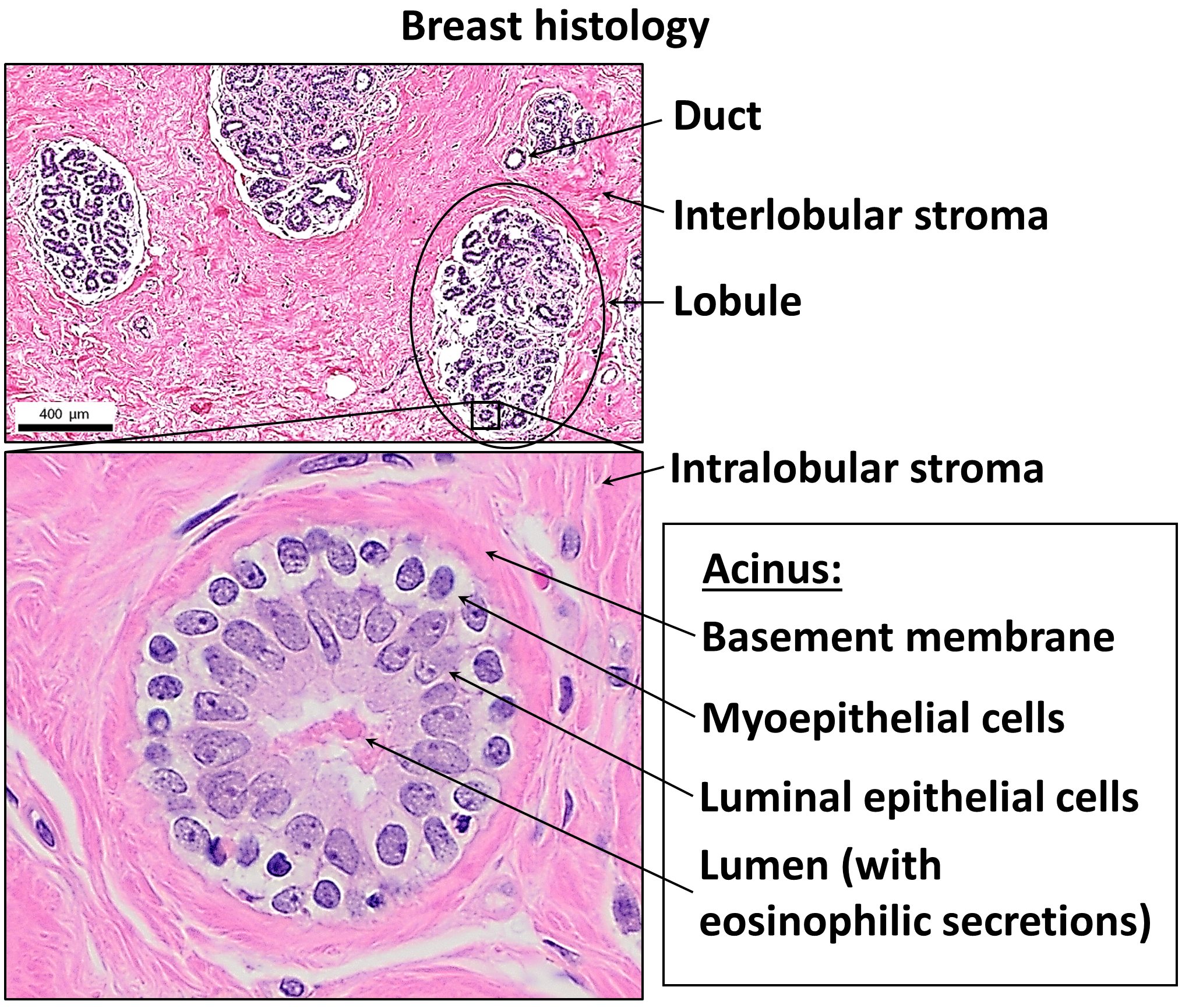
The basement membrane, also known as base membrane, is a thin, pliable sheet-like type of extracellular matrix that provides cell and tissue support and acts as a platform for complex signalling. The basement membrane sits between epithelial tissues including mesothelium and endothelium, and the underlying connective tissue. Structure As seen with the electron microscope, the basement membrane is composed of two layers, the basal lamina and the reticular lamina. The underlying connective tissue attaches to the basal lamina with collagen VII anchoring fibrils and fibrillin microfibrils. The basal lamina layer can further be subdivided into two layers based on their visual appearance in electron microscopy. The lighter-colored layer closer to the epithelium is called the lamina lucida, while the denser-colored layer closer to the connective tissue is called the lamina densa. The electron-dense lamina densa layer is about 30–70 nanometers thick and consists of an und ... [...More Info...] [...Related Items...] OR: [Wikipedia] [Google] [Baidu] |
Glycosaminoglycan
Glycosaminoglycans (GAGs) or mucopolysaccharides are long, linear polysaccharides consisting of repeating disaccharide units (i.e. two-sugar units). The repeating two-sugar unit consists of a uronic sugar and an amino sugar, except in the case of the sulfated glycosaminoglycan keratan, where, in place of the uronic sugar there is a galactose unit. GAGs are found in vertebrates, invertebrates and bacteria. Because GAGs are highly polar molecules and attract water; the body uses them as lubricants or shock absorbers. Mucopolysaccharidoses are a group of metabolic disorders in which abnormal accumulations of glycosaminoglycans occur due to enzyme deficiencies. Production Glycosaminoglycans vary greatly in molecular mass, disaccharide structure, and sulfation. This is because GAG synthesis is not template driven, as are proteins or nucleic acids, but constantly altered by processing enzymes. GAGs are classified into four groups, based on their core disaccharide structures: # H ... [...More Info...] [...Related Items...] OR: [Wikipedia] [Google] [Baidu] |
Neuroepithelium
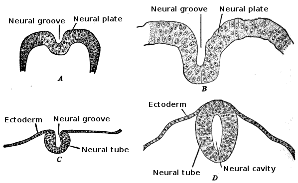
Neuroepithelial cells, or neuroectodermal cells, form the wall of the closed neural tube in early embryonic development. The neuroepithelial cells span the thickness of the tube's wall, connecting with the pial surface and with the ventricular or lumenal surface. They are joined at the lumen of the tube by junctional complexes, where they form a pseudostratified layer of epithelium called neuroepithelium. Neuroepithelial cells are the stem cells of the central nervous system, known as neural stem cells, and generate the intermediate progenitor cells known as radial glial cells, that differentiate into neurons and glia in the process of neurogenesis. Embryonic neural development Brain development During the third week of embryonic growth, the brain begins to develop in the early fetus in a process called morphogenesis. Neuroepithelial cells of the ectoderm begin multiplying rapidly and fold in forming the neural plate, which invaginates during the fourth week of embryonic grow ... [...More Info...] [...Related Items...] OR: [Wikipedia] [Google] [Baidu] |
Arachnoid Mater
The arachnoid mater (or simply arachnoid) is one of the three meninges, the protective membranes that cover the brain and spinal cord. It is so named because of its resemblance to a spider web. The arachnoid mater is a derivative of the neural crest mesoectoderm in the embryo. Structure The arachnoid mater is interposed between the two other meninges, the more superficial (closer to the surface) and much thicker dura mater and the deeper pia mater, from which it is separated by the subarachnoid space. The delicate arachnoid layer is not attached to the inside of the dura but against it, and surrounds the brain and spinal cord. It does not line the brain down into its sulci (folds), as does the pia mater, with the exception of the longitudinal fissure, which divides the left and right cerebral hemispheres. Cerebrospinal fluid (CSF) flows under the arachnoid in the subarachnoid space, within a meshwork of trabeculae which span between the arachnoid and the pia. The arachnoid ma ... [...More Info...] [...Related Items...] OR: [Wikipedia] [Google] [Baidu] |


