|
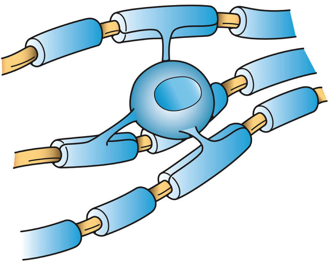
Oligodendrocyte
Oligodendrocytes (), also known as oligodendroglia, are a type of neuroglia whose main function is to provide the myelin sheath to neuronal axons in the central nervous system (CNS). Myelination gives metabolic support to, and insulates the axons of most vertebrates. A single oligodendrocyte can extend its Cellular extensions, processes to cover up to 40 axons, that can include multiple adjacent axons. The myelin sheath is segmented along the axon's length at gaps known as the nodes of Ranvier. In the peripheral nervous system the myelination of axons is carried out by Schwann cells. Oligodendrocytes are found exclusively in the CNS, which comprises the brain and spinal cord. They are the most widespread cell lineage, including oligodendrocyte progenitor cells, pre-myelinating cells, and mature myelinating oligodendrocytes in the CNS white matter. Non-myelinating oligodendrocytes are found in the grey matter surrounding and lying next to neuronal cell bodies. They are known as neu ... [...More Info...] [...Related Items...] OR: [Wikipedia] [Google] [Baidu] |
Oligodendrocyte Progenitor Cell
Oligodendrocyte progenitor cells (OPCs), also known as oligodendrocyte precursor cells, NG2-glia, O2A cells, or polydendrocytes, are a subtype of glia in the central nervous system named for their essential role as precursors to oligodendrocytes and myelin. They are typically identified in the human by co-expression of PDGFRA and CSPG4. OPCs play a critical role in developmental and adult myelinogenesis. They give rise to oligodendrocytes, which then wrap around axons and provide electrical insulation by forming a myelin sheath. This enables faster action potential propagation and high fidelity transmission without a need for an increase in axonal diameter. The loss or lack of OPCs, and consequent lack of differentiated oligodendrocytes, is associated with a loss of myelination and subsequent impairment of neurological functions. In addition, OPCs express receptors for various neurotransmitters and undergo membrane depolarization when they receive synaptic inputs from neurons. ... [...More Info...] [...Related Items...] OR: [Wikipedia] [Google] [Baidu] |
Glial Cell
Glia, also called glial cells (gliocytes) or neuroglia, are non-neuronal cells in the central nervous system (the brain and the spinal cord) and in the peripheral nervous system that do not produce electrical impulses. The neuroglia make up more than one half the volume of neural tissue in the human body. They maintain homeostasis, form myelin, and provide support and protection for neurons. In the central nervous system, glial cells include oligodendrocytes (that produce myelin), astrocytes, ependymal cells and microglia, and in the peripheral nervous system they include Schwann cells (that produce myelin), and satellite cells. Function They have four main functions: * to surround neurons and hold them in place * to supply nutrients and oxygen to neurons * to insulate one neuron from another * to destroy pathogens and remove dead neurons. They also play a role in neurotransmission and synaptic connections, and in physiological processes such as breathing. While glia w ... [...More Info...] [...Related Items...] OR: [Wikipedia] [Google] [Baidu] |
Neuroglia
Glia, also called glial cells (gliocytes) or neuroglia, are non- neuronal cells in the central nervous system (the brain and the spinal cord) and in the peripheral nervous system that do not produce electrical impulses. The neuroglia make up more than one half the volume of neural tissue in the human body. They maintain homeostasis, form myelin, and provide support and protection for neurons. In the central nervous system, glial cells include oligodendrocytes (that produce myelin), astrocytes, ependymal cells and microglia, and in the peripheral nervous system they include Schwann cells (that produce myelin), and satellite cells. Function They have four main functions: * to surround neurons and hold them in place * to supply nutrients and oxygen to neurons * to insulate one neuron from another * to destroy pathogens and remove dead neurons. They also play a role in neurotransmission and synaptic connections, and in physiological processes such as breathing. While ... [...More Info...] [...Related Items...] OR: [Wikipedia] [Google] [Baidu] |
Myelination
Myelination, or myelinogenesis, is the formation and development of myelin sheaths in the nervous system, typically initiated in late prenatal neurodevelopment and continuing throughout postnatal development. The term ''myelinogenesis'' is also sometimes used to differentiate the very early stages of embryonic myelination. Myelin is formed by oligodendrocytes in the central nervous system and Schwann cells in the peripheral nervous system. Myelination continues throughout the lifespan to support learning and memory via neural circuit plasticity as well as remyelination following injury. Successful myelination of axons increases action potential speed by enabling saltatory conduction, which is essential for timely signal conduction between spatially separate brain regions, as well as provides metabolic support to neurons. Stages Myelin is formed by oligodendrocytes in the central nervous system and Schwann cells in the peripheral nervous system. Therefore, the first stage of m ... [...More Info...] [...Related Items...] OR: [Wikipedia] [Google] [Baidu] |
Cellular Extensions
Cellular extensions also known as cytoplasmic protrusions and cytoplasmic processes are those structures that project from different cells, in the body, or in other organisms. Many of the extensions are cytoplasmic protrusions such as the axon and dendrite of a neuron, known also as cytoplasmic processes. Different glial cells project cytoplasmic processes. In the brain, the processes of astrocytes form terminal endfeet, foot processes that help to form protective barriers in the brain. In the kidneys specialised cells called podocytes extend processes that terminate in podocyte foot processes that cover capillaries in the nephron. End-processes may also be known as ''vascular footplates'', and in general may exhibit a pyramidal or finger-like morphology. Mural cells such as pericytes extend processes to wrap around capillaries. Foot-like processes are also present in Müller glia (modified astrocytes of the retina), pancreatic stellate cells, dendritic cells, oligodendro ... [...More Info...] [...Related Items...] OR: [Wikipedia] [Google] [Baidu] |
OLIG2
Oligodendrocyte transcription factor (OLIG2) is a basic helix-loop-helix ( bHLH) transcription factor encoded by the ''OLIG2'' gene. The protein is of 329 amino acids in length, 32 kDa in size and contains one basic helix-loop-helix DNA-binding domain. It is one of the three members of the bHLH family. The other two members are OLIG1 and OLIG3. The expression of OLIG2 is mostly restricted in central nervous system, where it acts as both an anti-neurigenic and a neurigenic factor at different stages of development. OLIG2 is well known for determining motor neuron and oligodendrocyte differentiation, as well as its role in sustaining replication in early development. It is mainly involved in diseases such as brain tumor and Down syndrome. Function OLIG2 is mostly expressed in restricted domains of the brain and spinal cord ventricular zone which give rise to oligodendrocytes and specific types of neurons. In the spinal cord, the pMN region sequentially generates motor neurons an ... [...More Info...] [...Related Items...] OR: [Wikipedia] [Google] [Baidu] |
Central Nervous System
The central nervous system (CNS) is the part of the nervous system consisting primarily of the brain, spinal cord and retina. The CNS is so named because the brain integrates the received information and coordinates and influences the activity of all parts of the bodies of bilateria, bilaterally symmetric and triploblastic animals—that is, all multicellular animals except sponges and Coelenterata, diploblasts. It is a structure composed of nervous tissue positioned along the Anatomical_terms_of_location#Rostral,_cranial,_and_caudal, rostral (nose end) to caudal (tail end) axis of the body and may have an enlarged section at the rostral end which is a brain. Only arthropods, cephalopods and vertebrates have a true brain, though precursor structures exist in onychophorans, gastropods and lancelets. The rest of this article exclusively discusses the vertebrate central nervous system, which is radically distinct from all other animals. Overview In vertebrates, the brain and spinal ... [...More Info...] [...Related Items...] OR: [Wikipedia] [Google] [Baidu] |
Nodes Of Ranvier
Nodes of Ranvier ( ), also known as myelin-sheath gaps, occur along a myelinated axon where the axolemma is exposed to the extracellular space. Nodes of Ranvier are uninsulated axonal domains that are high in sodium and potassium ion channels complexed with cell adhesion molecules, allowing them to participate in the exchange of ions required to regenerate the action potential. Nerve conduction in myelinated axons is referred to as saltatory conduction () due to the manner in which the action potential seems to "jump" from one node to the next along the axon. This results in faster conduction of the action potential. The nodes of Ranvier are present in both the peripheral and central nervous systems. Overview The nodes are primarily composed of sodium and potassium voltage-gated ion channels; CAMs such as neurofascin-186 and NrCAM; and cytoskeletal adaptor proteins such as ankyrin-G and spectrinβIV. Many vertebrate axons are surrounded by a myelin sheath, allowing rap ... [...More Info...] [...Related Items...] OR: [Wikipedia] [Google] [Baidu] |
CSPG4
Chondroitin sulfate proteoglycan 4, also known as melanoma-associated chondroitin sulfate proteoglycan (MCSP) or neuron-glial antigen 2 (NG2), is a chondroitin sulfate proteoglycan that in humans is encoded by the ''CSPG4'' gene. Function CSPG4 plays a role in stabilizing cell-substratum interactions during early events of melanoma cell spreading on endothelial basement membranes. It represents an integral membrane chondroitin sulfate proteoglycan expressed by human malignant melanoma cells. Implications in disease CSPG4/NG2 is also a hallmark protein of oligodendrocyte progenitor cells (OPCs) and OPC dysfunction has been implicated as a candidate pathophysiological mechanism of familial schizophrenia. A research group investigating the role of genetics in schizophrenia, reported, two rare missense mutations in ''CSPG4'' gene'','' segregating within families (''CSPG4A131T'' and ''CSPG4V901G'' mutations). The researchers also demonstrate that the induced pluripotent stem ce ... [...More Info...] [...Related Items...] OR: [Wikipedia] [Google] [Baidu] |
Axon
An axon (from Greek ἄξων ''áxōn'', axis) or nerve fiber (or nerve fibre: see American and British English spelling differences#-re, -er, spelling differences) is a long, slender cellular extensions, projection of a nerve cell, or neuron, in Vertebrate, vertebrates, that typically conducts electrical impulses known as action potentials away from the Soma (biology), nerve cell body. The function of the axon is to transmit information to different neurons, muscles, and glands. In certain sensory neurons (pseudounipolar neurons), such as those for touch and warmth, the axons are called afferent nerve fibers and the electrical impulse travels along these from the peripheral nervous system, periphery to the cell body and from the cell body to the spinal cord along another branch of the same axon. Axon dysfunction can be the cause of many inherited and acquired neurological disorders that affect both the Peripheral nervous system, peripheral and Central nervous system, central ne ... [...More Info...] [...Related Items...] OR: [Wikipedia] [Google] [Baidu] |


