|
Anterior Choroidal Artery
The anterior choroidal artery is a bilaterally paired artery of the brain. It is typically a branch of the internal carotid artery which supplies the choroid plexus of lateral ventricle and third ventricle as well as numerous structures of the brain. Occlusion of the artery can result in loss of sensation, loss of part of the visual field, and impaired movement, all on the opposite side of the body as the occlusion. Structure Origin The anterior choroidal artery typically originates from the internal carotid artery. It may (rarely) instead arise from the middle cerebral artery. It originates from the distal internal carotid artery (ICA) 5 mm distal to the origin of the posterior communicating artery and just proximal to the terminal bifurcation of the ICA. Course It initially course posterolaterally on the inferior surface of the cerebral hemisphere alongside the optic tract, crossing the tract medial-to-lateral inferior to the tract. At the level of the lateral geniculat ... [...More Info...] [...Related Items...] OR: [Wikipedia] [Google] [Baidu] |
Circle Of Willis
The circle of Willis (also called Willis' circle, loop of Willis, cerebral arterial circle, and Willis polygon) is a circulatory anastomosis that supplies blood to the brain and surrounding structures in reptiles, birds and mammals, including humans. It is named after Thomas Willis (1621–1675), an English physician. Structure The circle of Willis is a part of the cerebral circulation and is composed of the following arteries: * Anterior cerebral artery (left and right) at their A1 segments * Anterior communicating artery * Internal carotid artery (left and right) at its distal tip (carotid terminus) * Posterior cerebral artery (left and right) at their P1 segments * Posterior communicating artery (left and right) The middle cerebral arteries, supplying the brain, are also considered part of the Circle of Willis Origin of arteries The left and right internal carotid arteries arise from the left and right common carotid arteries. The posterior communicating artery is given ... [...More Info...] [...Related Items...] OR: [Wikipedia] [Google] [Baidu] |
Internal Capsule
The internal capsule is a paired white matter structure, as a two-way nerve tract, tract, carrying afferent nerve fiber, ascending and efferent nerve fiber, descending axon, fibers, to and from the cerebral cortex. The internal capsule is situated in the Anatomical terms of location#Medial and lateral, inferomedial part of each cerebral hemisphere of the brain. It carries information past the subcortical basal ganglia. As it courses it separates the caudate nucleus and the thalamus from the putamen and the globus pallidus. It also separates the caudate nucleus and the putamen in the dorsal striatum, a brain region involved in motor and reward pathways. The internal capsule is V-shaped in transection forming an anterior and posterior limb, with the angle between them called the genu. The corticospinal tract constitutes a large part of the internal capsule, carrying motor information from the primary motor cortex to the lower motor neurons in the spinal cord. Above the basal gangli ... [...More Info...] [...Related Items...] OR: [Wikipedia] [Google] [Baidu] |
Ischemia
Ischemia or ischaemia is a restriction in blood supply to any tissue, muscle group, or organ of the body, causing a shortage of oxygen that is needed for cellular metabolism (to keep tissue alive). Ischemia is generally caused by problems with blood vessels, with resultant damage to or dysfunction of tissue, i.e., hypoxia and microvascular dysfunction. It also implies local hypoxia in a part of a body resulting from constriction (such as vasoconstriction, thrombosis, or embolism). Ischemia causes not only insufficiency of oxygen but also reduced availability of nutrients and inadequate removal of metabolic wastes. Ischemia can be partial (poor perfusion) or total blockage. The inadequate delivery of oxygenated blood to the organs must be resolved either by treating the cause of the inadequate delivery or reducing the oxygen demand of the system that needs it. For example, patients with myocardial ischemia have a decreased blood flow to the heart and are prescribe ... [...More Info...] [...Related Items...] OR: [Wikipedia] [Google] [Baidu] |
Hemiplegia
Hemiparesis, also called unilateral paresis, is the weakness of one entire side of the body ('' hemi-'' means "half"). Hemiplegia, in its most severe form, is the complete paralysis of one entire side of the body. Either hemiparesis or hemiplegia can result from a variety of medical causes, including congenital conditions, trauma, tumors, traumatic brain injury and stroke.Detailed article about hemiparesis at Disabled-World.com Signs and symptoms Different types of hemiparesis can impair different bodily functions. Some effects, such as weakness or partial paralysis of a limb on the affected side, are generally always to be expected. Other impairments can appear, upon external examination, to be unrelated to the limb weakness, but are nevertheless also caused by damage to t ...[...More Info...] [...Related Items...] OR: [Wikipedia] [Google] [Baidu] |
Hemianopsia
Hemianopsia, or hemianopia, is a loss of vision or blindness ( anopsia) in half the visual field, usually on one side of the vertical midline. The most common causes of this damage are stroke, brain tumor, and trauma. This article deals only with permanent hemianopsia, and not with transitory or temporary hemianopsia, as identified by William Wollaston PRS in 1824. Temporary hemianopsia can occur in the aura phase of migraine. Etymology The word ''hemianopsia'' is from Greek origins, where: * ''hemi'' means "half", * ''an'' means "without", and * ''opsia'' means "seeing". Types When the pathology involves both eyes, it is either homonymous or heteronymous. Homonymous hemianopsia Paris as seen with left homonymous hemianopsia A homonymous hemianopsia is the loss of half of the visual field on the same side in both eyes. The visual images that we see to the right side travel from both eyes to the left side of the brain, while the visual images we see to the left side i ... [...More Info...] [...Related Items...] OR: [Wikipedia] [Google] [Baidu] |
Contralateral
Standard anatomical terms of location are used to describe unambiguously the anatomy of humans and other animals. The terms, typically derived from Latin or Greek roots, describe something in its standard anatomical position. This position provides a definition of what is at the front ("anterior"), behind ("posterior") and so on. As part of defining and describing terms, the body is described through the use of anatomical planes and axes. The meaning of terms that are used can change depending on whether a vertebrate is a biped or a quadruped, due to the difference in the neuraxis, or if an invertebrate is a non-bilaterian. A non-bilaterian has no anterior or posterior surface for example but can still have a descriptor used such as proximal or distal in relation to a body part that is nearest to, or furthest from its middle. International organisations have determined vocabularies that are often used as standards for subdisciplines of anatomy. For example, '' Terminolog ... [...More Info...] [...Related Items...] OR: [Wikipedia] [Google] [Baidu] |
Crus Cerebri
The cerebral crus (crus cerebri. ''crus'' means ‘leg’ in Latin.) is the anterior portion of the cerebral peduncle which contains the motor tracts, traveling from the cerebral cortex to the pons and spine. The plural of which is cerebral crura. In some older texts, this is called the cerebral peduncle, but presently, it is usually limited to just the anterior white matter portion of it. Additional images File:Human brain frontal (coronal) section description 2.JPG, Human brain frontal (coronal) section, number 28 indicates the cerebral crus. See also * Efferent nerve fiber * Motor neuron (efferent neuron) * Motor nerve References External links * * NIF Search - Cerebral Crusvia the Neuroscience Information Framework The Neuroscience Information Framework is a repository of global neuroscience web resources, including experimental, clinical, and translational neuroscience databases, knowledge bases, atlases, and genetic/ genomic resources and provides many aut ... ... [...More Info...] [...Related Items...] OR: [Wikipedia] [Google] [Baidu] |
Red Nucleus
The red nucleus or nucleus ruber is a structure in the rostral midbrain involved in motor coordination. The red nucleus is pale pink, which is believed to be due to the presence of iron in at least two different forms: hemoglobin and ferritin. The structure is located in the midbrain tegmentum next to the substantia nigra and comprises caudal magnocellular and rostral parvocellular components. The red nucleus and substantia nigra are subcortical centers of the extrapyramidal motor system. Function In a vertebrate without a significant corticospinal tract, gait is mainly controlled by the red nucleus. However, in primates, where the corticospinal tract is dominant, the rubrospinal tract may be regarded as vestigial in motor function. Therefore, the red nucleus is less important in primates than in many other mammals. Nevertheless, the crawling of babies is controlled by the red nucleus, as is arm swinging in typical walking. The red nucleus may play an additional role ... [...More Info...] [...Related Items...] OR: [Wikipedia] [Google] [Baidu] |
Substantia Nigra
The substantia nigra (SN) is a basal ganglia structure located in the midbrain that plays an important role in reward and movement. ''Substantia nigra'' is Latin for "black substance", reflecting the fact that parts of the substantia nigra appear darker than neighboring areas due to high levels of neuromelanin in dopaminergic neurons. Parkinson's disease is characterized by the loss of dopaminergic neurons in the substantia nigra pars compacta. Although the substantia nigra appears as a continuous band in brain sections, anatomical studies have found that it actually consists of two parts with very different connections and functions: the pars compacta (SNpc) and the pars reticulata (SNpr). The pars compacta serves mainly as a projection to the basal ganglia circuit, supplying the striatum with dopamine. The pars reticulata conveys signals from the basal ganglia to numerous other brain structures. Structure The substantia nigra, along with four other nuclei, is ... [...More Info...] [...Related Items...] OR: [Wikipedia] [Google] [Baidu] |
Amygdala
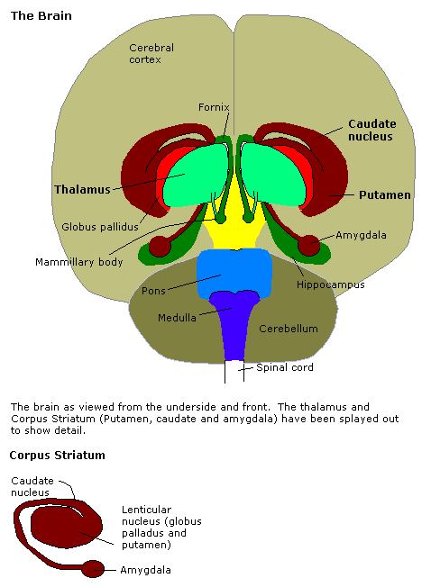
The amygdala (; : amygdalae or amygdalas; also '; Latin from Greek language, Greek, , ', 'almond', 'tonsil') is a paired nucleus (neuroanatomy), nuclear complex present in the Cerebral hemisphere, cerebral hemispheres of vertebrates. It is considered part of the limbic system. In Primate, primates, it is located lateral and medial, medially within the temporal lobes. It consists of many nuclei, each made up of further subnuclei. The subdivision most commonly made is into the Basolateral amygdala, basolateral, Central nucleus of the amygdala, central, cortical, and medial nuclei together with the intercalated cells of the amygdala, intercalated cell clusters. The amygdala has a primary role in the processing of memory, decision making, decision-making, and emotions, emotional responses (including fear, anxiety, and aggression). The amygdala was first identified and named by Karl Friedrich Burdach in 1822. Structure Thirteen Nucleus (neuroanatomy), nuclei have been identif ... [...More Info...] [...Related Items...] OR: [Wikipedia] [Google] [Baidu] |
Hippocampus
The hippocampus (: hippocampi; via Latin from Ancient Greek, Greek , 'seahorse'), also hippocampus proper, is a major component of the brain of humans and many other vertebrates. In the human brain the hippocampus, the dentate gyrus, and the subiculum are components of the hippocampal formation located in the limbic system. The hippocampus plays important roles in the Memory consolidation, consolidation of information from short-term memory to long-term memory, and in spatial memory that enables Navigation#Navigation in spatial cognition, navigation. In humans, and other primates the hippocampus is located in the archicortex, one of the three regions of allocortex, in each cerebral hemisphere, hemisphere with direct neural projections to, and reciprocal indirect projections from the neocortex. The hippocampus, as the medial pallium, is a structure found in all vertebrates. In Alzheimer's disease (and other forms of dementia), the hippocampus is one of the first regions of th ... [...More Info...] [...Related Items...] OR: [Wikipedia] [Google] [Baidu] |
Tuber Cinereum
The tuber cinereum is the portion of hypothalamus forming the floor of the third ventricle situated between the optic chiasm, and the mammillary bodies. The tuberal region is one of the three regions of the hypothalamus, the other two being the chiasmatic region and the mamillary region. Structure The tuber cinereum is a convex mass of grey matter, a ventral/inferior distention of the hypothalamus forming the floor of the third ventricle. The portion of the tuber cinereum at the base of the infundibulum (pituitary stalk) is the median eminence; the infundibulum extends ventrally/inferiorly from the median eminence to become continuous with the infundibulum. The arcuate nucleus is a part of the tuber cinereum. The lateral portions of tuber cinereum lodge the lateral tuberal nucleus, and tuberomammillary nucleus. The basolateral aspect of the tuber cinereum often presents slight elevations produced by the underlying lateral tuberal nucleus - the lateral eminence. Relations ... [...More Info...] [...Related Items...] OR: [Wikipedia] [Google] [Baidu] |


