|
Acid Fuchsin
Acid fuchsin or fuchsine acid, (also called Acid Violet 19 and C.I. 42685) is an acidic magenta dye with the chemical formula C20H17N3Na2O9S3. It is a sodium sulfonate derivative of fuchsine. Acid fuchsin has wide use in histology, and is one of the dyes used in Masson's trichrome stain. This method is commonly used to stain cytoplasm and nuclei of tissue sections in the histology laboratory in order to distinguish muscle from collagen. The muscle stains red with the acid fuchsin, and the collagen is stained green or blue with Light Green SF yellowish or methyl blue. It can also be used to identify growing bacteria. See also * New fuchsine * Pararosanilin * Verhoeff’s Stain Verhoeff's stain, also known as Verhoeff's elastic stain (VEG) or Verhoeff–Van Gieson stain (VVG), is a staining protocol used in histology, developed by American ophthalmic surgeon and pathologist Frederick Herman Verhoeff (1874–1968) in 190 ... * Pollen grain staining (Alexander's stain) ... [...More Info...] [...Related Items...] OR: [Wikipedia] [Google] [Baidu] |
Colour Index International
Colour Index International (CI) is a reference database jointly maintained bSDC Enterprisesand the American Association of Textile Chemists and Colorists. It currently contains over 27,000 individual products listed under 13,000 Colour Index Generic Names. It was first printed in 1924 but is now published solely on the Internet. The index serves as a common reference database of manufactured colour products and is used by manufacturers and consumers, such as artists and decorators. Colourants (both dyes and pigments) are listed using a dual classification which use the Colour Index Generic Name the prime identifier and Colour Index Constitution Numbers. These numbers are prefixed with C.I. for example, C.I. Acid Orange 7 or C.I. 15510. (This abbreviation is sometimes mistakenly thought to be CL, due to the font used to display it.) The generic name lists first the class of dye (acid dye, disperse dye, etc.), then its hue (e.g., orange), followed by a number assigned by the Col ... [...More Info...] [...Related Items...] OR: [Wikipedia] [Google] [Baidu] |
Light Green SF Yellowish
Light, visible light, or visible radiation is electromagnetic radiation that can be perceived by the human eye. Visible light spans the visible spectrum and is usually defined as having wavelengths in the range of 400–700 nanometres (nm), corresponding to frequencies of 750–420 terahertz. The visible band sits adjacent to the infrared (with longer wavelengths and lower frequencies) and the ultraviolet (with shorter wavelengths and higher frequencies), called collectively '' optical radiation''. In physics, the term "light" may refer more broadly to electromagnetic radiation of any wavelength, whether visible or not. In this sense, gamma rays, X-rays, microwaves and radio waves are also light. The primary properties of light are intensity, propagation direction, frequency or wavelength spectrum, and polarization. Its speed in vacuum, , is one of the fundamental constants of nature. All electromagnetic radiation exhibits some properties of both particles and waves. Si ... [...More Info...] [...Related Items...] OR: [Wikipedia] [Google] [Baidu] |
Staining Dyes
Staining is a technique used to enhance contrast in samples, generally at the microscopic level. Stains and dyes are frequently used in histology (microscopic study of biological tissues), in cytology (microscopic study of cells), and in the medical fields of histopathology, hematology, and cytopathology that focus on the study and diagnoses of diseases at the microscopic level. Stains may be used to define biological tissues (highlighting, for example, muscle fibers or connective tissue), cell populations (classifying different blood cells), or organelles within individual cells. In biochemistry, it involves adding a class-specific (DNA, proteins, lipids, carbohydrates) dye to a substrate to qualify or quantify the presence of a specific compound. Staining and fluorescent tagging can serve similar purposes. Biological staining is also used to mark cells in flow cytometry, and to flag proteins or nucleic acids in gel electrophoresis. Light microscopes are used for viewing s ... [...More Info...] [...Related Items...] OR: [Wikipedia] [Google] [Baidu] |
Pollen
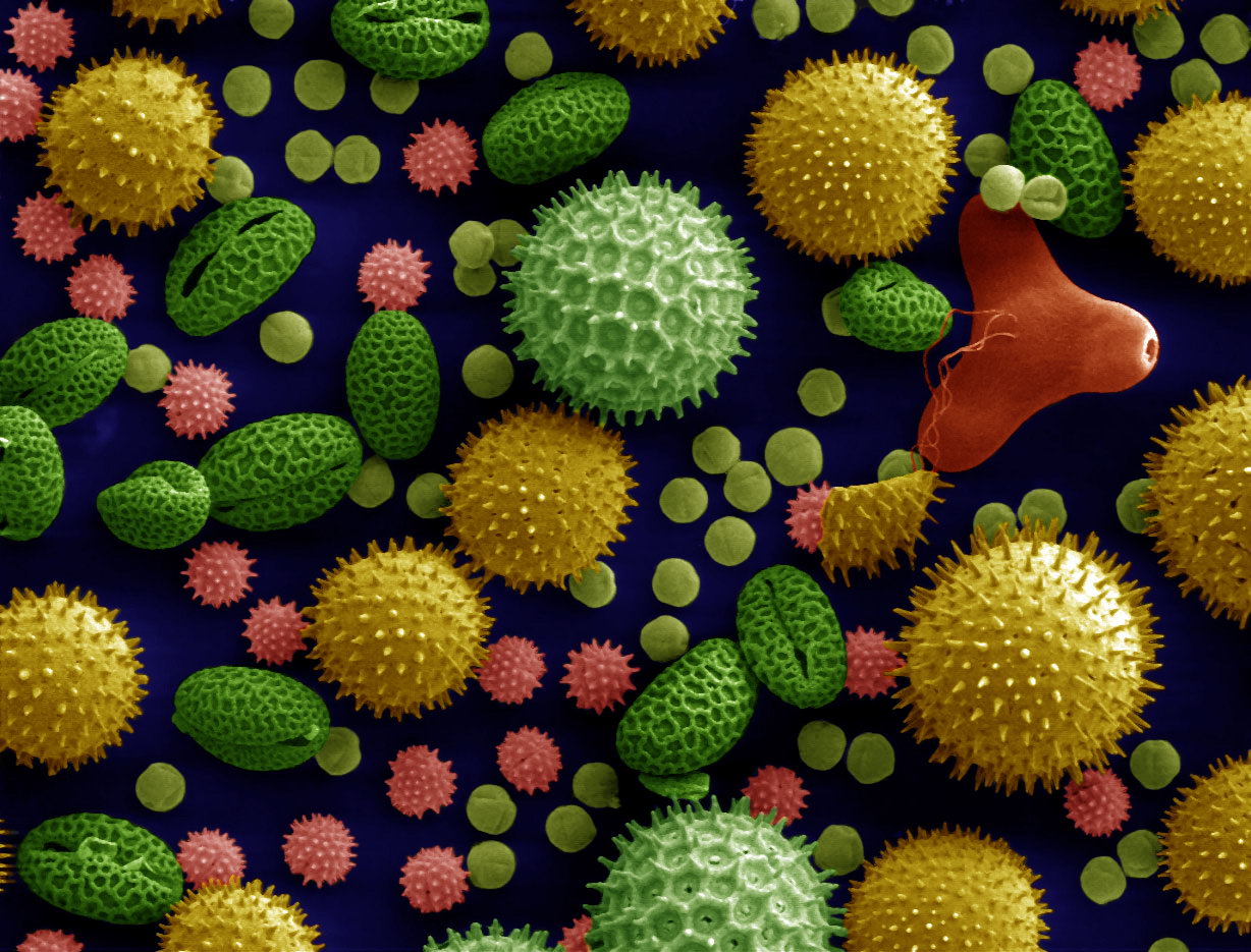
Pollen is a powdery substance produced by most types of flowers of seed plants for the purpose of sexual reproduction. It consists of pollen grains (highly reduced Gametophyte#Heterospory, microgametophytes), which produce male gametes (sperm cells). Pollen grains have a hard coat made of sporopollenin that protects the gametophytes during the process of their movement from the stamens to the pistil of flowering plants, or from the male Conifer cone, cone to the female cone of gymnosperms. If pollen lands on a compatible pistil or female cone, it Germination, germinates, producing a pollen tube that transfers the sperm to the ovule containing the female gametophyte. Individual pollen grains are small enough to require magnification to see detail. The study of pollen is called palynology and is highly useful in paleoecology, paleontology, archaeology, and Forensic science, forensics. Pollen in plants is used for transferring Ploidy#Haploid and monoploid, haploid male genetic ma ... [...More Info...] [...Related Items...] OR: [Wikipedia] [Google] [Baidu] |
Verhoeff’s Stain
Verhoeff's stain, also known as Verhoeff's elastic stain (VEG) or Verhoeff–Van Gieson stain (VVG), is a staining protocol used in histology, developed by American ophthalmic surgeon and pathologist Frederick Herman Verhoeff (1874–1968) in 1908. The formulation is used to demonstrate normal or pathologic elastic fibers. Verhoeff's stain forms a variety of cationic, anionic and non-ionic bonds with elastin, the main constituent of elastic fiber tissue. Elastin has a strong affinity for the iron-hematoxylin complex formed by the reagents in the stain and will hence retain dye longer than other tissue elements. This allows elastin to remain stained while the remaining tissue elements are decolorized. Sodium thiosulfate is used to remove excess iodine and a counterstain (most often Van Gieson's stain) is used to contrast the principal stain. Elastic fibers and cell nuclei are stained black, collagen fibers are stained red, and other tissue elements including cytoplasm are st ... [...More Info...] [...Related Items...] OR: [Wikipedia] [Google] [Baidu] |
Pararosanilin
Pararosaniline, pararosaniline free base, Basic Red 9, or C.I. 42500 is an organic compound with the formula . It is the free base form of pararosaniline hydrochloride, , a magenta solid with a variety of uses as a dye. It is one of the four components of basic fuchsine. It is structurally related to other triarylmethane dyes called methyl violets (e.g. crystal violet) which feature methyl groups on nitrogen. It is prepared by the condensation of aniline and . Alternatively, it arises from the oxidation of 4,4'-bis(aminophenyl)methane in the presence of aniline. Uses *It is used to dye polyacrylonitrile fibers. *It is used to detect sulfur dioxide. *Pararosaniline is used as a colorimetric test for aldehydes, in the Schiff test. It is the only basic fuchsine component suitable for making the aldehyde-fuchsine stain for pancreatic islet beta cells. *It has use as an antischistosomal. Related compounds * 4,4'-Thiodianiline * 4,4'-Methylenedianiline * 4,4'-Oxydianiline * Dapso ... [...More Info...] [...Related Items...] OR: [Wikipedia] [Google] [Baidu] |
New Fuchsine
New fuchsine is an organic compound with the formula H2N(CH3)C6H3)3Cl. It is a green-colored solid that is used as a dye of the triarylmethane class. It is one of the four components of basic fuchsine, and one of the two that are available as single dyes. The other is pararosaniline. It is prepared by condensation of ortho-toluidine with formaldehyde. This process initially gives the benzhydrol 4,4'-bis(dimethylamino)benzhydrol, which is further condensed to give the leuco (colorless) tertiary alcohol [(H2N(CH3)C6H3)3COH, which is oxidized in acid to give the dye. Use as dye and stain New fuchsine is used to dye polyacrylonitrile, paper, and leather. In biology, it can be used for staining (biology), staining acid-fast organisms, e.g. by Ziehl–Neelsen stain, and for making Schiff's reagent. As a primary amine, the dye can be diazotized in the laboratory, and the resulting diazonium salt used as a trapping agent in enzyme histochemistry.Lojda Z, Gossrau R, Schiebler TH (1 ... [...More Info...] [...Related Items...] OR: [Wikipedia] [Google] [Baidu] |
Methyl Blue
Methyl blue is a chemical compound with the molecular formula C37H27N3Na2O9S3. It is used as a stain in histology, and stains collagen blue in tissue sections. It can be used in some differential staining techniques such as Mallory's trichrome stain and Gömöri trichrome stain, and can be used to mediate electron transfer in microbial fuel cells. Fungal cell walls are also stained by methyl blue. Methyl blue is also available in mixture with water blue, under name Aniline Blue WS, Aniline blue, China blue, or Soluble blue; and in a solution of phenol, glycerol, and lactic acid under the name Lactophenol cotton blue (LPCB), which is used for microscopic visualization of fungi. Chemistry Methyl blue ( is[4-[(sulfophenyl)amino.html" ;"title="4-[Bis[4-[(sulfophenyl)amino">4-[Bis[4-[(sulfophenyl)aminohenylethylene]-2,5-cyclohexadien-1-ylidene]amino]-benzenesulfonic acid disodium salt) is distinctly different to methylene blue ([7-(dimethylamino)phenothiazin-3-ylidene]-dimethy ... [...More Info...] [...Related Items...] OR: [Wikipedia] [Google] [Baidu] |
Collagen
Collagen () is the main structural protein in the extracellular matrix of the connective tissues of many animals. It is the most abundant protein in mammals, making up 25% to 35% of protein content. Amino acids are bound together to form a triple helix of elongated fibril known as a collagen helix. It is mostly found in cartilage, bones, tendons, ligaments, and skin. Vitamin C is vital for collagen synthesis. Depending on the degree of biomineralization, mineralization, collagen tissues may be rigid (bone) or compliant (tendon) or have a gradient from rigid to compliant (cartilage). Collagen is also abundant in corneas, blood vessels, the Gut (anatomy), gut, intervertebral discs, and the dentin in teeth. In muscle tissue, it serves as a major component of the endomysium. Collagen constitutes 1% to 2% of muscle tissue and 6% by weight of skeletal muscle. The fibroblast is the most common cell creating collagen in animals. Gelatin, which is used in food and industry, is collagen t ... [...More Info...] [...Related Items...] OR: [Wikipedia] [Google] [Baidu] |
Acid
An acid is a molecule or ion capable of either donating a proton (i.e. Hydron, hydrogen cation, H+), known as a Brønsted–Lowry acid–base theory, Brønsted–Lowry acid, or forming a covalent bond with an electron pair, known as a Lewis acid. The first category of acids are the proton donors, or Brønsted–Lowry acid–base theory, Brønsted–Lowry acids. In the special case of aqueous solutions, proton donors form the hydronium ion H3O+ and are known as Acid–base reaction#Arrhenius theory, Arrhenius acids. Johannes Nicolaus Brønsted, Brønsted and Martin Lowry, Lowry generalized the Arrhenius theory to include non-aqueous solvents. A Brønsted–Lowry or Arrhenius acid usually contains a hydrogen atom bonded to a chemical structure that is still energetically favorable after loss of H+. Aqueous Arrhenius acids have characteristic properties that provide a practical description of an acid. Acids form aqueous solutions with a sour taste, can turn blue litmus red, and ... [...More Info...] [...Related Items...] OR: [Wikipedia] [Google] [Baidu] |
Cytoplasm
The cytoplasm describes all the material within a eukaryotic or prokaryotic cell, enclosed by the cell membrane, including the organelles and excluding the nucleus in eukaryotic cells. The material inside the nucleus of a eukaryotic cell and contained within the nuclear membrane is termed the nucleoplasm. The main components of the cytoplasm are the cytosol (a gel-like substance), the cell's internal sub-structures, and various cytoplasmic inclusions. In eukaryotes the cytoplasm also includes the nucleus, and other membrane-bound organelles.The cytoplasm is about 80% water and is usually colorless. The submicroscopic ground cell substance, or cytoplasmic matrix, that remains after the exclusion of the cell organelles and particles is groundplasm. It is the hyaloplasm of light microscopy, a highly complex, polyphasic system in which all resolvable cytoplasmic elements are suspended, including the larger organelles such as the ribosomes, mitochondria, plant plasti ... [...More Info...] [...Related Items...] OR: [Wikipedia] [Google] [Baidu] |


