|
Vitreous Humor
The vitreous body (''vitreous'' meaning "glass-like"; , ) is the clear gel that fills the space between the lens and the retina of the eyeball (the vitreous chamber) in humans and other vertebrates. It is often referred to as the vitreous humor (also spelled humour), from Latin meaning liquid, or simply "the vitreous". Vitreous fluid or "liquid vitreous" is the liquid component of the vitreous gel, found after a vitreous detachment. It is not to be confused with the aqueous humor, the other fluid in the eye that is found between the cornea and lens. Structure The vitreous humor is a transparent, colorless, gelatinous mass that fills the space in the eye between the lens and the retina. It is surrounded by a layer of collagen called the vitreous membrane (or hyaloid membrane or vitreous cortex) separating it from the rest of the eye. It makes up four-fifths of the volume of the eyeball. The vitreous humour is fluid-like near the centre, and gel-like near the edges. The vitreou ... [...More Info...] [...Related Items...] OR: [Wikipedia] [Google] [Baidu] |
Human Eye
The human eye is a sensory organ in the visual system that reacts to light, visible light allowing eyesight. Other functions include maintaining the circadian rhythm, and Balance (ability), keeping balance. The eye can be considered as a living optics, optical device. It is approximately spherical in shape, with its outer layers, such as the outermost, white part of the eye (the sclera) and one of its inner layers (the pigmented choroid) keeping the eye essentially stray light, light tight except on the eye's optic axis. In order, along the optic axis, the optical components consist of a first lens (the cornea, cornea—the clear part of the eye) that accounts for most of the optical power of the eye and accomplishes most of the Focus (optics), focusing of light from the outside world; then an aperture (the pupil) in a Diaphragm (optics), diaphragm (the Iris (anatomy), iris—the coloured part of the eye) that controls the amount of light entering the interior of the eye; then an ... [...More Info...] [...Related Items...] OR: [Wikipedia] [Google] [Baidu] |
Vitreous Membrane
The vitreous membrane (or hyaloid membrane or vitreous cortex) is a layer of collagen separating the vitreous humour from the rest of the eye. At least two parts have been identified anatomically. The posterior hyaloid membrane separates the rear of the vitreous from the retina. It is a false anatomical membrane. The anterior hyaloid membrane separates the front of the vitreous from the lens (anatomy), lens. Andres Bernal, Jean-Marie Parel, Fabrice MannsEvidence for posterior zonular fiber attachment on the anterior hyaloid membrane "Investigative Ophthalmology and Visual Science" 2006, 47, 4708-4713. Bernal et al. describe it "as a delicate structure in the form of a thin layer that runs from the pars plana to the posterior lens, where it shares its attachment with the posterior zonule via Weigert's ligament, also known as Egger's line". References External links Image at ivy-rose.co.uk Human eye anatomy {{eye-stub ... [...More Info...] [...Related Items...] OR: [Wikipedia] [Google] [Baidu] |
Glycosaminoglycan
Glycosaminoglycans (GAGs) or mucopolysaccharides are long, linear polysaccharides consisting of repeating disaccharide units (i.e. two-sugar units). The repeating two-sugar unit consists of a uronic sugar and an amino sugar, except in the case of the sulfated glycosaminoglycan keratan, where, in place of the uronic sugar there is a galactose unit. GAGs are found in vertebrates, invertebrates and bacteria. Because GAGs are highly polar molecules and attract water; the body uses them as lubricants or shock absorbers. Mucopolysaccharidoses are a group of metabolic disorders in which abnormal accumulations of glycosaminoglycans occur due to enzyme deficiencies. Production Glycosaminoglycans vary greatly in molecular mass, disaccharide structure, and sulfation. This is because GAG synthesis is not template driven, as are proteins or nucleic acids, but constantly altered by processing enzymes. GAGs are classified into four groups, based on their core disaccharide structures: # H ... [...More Info...] [...Related Items...] OR: [Wikipedia] [Google] [Baidu] |
Hyaluronan
Hyaluronic acid (; abbreviated HA; conjugate acid, conjugate base hyaluronate), also called hyaluronan, is an anion#Anions and cations, anionic, Sulfation, nonsulfated glycosaminoglycan distributed widely throughout connective tissue, connective, epithelial tissue, epithelial, and neural tissues. It is unique among glycosaminoglycans as it is non-sulfated, forms in the plasma membrane instead of the Golgi apparatus, and can be very large: human Synovial fluid, synovial HA averages about per molecule, or about 20,000 disaccharide monomers, while other sources mention . Medically, hyaluronic acid is used to treat osteoarthritis of the knee and dry eye, for wound repair, and as a cosmetic filler. The average 70 kg (150 lb) person has roughly 15 grams of hyaluronan in the body, one third of which is turned over (i.e., degraded and synthesized) per day. As one of the chief components of the extracellular matrix, it contributes significantly to cell proliferation and ... [...More Info...] [...Related Items...] OR: [Wikipedia] [Google] [Baidu] |
Hyalocyte
Hyalocytes, also known as vitreous cells, are cells of the vitreous body, which is the clear gel that fills the space between the lens and the retina of the eye. Hyalocytes occur in the peripheral part of the vitreous body, and may produce hyaluronic acid and collagen fibrils, Hyalocytes are star-shaped (stellate) cells with oval nuclei. The development of the vitreous is organized into three stages: primary, secondary, and tertiary. During the primary stage, which occurs from 3–6 weeks, the basic components of the vitreous begin to form from the mesenchyme embryonic cell layer. Hyalocytes likely develop from the vascular primary vitreous. See also List of distinct cell types in the adult human body The list of human cell types provides an enumeration and description of the various specialized cells found within the human body, highlighting their distinct functions, characteristics, and contributions to overall physiological processes. Cell ... References Eye { ... [...More Info...] [...Related Items...] OR: [Wikipedia] [Google] [Baidu] |
Visual Field
The visual field is "that portion of space in which objects are visible at the same moment during steady fixation of the gaze in one direction"; in ophthalmology and neurology the emphasis is mostly on the structure inside the visual field and it is then considered “the field of functional capacity obtained and recorded by means of perimetry”.Strasburger, Hans; Pöppel, Ernst (2002). Visual Field. In G. Adelman & B.H. Smith (Eds): ''Encyclopedia of Neuroscience''; 3rd edition, on CD-ROM. Elsevier Science B.V., Amsterdam, New York. However, the visual field can also be understood as a predominantly ''perceptual'' concept and its definition then becomes that of the "spatial array of visual sensations available to observation in introspectionist psychological experiments" (for example in van Doorn et al., 2013). The corresponding concept for optical instruments and image sensors is the field of view (FOV). In humans and animals, the FOV refers to the area visible when eye mov ... [...More Info...] [...Related Items...] OR: [Wikipedia] [Google] [Baidu] |
Phagocyte
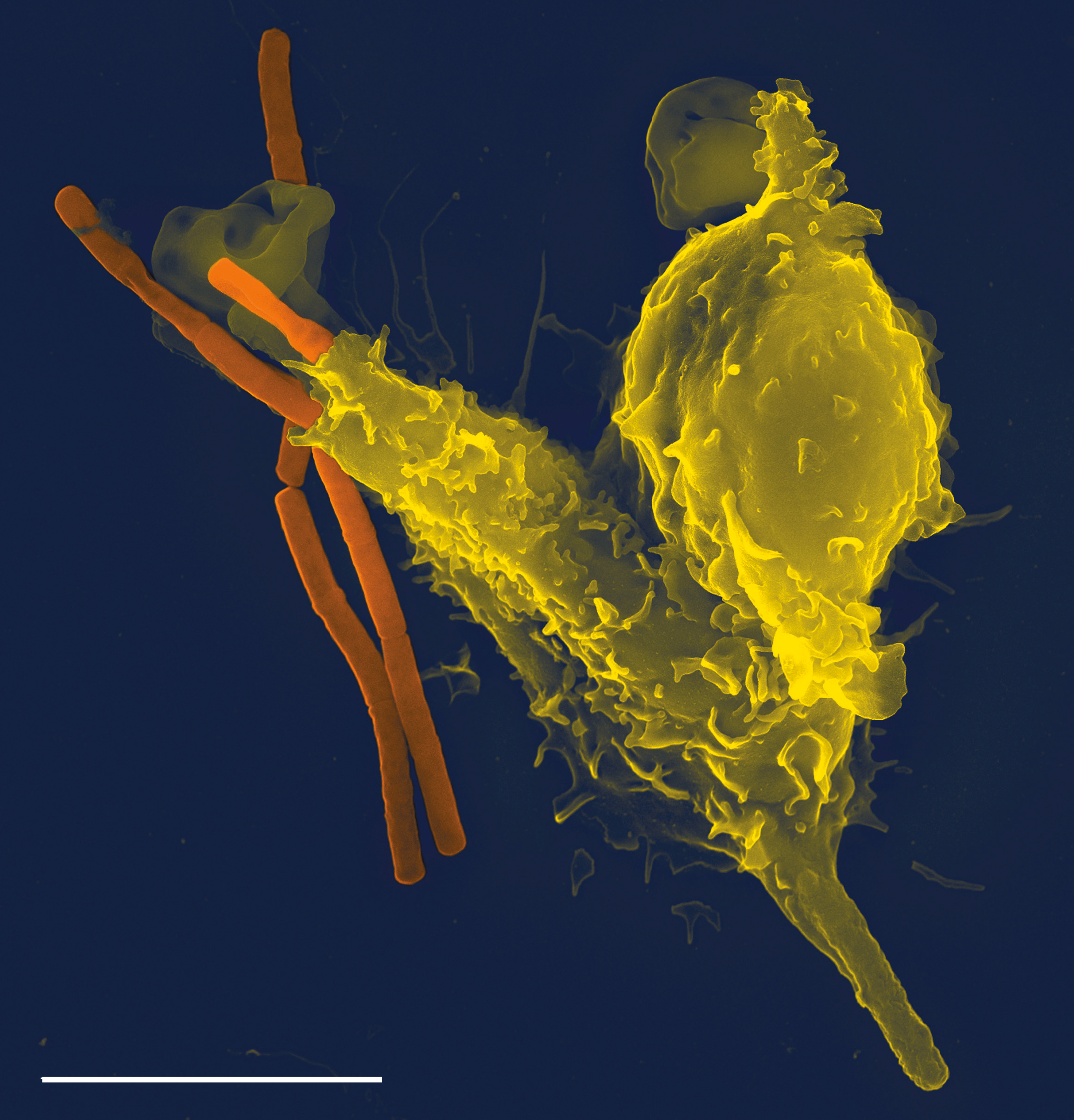
Phagocytes are cells that protect the body by ingesting harmful foreign particles, bacteria, and dead or dying cells. Their name comes from the Greek ', "to eat" or "devour", and "-cyte", the suffix in biology denoting "cell", from the Greek ''kutos,'' "hollow vessel". They are essential for fighting infections and for subsequent immunity. Phagocytes are important throughout the animal kingdom and are highly developed within vertebrates. One litre of human blood contains about six billion phagocytes. They were discovered in 1882 by Ilya Ilyich Mechnikov while he was studying starfish larvae.Ilya Mechnikov retrieved on November 28, 2008. Fro ''Physiology or Medicine 1901–192 ... [...More Info...] [...Related Items...] OR: [Wikipedia] [Google] [Baidu] |
Ciliary Processes
In the anatomy of the eye, the ciliary processes are formed by the inward folding of the various layers of the choroid, viz. the choroid proper and the lamina basalis, and are received between corresponding foldings of the suspensory ligament of the lens. Anatomy They are arranged in a circle, and form a sort of frill behind the iris, around the margin of the lens A lens is a transmissive optical device that focuses or disperses a light beam by means of refraction. A simple lens consists of a single piece of transparent material, while a compound lens consists of several simple lenses (''elements'') .... They vary from sixty to eighty in number, lie side by side, and may be divided into large and small; the former are about 2.5 mm. in length, and the latter, consisting of about one-third of the entire number, are situated in spaces between them, but without regular arrangement. They are attached by their periphery to three or four of the ridges of the orb ... [...More Info...] [...Related Items...] OR: [Wikipedia] [Google] [Baidu] |
Bergmeister's Papilla
Bergmeister's papilla arises from the centre of the optic disc, consists of a small tuft of fibrous tissue and represents a remnant of the fetal hyaloid artery. The hyaloid artery provides nutrition to the lens A lens is a transmissive optical device that focuses or disperses a light beam by means of refraction. A simple lens consists of a single piece of transparent material, while a compound lens consists of several simple lenses (''elements'') ... during development in the fetus, and runs forward to the lens from the optic disc. The optic disc is covered by a plaque of fibrous cells called the central supporting tissue meniscus of Kuhnt. This plaque forms a fibrous sheath around the hyaloid artery where it leaves the optic disc. At birth the hyaloid artery regresses, and is normally completely regressed by the time of eyelid opening. Bergmeister's papilla is a remnant of the hyaloid artery fibrous sheath and is frequently observed as an incidental clinical fin ... [...More Info...] [...Related Items...] OR: [Wikipedia] [Google] [Baidu] |
Mittendorf's Dot
The hyaloid artery is a branch of the ophthalmic artery, which is itself a branch of the internal carotid artery. It is contained within the optic stalk of the eye and extends from the optic disc through the vitreous humor to the lens. Usually fully regressed before birth, its purpose is to supply nutrients to the developing lens in the growing fetus. During the tenth week of development in humans (time varies depending on species), the lens grows independent of a blood supply and the hyaloid artery usually regresses. Its proximal portion remains as the central artery of the retina. Regression of the hyaloid artery leaves a clear central zone through the vitreous humor, called the hyaloid canal or Cloquet's canal. Cloquet's canal is named after the French physician Jules Germain Cloquet (1790–1883) who first described it. Occasionally the artery may not fully regress, resulting in the condition ''persistent hyaloid artery''. More commonly, small remnants of the artery may re ... [...More Info...] [...Related Items...] OR: [Wikipedia] [Google] [Baidu] |
Optic Disc
The optic disc or optic nerve head is the point of exit for ganglion cell axons leaving the eye. Because there are no rods or cones overlying the optic disc, it corresponds to a small blind spot in each eye. The ganglion cell axons form the optic nerve after they leave the eye. The optic disc represents the beginning of the optic nerve and is the point where the axons of retinal ganglion cells come together. The optic disc in a normal human eye carries 1–1.2 million afferent nerve fibers from the eye toward the brain. The optic disc is also the entry point for the major arteries that supply the retina with blood, and the exit point for the veins from the retina. Structure The optic disc is located 3 to 4 mm to the nasal side of the fovea. It is a vertical oval, with average dimensions of 1.76mm horizontally by 1.92mm vertically. There is a central depression, of variable size, called the optic cup. This depression can be a variety of shapes from a shallow indent ... [...More Info...] [...Related Items...] OR: [Wikipedia] [Google] [Baidu] |
Inner Limiting Membrane
The internal limiting membrane, or inner limiting membrane, is the boundary between the retina and the vitreous body, formed by astrocytes and the end feet of Müller cells Müller may refer to: Companies * Müller (company), a German multinational dairy company ** Müller Milk & Ingredients, a UK subsidiary of the German company * Müller (store), a German retail chain * GMD Müller, a Swiss aerial lift manufact .... It is separated from the vitreous body by a basal lamina. External links * Human eye anatomy {{eye-stub ... [...More Info...] [...Related Items...] OR: [Wikipedia] [Google] [Baidu] |


